1
Rita Valador Fernandes
Licenciada em Biologia Celular e Molecular
Study of the Long Non-Coding RNA
C/EBPβ-AS in Cutaneous Melanoma
Dissertação para obtenção do Grau de Mestre em Genética Molecular e Biomedicina
Orientador: Dan Grandér, MD, PhD, Professor, Karolinska Institutet
Setembro de 2017
I
Rita Valador Fernandes
Licenciada em Biologia Celular e Molecular
Study of the Long Non-Coding RNA
C/EBPβ-AS in Cutaneous Melanoma
Dissertação para obtenção do Grau de Mestre em Genética Molecular e Biomedicina
Orientador: Dan Grandér, MD, PhD, Professor, Karolinska Institutet
Setembro de 2017
II
Study of the Long Non-Coding RNA C/EBPβ-AS in Cutaneous Melanoma
Copyright © Rita Valador Fernandes, FCT/UNL, UNL
A Faculdade de Ciências e Tecnologia e a Universidade Nova de Lisboa têm o direito, perpétuo e sem limites geográficos, de arquivar e publicar esta dissertação através de exemplares impressos reproduzidos em papel ou de forma digital, ou por qualquer outro meio conhecido ou que venha a ser inventado, e de a divulgar através de repositórios científicos e de admitir a sua cópia e distribuição com objetivos educacionais ou de investigação, não comerciais, desde que seja dado crédito ao autor e editor.
III
Aknowledgments (Agradecimentos)
I wish to thank Dan Grandér and Katja Pokrovskaja Tamm for accepting me to their lab and experience what was one of the most important and enriching years in all of my academic education, so far. Also, for their kind and amusing personalities.
I would like to express my sincere gratitude to Linda Vidarsdóttir, who very patiently taught me almost everything I learnt throughout this year. From very basic to more excitingly complex scientific contents, and a great deal of laboratorial techniques with all the tips that come along. All the honest conversations on how the scientific world spins around and the sincere advice (that I very much cherished) for my future career and life. Thank you for your great humour, time, patience and care.
I would also like to leave my appreciation words to all the (current and previous) members of Grandér’s group Elin, Matheus, Sander, Nathan, Vasilios, Martin, Nicolò, Kristina and Henri, for making life around the lab so amusing and fun. With them, monotony and boredom were never around.
I am very grateful for having Lotte’s calmness and experience nearby, crucial in overcoming some initial laboratorial anxiety.
I want to thank Christos and Matheus for all the precious help and patience with confocal microscopy.
Also, Alireza, Rainer and Ishani shared a great deal of melanoma-related knowledge, and revealed to be very kind and nice people. I want to specially thank Ishani for the enriching collaboration.
Life outside the lab wouldn’t be the same without Nathan, Bruce, Nicolò and Ernesto. Thank you for all the field trips around Stockholm and the funny greasy-pizza gatherings.
Muitos foram os que contribuíram, directa ou indirectamente, para que há um ano uma grande etapa estivesse a começar e para que aqui tenha chegado, desde amigos a professores que me inspiraram pelo percurso. O meu agradecimento a todos, especialmente:
Obrigada Pedro Fonseca, pela disponibilidade e partilha de experiências, essenciais à candidatura ao mestrado e à posição no Instituto Karolinska.
Obrigada Martocas, não teria superado este louco, mas divertido ano sem a tua companhia, a quase 3000 km de casa. Todas a conversas ao pequeno-almoço e jantares tardios e todas as visitas a Estocolmo e arredores (incluindo obviamente as frequentes viagens ao Gröna Lund) ajudaram a aproveitar e apreciar estes meses.
Péliculi, Péliculá.
Obrigada Fernanda e Andreia, pela vossa amizade. As viagens casa-laboratório pelas geladas manhãs de Estocolmo não seriam a mesma coisa sem as divertidas mensagens da Fernanda, certamente.
Obrigada aos meus tios Tópê e Ana pelas palavras de ânimo a longa-distância. Obrigada aos meus primos Simão e Vasco – essas carinhas larocas sempre me encherão o coração de boa disposição.
Obrigada ao meu irmão Guilherme, pelo seu carinho e amizade revelados através das formas mais estrambólicas. Um profundo obrigada aos meus avós que sempre me irão tratar com todo o afeto e preocupação, onde quer que estejam e para onde quer que eu vá. Obrigada por sempre me transmitirem valores de humildade e gratidão. Quero ainda agradecer ao meu melhor amigo e namorado – obrigada Pedro, nada seria possível sem o teu infinito e encorajador apoio e amor, permitindo superar toda e qualquer crise-existencial.
Tudo seria impossível sem a ajuda, carinho, amor e apoio incondicional dos meus pais, que sempre me ensinaram a não desistir. Obrigada Mamã. Obrigada Papá.
V
Resumo
Melanoma Maligno Cutâneo (CMM) é o nono tipo de cancro mais frequentemente diagnosticado em regiões de maior desenvolvimento humano. Apesar de representar menos de 5% de todos os casos de cancro de pele, CMM é a neoplasia de pele com maior taxa de mortalidade, tendo-se detetado um aumento de incidência nas últimas décadas. Vemurafenib é um inibidor de B-Rapidly Accelerated Fibrosarcoma (BRAF) com eficácia demonstrada em cerca de 80% dos doentes com CMM portadores da mutação BRAFV600E. No entanto, a maioria destes doentes tende a desenvolver resistência ao
tratamento, o que torna imperativo investigar novas estratégias terapêuticas.
Os RNAs longos não-codificantes (lncRNAs) representam uma classe diversificada de transcritos funcionais que geralmente não codificam proteínas e possuem mais de 200 nucleótidos de extensão. O progressivo aumento de sensibilidade de métodos de sequenciação de RNA, bem como de técnicas computacionais preditivas, permite a identificação de um crescente número de lncRNAs. Entre os poucos lncRNAs já caracterizados funcionalmente, vários foram relacionados com diversos aspetos da carcinogénese, tendo um papel evidente na regulação da expressão génica.
CCAAT/Enhancer-Binding Protein β (C/EBPβ) é um fator de transcrição envolvido em diversos processos celulares, designadamente senescência e proliferação celular. CCAAT/Enhancer-Binding Protein β Antisense (C/EBPβ-AS) é um lncRNA antisense, transcrito da cadeia complementar à de C/EBPβ, com uma sobreposição genómica na região 5’ com o gene C/EBPβ. Este lncRNA não foi, até ao momento, caracterizado.
Neste estudo identificamos características biologicamente relevantes de C/EBPβ-AS e propomos um papel para este lncRNA na regulação epigenética da expressão de C/EBPβ em linhas celulares de melanoma. Demonstramos ainda que a modulação da expressão de C/EBPβ-AS ressensibiliza células de melanoma resistentes a vemurafenib. Finalmente, investigamos o impacto da modulação da expressão de C/EBPβ-AS nas vias de sinalização MAPK/ERK e PI3K/AKT, ambas frequentemente desreguladas em CMM.
Desta forma, este trabalho revela um novo mecanismo de regulação génica mediado por um lncRNA com implicações na resistência à terapia direcionada em CMM.
Palavras-chave
Melanoma Maligno Cutâneo
RNA Longo Não-Codificante Antisense C/EBPβ-AS
C/EBPβ
VII
Abstract
Cutaneous malignant melanoma (CMM) is the ninth most common cancer type in more developed regions. Despite comprehending less than 5% of all skin cancer cases, CMM stands as the most lethal skin neoplasm, with a detectable increase in incidence throughout recent decades. While the B-rapidly accelerated fibrosarcoma (BRAF) inhibitor vemurafenib appears to be effective in ~80% of CMM patients carrying the BRAFV600E mutation, the vast majority of patients becomes resistant to treatment.
Given that, it is imperative to seek new therapeutic strategies.
Long non-coding RNAs (lncRNAs) are a functionally diverse class of transcripts that lack an evident protein-coding function and have over 200 nucleotides of length. The advent of growing sensitivity of RNA sequencing methods, as well as computational prediction techniques is enabling the increasing identification of such RNA transcripts. Among the few that have been functionally characterized, several have been linked to numerous aspects of carcinogenesis, with an evident role in gene expression regulation.
CCAAT/Enhancer-Binding Protein β (C/EBPβ) is a transcription factor implicated in many fundamental cellular processes, including cellular senescence and proliferation. CCAAT/Enhancer-Binding Protein β Antisense (C/EBPβ-AS) is an antisense lncRNA transcribed from the reverse strand of C/EBPβ, with a genomic 5’ overlap with C/EBPβ gene, which has not previously been studied.
Here we characterize biologically relevant features of AS and propose a role for C/EBPβ-AS in epigenetic regulation of C/EBPβ expression in melanoma cell lines. Moreover, we show that modulation of C/EBPβ-AS expression resensitizes vemurafenib-resistant melanoma cells to vemurafenib. Finally, we investigate the impact of modulation of C/EBPβ-AS expression in MAPK/ERK and PI3K/AKT pathways, both commonly found to be dysregulated in CMM.
Taken together, our research provides new insights on an antisense lncRNA-mediated mechanism of gene regulation, with implications on CMM targeted-therapy resistance.
Keywords
Cutaneous Malignant Melanoma Antisense Long Non-Coding RNA C/EBPβ-AS
C/EBPβ
IX
Contents
1. Introduction
... 11.1. Cutaneous Malignant Melanoma ... 1
1.1.1. Epidemiologic Scenario of Cutaneous Malignant Melanoma: Incidence, Geographical Distribution and Mortality ... 1
1.1.2. Cutaneous Malignant Melanoma: Development, Staging and Risk Factors ... 1
1.1.3. Dysregulation of Signalling Pathways in Cutaneous Malignant Melanoma ... 3
1.1.3.1. Dysregulation of the MAPK/ERK Pathway in Cutaneous Malignant Melanoma ... 4
1.1.3.2. Dysregulation of the PI3K/AKT Pathway in Cutaneous Malignant Melanoma ... 5
1.1.4. Therapeutic Options for Cutaneous Malignant Melanoma Patients ... 5
1.1.5. Therapy Resistance in Cutaneous Malignant Melanoma ... 7
1.2. Non-Coding RNAs ... 8
1.2.1. A Class of Functional RNAs: Non-Coding RNAs ... 8
1.2.2. Long Non-Coding RNAs ... 8
1.2.2.1. Subclasses of Long Non-Coding RNAs ... 9
1.2.2.2. Features of Long Non-Coding RNAs ... 9
1.2.2.3. Mechanisms of Regulation Mediated by Antisense Non-Coding RNAs: an Overview ... 9
1.2.3. Biological Settings of Long Non-Coding RNAs ... 13
1.2.3.1. Long Non-Coding RNAs in Melanoma ... 14
1.2.4. Therapeutic Aspects of Long Non-Coding RNAs ... 14
1.3. C/EBPβ and C/EBPβ-AS ... 15
1.3.1. C/EBPβ ... 15
1.3.2. The Long Non-Coding RNA C/EBPβ-AS ... 16
1.3.3. C/EBPβ and C/EBPβ-AS in Cutaneous Malignant Melanoma ... 17
1.4. Aims ... 18
2. Materials and Methods
... 192.1. Cell Culture ... 19
2.1.1. Cell Lines ... 19
2.1.2. Cell Culture Conditions ... 19
2.2. Transfection of Melanoma Cell Lines; siRNAs ... 19
2.3. RNA Extraction, DNase Treatment and cDNA Synthesis ... 20
2.3.1. RNA Extraction ... 20
2.3.2. DNase Treatment ... 20
2.3.3. cDNA Synthesis ... 20
2.4. Assessment of Gene Expression at RNA Level ... 20
2.4.1. Semi-Quantitative Polymerase Chain Reaction and Agarose Gel Electrophoresis ... 20
2.4.2. Real-Time Quantitative Polymerase Chain Reaction ... 21
2.4.3. Primers ... 21
2.5. Protein Analysis ... 21
X
2.5.2. Western Blot ... 22
2.6. Assessment of RNA Cellular Localization ... 22
2.6.1. Subcellular Fractionation ... 22
2.6.2. Single-Molecule RNA Fluorescence in Situ Hybridization ... 23
2.7. Actinomycin D Treatment: Assessment of RNA Stability ... 23
2.8. Polyadenylated RNA Depletion from Total Extracted RNA ... 23
2.9. Chromatin Immunoprecipitation ... 24
2.10. MrcBC Treatment: Evaluation of Promoter Methylation Status ... 24
2.11. Cell Viability Assessment ... 24
2.11.1. Vemurafenib Treatment ... 24
2.11.2. Fluorescence Activated Cell Sorting ... 25
2.12. Statistical Analysis ... 25
3. Results
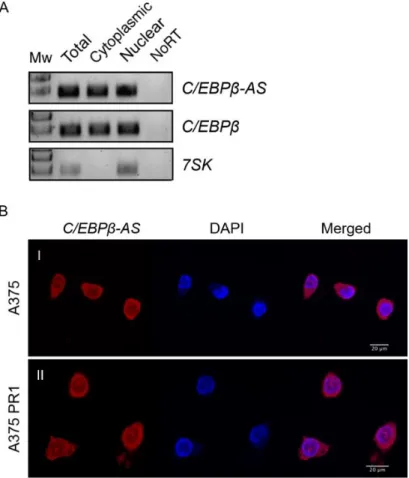
... 273.1. Characterization of the Long Non-Coding RNA C/EBPβ-AS ... 27
3.1.1. Assessment of the Subcellular Localization of C/EBPβ-AS ... 27
3.1.2. Assessment of the Stability of C/EBPβ-AS and C/EBPβ Transcripts ... 28
3.1.3. Assessment of the Polyadenylation Status of C/EBPβ-AS Transcript ... 29
3.1.4. Assessment of the Extent of C/EBPβ-AS and C/EBPβ 5’ End Overlap ... 29
3.2. Study of the Regulation of C/EBPβ-AS and C/EBPβ Expression ... 30
3.2.1. Evaluation of C/EBPβ-AS Role on C/EBPβ Expression ... 30
3.2.2. Evaluation of the Role of Epigenetic Modulators on C/EBPβ Expression ... 33
3.2.3. Evaluation of C/EBPβ-AS Role on Epigenetic Regulation of the C/EBPβ Promoter ... 34
3.2.4. Evaluation of C/EBPβ-AS Impact on C/EBPβ Recruitment to the C/EBPβ Promoter ... 36
3.2.5. Evaluation of C/EBPβ Role on C/EBPβ-AS Expression ... 37
3.3. Evaluation of C/EBPβ-AS Role on Vemurafenib Sensitivity ... 38
3.4. Evaluation of the Impact of C/EBPβ-AS Knockdown on MAPK/ERK1/2 and PI3K/AKT Pathways ... 39
4. Discussion
... 415. Conclusion
... 516. References
... 53XI
List of Figures
Figure 1.1. Incidence of Melanoma in more developed regions ... 1
Figure 1.2. The Clark Model of development and progression of melanoma ... 2
Figure 1.3. Diagram of the MAPK/ERK and PI3K/AKT pathways and common genetic alterations in cutaneous melanoma ... 3
Figure 1.4. Categorization of antisense transcripts according to genomic origin ... 10
Figure 1.5. Antisense lncRNA-mediated regulatory mechanisms found at every level of gene regulation ... 11
Figure 1.6. Representation of the genomic arrangement of C/EBPβ and C/EBPβ-AS locus ... 16
Figure 1.7. C/EBPβ and C/EBPβ-AS in melanoma ... 17
Figure 3.1. Subcellular localization of C/EBPβ-AS and C/EBPβ transcripts in cutaneous melanoma cell lines ... 27
Figure 3.2. Stability analysis of C/EBPβ-AS and C/EBPβ transcripts ... 28
Figure 3.3. Polyadenylation status of C/EBPβ-AS transcript ... 29
Figure 3.4. Characterization of C/EBPβ-AS and C/EBPβ overlap ... 29
Figure 3.5. C/EBPβ-AS knockdown impacts C/EBPβ mRNA levels ... 31
Figure 3.6. C/EBPβ-AS knockdown impacts RNA Pol II recruitment to the C/EBPβ promoter ... 32
Figure 3.7. C/EBPβ-AS knockdown impacts C/EBPβ protein levels ... 32
Figure 3.8. Impact of EZH2, G9a or DNMT3A knockdown on C/EBPβ mRNA levels ... 33
Figure 3.9. C/EBPβ-AS knockdown impacts enrichment of EZH2 and H3K27me3 at the C/EBPβ promoter ... 35
Figure 3.10. C/EBPβ-AS knockdown impacts methylation of the C/EBPβ promoter ... 35
Figure 3.11. C/EBPβ-AS knockdown impacts enrichment of C/EBPβ at the C/EBPβ promoter ... 36
Figure 3.12. C/EBPβ knockdown impacts C/EBPβ-AS RNA levels ... 37
Figure 3.13. C/EBPβ-AS knockdown impacts enrichment of C/EBPβ at the C/EBPβ-AS promoter ... 38
Figure 3.14. C/EBPβ-AS knockdown impacts cutaneous melanoma cell sensitivity to vemurafenib .. 39
Figure 3.15. C/EBPβ-AS knockdown impacts MAPK/ERK and PI3K/AKT signalling pathways ... 40
XIII
List of Tables
Table 2.1. siRNAs used in cationic lipid-mediated transfection of cells ... 20
Table 2.2. Primary antibodies used for protein immunoblotting ... 22
Table 2.3. Antibodies used for ChIP ... 24
Supplementary Table S.1. siRNAs used in cationic lipid-mediated transfection of cells ... i
Supplementary Table S.2. Primer sets used in RT-qPCR or semi-quantitative PCR reactions ... ii
XV
List of Abreviations
AIRN Antisense Of IGF2R Non-Protein Coding RNA
AJCC American Joint Committee on Cancer
AKT Protein Kinase B
ANRIL CDKN2B Antisense RNA 1
ARAF A Rapidly Accelerated Fibrosarcoma
asRNAs antisense RNAs
ATCC American Type Culture Collection
BANCR BRAF-Regulated lncRNA 1
BRAF B-Rapidly Accelerated Fibrosarcoma
BRAFV600E BRAF muteted form (substitution of valine to glutamic acid at amino acid 600)
bZIP domain basic leucine zipper domain
C/EBPβ CCAAT/Enhancer-Binding Protein β
C/EBPβ-AS CCAAT/Enhancer-Binding Protein β Antisense CCAT1-L Colon Cancer Associated Transcript 1
CDKN2A Cyclin-Dependent Kinase Inhibitor 2A
cDNA complementary DNA
ChIP chromatin immunoprecipitation
ChIP-seq chromatin immunoprecipitation-sequencing
CMM Cutaneous Malignant Melanoma
CRAF C Rapidly Accelerated Fibrosarcoma
CRE cAMP Response Element
CTLA-4 Cytotoxic T Lymphocyte Antigen-4
DMSO dimethyl sulfoxide
DNA deoxyribonucleic acid
DNMT3A DNA Methyltransferase 3A
dNTPS nucleotides for DNA synthesis
dsRNA double-stranded RNA
DTT dithiothreitol
ENCODE ENCyclopedia Of DNA Elements
ERK Extracellular Signal-Regulated Kinase FACS Fluorescence Activated Cell Sorting GAS5 Growth Arrest-Specific Transcript 5
GDP guanosine diphosphate
Grb2 Growth Factor Receptor-Bound Protein 2
GRCh37/hg17 Genome Reference Consortium Human Build 37 GRCh38/hg38 Genome Reference Consortium Human Build 38
GTP guanosine triphosphate
XVI
H3K27me3 Histone H3 Lysine 27 Trimethylation
H3K9 Histone H3 Lysine 9
HOTAIR HOX Antisense Intergenic RNA
HOX Homeobox
HOXC Homeobox Protein C
HOXD Homeobox Protein D
IFNα Interferon Alpha
lincRNAs large intergenic ncRNAs
lncRNAs long ncRNAs
MALAT1 Metastasis Associated Lung Adenocarcinoma Transcript 1 MAP2K1 Mitogen-Activated Protein Kinase Kinase 1
MAP2K2 Mitogen-Activated Protein Kinase Kinase 2 MAPK Mitogen-Activated Protein Kinase
MEFs Mouse Embryo Fibroblasts
MEK MAPK/ERK Kinase
MITF Melanogenesis-Associated Transcription Factor MMP Matrix Mettaloproteinase 2 Protein
mTORC2 Mechanistic Target of Rapamycin Complex 2
NCI National Cancer Institute
ncRNAs non-coding RNAS
NHGRI National Human Genome Research Institute
NORAD Non-Coding RNA Activated By DNA Damage
NRAS Neuroblastoma RAS Viral Oncogene Homolog
Oct4 Octamer-Binding Transcription Factor 4
ORF open reading frame
PD-1 Programmed Cell Death-1
PDK1 Kinase-3′-Phosphoinositide-Dependent Kinase 1
PI3K Phosphatidylinositol 3-Kinase
PIP3 Phosphatidylinositol-3,4,5-Triphosphate
piRNAs Piwi-Interacting RNAs
PRC1 Polycomb Repressive Complex 1
PTEN Phosphatase and Tensin Homologue
PTENpg1 asRNA α PTEN Pseudogene-Encoded Antisense RNA α PTENpg1 asRNA β PTEN Pseudogene-Encoded Antisense RNA β PTENpg1 sense PTEN Pseudogene Sense
RT-qPCR real-time quantitative polymerase chain reaction
RAF Rapidly Accelerated Fibrosarcoma
RAS Rat Sarcoma Proteins
XVII
RNA ribonucleic acid
RNA Pol II RNA Polymerase II
RNAi RNA interference
RTK Receptor Tyrosine Kinase
SAMMSON Survival-Associated Mitochondrial Melanoma-Specific Oncogenic Non-Coding RNA
SEM standard error of the mean
siRNAs small interfering RNAs
smFISH single-molecule RNA Fluorescence In Situ Hybridization
sncRNAs short ncRNAs
snoRNAs small nucleolar RNAs
snRNAs small nuclear RNAs
Sos Son of Sevenless
STAU1 Staufen 1
TAE buffer Tris-Acetate-EDTA buffer TBS-T buffer Tris-buffered saline-Tween 20
TCGA The Cancer Genome Atlas
tiRNAs transcription initiation RNAs
Tsix Reverse of Xist
UCSC University of California Santa Cruz
UVR Ultra-Violet Radiation
1
1. Introduction
1.1. Cutaneous Malignant Melanoma
1.1.1. Epidemiologic Scenario of Cutaneous Malignant Melanoma: Incidence, Geographical Distribution and Mortality
According to GLOBOCAN estimates, cutaneous malignant melanoma (CMM) was accounted for over 1,5% of all new diagnosed cancer cases, worldwide, in 2012, with 191 thousand new cases in more developed regions, both sexes combined, in 2012 (Figure 1.1.), being the ninth most common cancer type in more developed regions (Ferlay et al., 2015).
Throughout recent decades the incidence of CMM has been increasing, with highest reported incidence areas worldwide being Northern Europe, Australia and North America (Erdmann et al., 2013). In these regions, Caucasians represent the sub-population that is more prone to develop such cancer type.
Although CMM comprehends less than 5% of all skin cancer cases, it stands as the most lethal skin neoplasm, because of its high mortality when identified at advanced stages, being responsible for about 80% of dermatological cancer related deaths (Miller and Mihm, 2006).
1.1.2. Cutaneous Malignant Melanoma: Development, Staging and Risk Factors
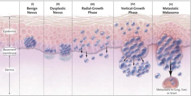
CMM is a type of skin cancer that arises from malignant transformation of melanin-producing cells of the skin found in the basal layer of the epidermis, designated melanocytes. According to the Clark model of development and progression of CMM (Figure 1.2.), the first step is the development of benign melanocytic nevi (commonly designated by moles), resulting from controlled melanocyte proliferation and its transformation into atypical/dysplasic nevi (pre-malignant nevi with aberrant proliferation). Next,
Figure 1.1. Incidence of Melanoma in more developed regions.
Estimated global numbers of new cancer cases (in thousands) with proportions for more developed regions, both sexes combined, in 2012. The area of the pie is proportional to the number of new cases. Melanoma of skin appears as the ninth most common cancer type, with 191 thousand cases (3.1%) out of 6076 thousand total new cancer
2
is the Radial Growth Phase, in which transformed cells acquire the ability to intraepidermally proliferate, followed by the Vertical Growth Phase. In this phase, transformed cells acquire the ability to invade the dermis (inner layer of the skin) through the basement membrane. Ultimately, the last step is the metastatic phase, in which malignant melanocytes successfully proliferate and spread to lymph nodes and other tissues (Clark et al., 1984).
CMM staging is determined by the American Joint Committee on Cancer (AJCC) system, that incorporates tumour thickness, ulceration (defined as the interruption of the surface epithelium by tumour cells), mitotic index, the lymph node status and distant metastases (Balch et al., 2009). This staging system categorizes melanoma patients into three main groups: localized disease with no evidence of metastases (stage I–II), regional disease (stage III) and distant metastatic disease (stage IV).
The etiology of CMM is multifactorial, with the most relevant risk factors being Ultra-Violet Radiation (UVR) exposure, genetic predisposition, light sensitivity – including low skin-phototype (fair skin), multiple benign or atypical nevi –, and immunosuppression of the host (Lo and Fisher, 2014). Intermittent UVR exposure as well as history of severe sunburns in childhood or adolescence have been implicated in epidemiologic studies as conferring the highest risk (Whiteman, et al., 2001).
Figure 1.2. The Clark Model of development and progression of melanoma.
The Clark model describes the histological changes that accompany the progression from normal melanocytes to
malignant melanoma. The model depicts the proliferation of melanocytes in the process of (I) forming benign nevi (resulting from controlled melanocyte proliferation in normal melanocytes), (II) the subsequent transformation into atypical/dysplasic nevi (pre-malignant nevi with aberrant proliferation), (III) the Radial Growth Phase, in which transformed cells acquire the ability to intraepidermally proliferate, followed by (IV) the Vertical Growth Phase, during which transformed cells acquire the ability to invade the dermis (inner layer of the skin) through the basement membrane and ultimately and (V) the metastatic phase, in which malignant melanocytes successfully proliferate and spread to lymph nodes and other tissues. Adapted from (Miller and Mihm, 2006).
3
UVR promotes malignant transformation of melanocytes by UVR-induced DNA damage, which is a known fundamental event in photocarcinogenesis, highly connected to CMM development (Sarasin, 1999), by having a systemic as well as local (cutaneous) immunosuppressive effect (Schwarz, 2005) and by promoting reactive oxygen species of melanin that cause DNA damage and suppress apoptosis (Meyskens, et al., 2004).
Prolonged UVR exposure is a known fundamental event in photocarcinogenesis and gives rise to characteristic UVR signature-mutations: mainly C-to-T substitutions (Cytosines to Thymines pyrimidine bases). Such signature-mutation is described to be extensively accountable for the high mutation rate in melanoma (Pleasance et al., 2010; Lawrence et al., 2013).
1.1.3. Dysregulation of Signalling Pathways in Cutaneous Malignant Melanoma
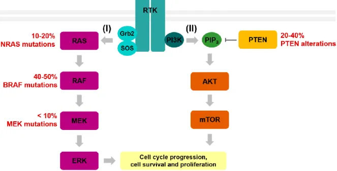
Melanoma development results from accumulated genetic alterations and activation of main signalling pathways in melanocytes (Thompson et al., 2005), including the mitogen-activated protein kinase (MAPK)/extracellular signal-regulated kinase (ERK) pathway and the phosphatidylinositol 3-kinase (PI3K)/ protein 3-kinase B (AKT) pathway, summarized below.
Figure 1.3. Diagram of the MAPK/ERK and PI3K/AKT pathways and common genetic alterations in cutaneous melanoma.
(I) Upon Receptor Tyrosine Kinase (RTK) activation, association of the adaptors Grb2 and SOS leads to RAS activation, which in turn results in subsequent activation of a downstream cascade core module. This core modules consists of three kinases: RAF (which exists in 3 isoforms: ARAF, BRAF and CRAF) that phosphorylates and activates MEK, which in turn activates ERK. This leads to transcription factor activation, which can ultimately lead to cell proliferation and survival. (II) RTK activation also leads to recruitment of PI3K to the cellular membrane, resulting in the production of PIP3, which in turn drives the recruitment of a subset of signalling proteins, including AKT. Activation of AKT and mTOR leads to activation of downstream targets, which can ultimately result in cell cycle progression and cell survival. The tumour suppressor PTEN counteracts the PI3K/AKT pathway through PIP3 dephosphorylation, thus inhibiting recruitment and activation of AKT, inactivating the pathway. The most common alterations in components of MAPK/ERK and PI3K/AKT pathways in cutaneous melanoma are shown in red. Adapted from (Solus and Kraft, 2013).
4
1.1.3.1. Dysregulation of the MAPK/ERK Pathway in Cutaneous Malignant Melanoma
The MAPK/ERK pathway is one of the primordial signalling systems present in all eukaryotes, controlling such fundamental cellular processes as cell proliferation, differentiation, senescence, survival and apoptosis (Kolch, 2000). The basic arrangement initiates with the interaction of an extracellular ligand with a receptor tyrosine kinase (RTK) and association of the adaptors growth factor receptor-bound protein 2 (Grb2) and son of sevenless (Sos), which activates rat sarcoma proteins (RAS) – members of the small GTPase family of proteins, whose activation depends on guanosine diphosphate (GDP) switch to guanosine triphosphate (GTP) –, that activate a downstream cascade core module. The core module consists of three kinases: rapidly accelerated fibrosarcoma (RAF) (which exists in 3 isoforms: ARAF, BRAF and CRAF), that phosphorylates and activates MAPK/ERK kinase (MEK), which in turn activates ERK (Kolch, 2000). Upon activation, ERK translocates to the nucleus, phosphorylating and activating downstream targets, such as transcription factors (Smalley, 2003). This signalling cascade can ultimately induce cell cycle progression (Katz et al., 2007) (Figure 1.3.).
The molecular pathogenesis of CMM is strongly correlated with a constitutive activation of the MAPK/ERK pathway, that appears early in tumorigenesis and is preserved through progression (Omholt et al., 2003). Such phenomenon is probably a consequence of mutations in upstream components of this pathway.
An evaluation of mutations in components of the MAPK/ERK pathway in a large panel of common cancers showed that 40 to 50% of melanomas, and 7 to 8% of all cancers, carry an activating mutation in the gene encoding the protein kinase BRAF (a protein that shows higher kinase activity compared to the other RAFs – ARAF and CRAF). Additionally, 90% of reported BRAF mutations result in a substitution of valine to glutamic acid at amino acid 600 (the V600E mutation, giving rise to the designated BRAFV600E protein). This mutation increases catalytic activity of BRAF, constitutively
activating it, leading to downstream propagation of the MAPK/ERK pathway signalling (Flaherty et al., 2010). Mutations in ARAF and CRAF are uncommon.
In CMM, Neuroblastoma RAS Viral Oncogene Homolog (NRAS) mutations stand as the most common in RAS-family members – with mutations in other RAS-family members, KRAS and HRAS, being relatively rare (Smalley, 2003). Additionally, melanomas with wild-type BRAF often display activating mutations in NRAS gene (Solus and Kraft, 2013). The prevalence of activating mutations in the gene coding for NRAS protein, predominantly at codon 61, is close to 30% in CMM (Omholt et al., 2002). Such mutation constitutively activates NRAS, leading to downstream signal transduction in the MAPK/ERK pathway.
Mutations in other components of the MAPK/ERK pathway were also identified, including MEK1 and MEK2 coding genes – Dual Specificity Mitogen-Activated Protein Kinase Kinase 1 (MAP2K1) and Dual Specificity Mitogen-Activated Protein Kinase Kinase 2 (MAP2K2), although found to be less frequent in CMM cases (Nikolaev et al., 2011). These mutations also result in constitutive downstream signalling.
In melanocytes presence of mutations in components of MAPK/ERK pathway, such as in BRAF or NRAS genes, appears to induce oncogene-induced senescence (an irreversible form of cell cycle arrest). Generally, to induce malignant transformation of melanocytes to melanoma, additional gene alterations are required, such as in Cyclin-Dependent Kinase Inhibitor 2A (CDKN2A) (leading to
5
inactivation of the CDKN2A-encoded protein p16INK4A), p53 and/or Phosphatase and Tensin Homologue (PTEN) gene, affecting the tumour suppressor activity of respectively encoded proteins (Ko et al., 2010).
1.1.3.2. Dysregulation of the PI3K/AKT Pathway in Cutaneous Malignant Melanoma
Similarly to the MAPK/ERK pathway, the PI3K/AKT pathway is initiated by activation of a receptor tyrosine kinase through its interaction with an extracellular ligand. Activation of receptor tyrosine kinases leads to recruitment of PI3K to the cellular membrane, which results in the production of the second messenger phosphatidylinositol-3,4,5-triphosphate (PIP3). In turn, PIP3 drives the recruitment of a
subset of signalling proteins, including kinase-3′-phosphoinositide-dependent kinase 1 (PDK1) and AKT. AKT can then be phosphorylated and activated by PDK1 and mechanistic target of rapamycin complex 2 (mTORC2) and thereafter activate downstream targets, ultimately regulating several cell processes involved in cell cycle progression and cell survival (Chang et al., 2003) (Figure 1.3.).
The PI3K/AKT pathway is counteracted by the tumour suppressor PTEN. PTEN regulates PI3K signalling by dephosphorylating the lipid signalling intermediate PIP3, thus inhibiting recruitment and
activation of AKT, inactivating the pathway (Simpson and Parsons, 2001). PTEN mutations, deletions or methylation of its promoter result in PTEN loss, ultimately leading to PI3K/AKT signalling activation and cell survival. The rate of such alterations in metastatic CMM is over 20% (Aguissa-Touré and Li, 2012) and is correlated with increased melanoma invasive capacity and decreased overall survival of CMM patients carrying the BRAFV600E mutation (Bucheit et al., 2014).
1.1.4. Therapeutic Options for Cutaneous Malignant Melanoma Patients
A brief overview of the main current therapeutic options for cutaneous melanoma patients is presented below, focusing on surgery, chemotherapy, targeted-therapy, immunotherapy and immunostimulants.
The gold standard treatment option for patients with local CMM consists of surgical excision of the primary tumour, with safety margins. Treatment with this method leads to a very good prognosis. Surgery may also be performed in advanced metastatic disease, for complete lymph node dissection – in cases where the sentinel lymph node (first lymph node to which transformed cells are most likely to spread from a primary tumour) is found positive for metastases. However, after this treatment patients will still have a poor prognosis (Schadendorf et al., 2015). In some more advanced stages surgery can also be performed, solely as a palliative measure (e.g. to remove obstruction in the bowel), but systemic drug treatment is often used (Schadendorf et al., 2015).
Chemotherapy has been, for several decades, used as palliative treatment of patients with metastatic melanoma, either as a systemic mono-therapy – commonly single agent dacarbazine, temozolomide and fotemustine – or combination therapy of chemotherapeutic agents (Thompson et al., 2005). However, such regimens result in low therapy response rates (5-12%), with a median overall survival inferior to one year (Garbe et al., 2011).
The molecular pathways identified as being central to melanoma development and progression are subject of intense investigation for their potential in “targeted-therapy”. This is approached by design of small molecules, aiming to inhibit specific molecules present in cells driving aberrant proliferation and
6
growth. High specificity is often pursued, since it may represent elevated efficiency, with fewer side effects than those reported for cytotoxic chemotherapies.
The first targeted-therapies to demonstrate substantial efficacy against advanced CMM with the mutated form of BRAF protein BRAFV600E were two adenosine triphosphate-competitive inhibitors of
BRAFV600E – vemurafenib and dabrafenib –, both approved by drug regulatory authorities (Lo et al.,
2014). These compounds bind to BRAFV600E monomers, inhibiting their activity. Studies comparing the
two BRAF inhibitors with the chemotherapy agent dacarbazine showed improved response rates and improved progression-free and overall survival, with the targeted-therapies (Chapman et al., 2011; Hauschild et al., 2012).
Two inhibitors (cobimetinib and trametinib) targeting the wild type MEK protein (downstream of BRAF/CRAF in the MAPK/ERK pathway) have also been developed and approved, showing an overall survival benefit compared to dacarbazine, in the treatment of metastatic BRAF-mutant CMM (Carlino et al., 2015).
Interestingly, BRAF inhibitors possess the remarkable and paradoxical feature of triggering MAPK/ERK pathway reactivation (Heidorn et al., 2010) – briefly discussed below. Considering this paradoxical event, the two MEK inhibitors have also been approved for use in patients with mutated BRAF, in combination with vemurafenib or dabrafenib (Lo et al., 2014). Although BRAF inhibitors, as well as MEK inhibitors, can be used alone or in combination as described, studies point out that combination therapy is more advantageous than targeted-monotherapy in terms of toxicity and efficacy (Long et al., 2014a; Larkin et al., 2015).
Attempts to target the major defences that melanoma cells display against an effective immune response have been pursued, counteracting the development of host tolerance to melanoma antigens, mainly due to the production of immunosuppressive factors by melanoma cells. Given that, immunotherapy options continue to be investigated intensively, regarding both adjuvant and advanced CMM settings.
In short, parallel to antigen presentation, activation of T cells (one of the main classes of players in the anti-cancer immune response) requires a costimulatory interaction between T cells and antigen-presenting cells, which can be mediated by either stimulatory or inhibitory receptor-ligand pairs known as “immune-checkpoints” (Sharpe, 2009). Two of the best studied checkpoints involve cytotoxic T lymphocyte antigen-4 (CTLA-4), programmed cell death-1 (PD-1) and its ligand (PDL-1). The most successful immunotherapy approaches to date have been immune checkpoint inhibition, through the administration of antibodies blocking CTLA-4 in advanced CMM – with improved overall survival (Hodi et al., 2010) –, as well as antibodies blocking PD-1 and its interaction with PDL-1 – with increased response rate and overall survival in wild type BRAF CMM patients, compared to the chemotherapy agent dacarbazine (Robert et al., 2014; Weber et al., 2015).
Approaches designed to modulate the immune system to induce an anti-cancer response in CMM patients also include non-specific immunostimulants such as interleukin 2 (IL-2) and interferon alpha (IFNα). Compilation of studies comparing combination therapy of chemotherapy agents, IL-2 and low dose IFNα with the use of chemotherapy agents in monotherapy regimens shows that, despite
7
moderately improved response rates, increased toxicity and absence of increased overall survival are evident with combination therapy (Bhatia et al., 2009).
1.1.5. Therapy Resistance in Cutaneous Malignant Melanoma
Resistance to therapies consists a major problem for CMM treatment. Chemotherapy with dacarbazine and temozolomide has been unsuccessful mainly due to unclear innate and/or acquired resistance of melanoma cells to treatment. Suggested mechanisms that contribute to chemoresistant phenotypes include changes in drug transport and metabolism. This may occur through elevated expression and activity of cell membrane efflux pumps, enzymatic detoxification – e.g. with the involvement of enzyme glutathione-S-transferase, by conjugation of certain chemotherapeutic agents to glutathione –, or through disruption of drug-target interactions possibly due to alterations in the targets which results in reduced binding affinity (Helmbach et al., 2003). Anti-apoptotic pathways and enhanced DNA repair in cancer cells also play roles in unresponsiveness to chemotherapy in CMM (Grossman and Altieri, 2001).
Despite the encouraging response rate and improvement in progression-free survival in ~80% of patients carrying the BRAFV600E mutation treated with BRAF inhibitors vemurafenib and dabrafenib, the
majority of such patients exhibits disease progression following tumour regression, within 6-8 months (Chapman et al., 2011; Hauschild et al., 2012). Similarly, one-third of BRAF-mutant metastatic melanoma patients treated with combined BRAF and MEK inhibitors shows disease progression within 6 months (Long et al., 2014b). Treatment options for these patients remain limited, motivating the elucidation of underlying intrinsic and acquired resistance mechanisms.
Tumours with mutant BRAF are dependent on the MAPK/ERK signalling pathway for their growth. However, even though BRAF inhibitors prevent ERK signalling in cells with mutant BRAF, an unexpected enhancement effect of ERK signalling has been shown (Poulikakos et al., 2010). Given that, among the resistance mechanisms under investigation, reactivation of MAPK/ERK pathway in resistant tumours stands as one of the major phenomenon taking place. Gatekeeper mutations in BRAF, which would prevent vemurafenib from binding to BRAF, have not been observed as a causal event of this phenomenon (Nazarian et al., 2010). Instead, currently available data suggests other events namely, genetic alterations such as gene amplification (e.g. amplification of BRAF) (Shi et al., 2012), CRAF overexpression (leading to BRAF signalling bypass) (Poulikakos et al., 2011) mainly in the co-existence of RAS mutations (Dumaz et al., 2006) , secondary mutations in NRAS (Nazarian et al., 2010) or novel mutations in MEK (Wagle et al., 2011), as well as emergence of novel abnormal hyperactive forms of BRAF that dimerize in a RAS-independent manner (e.g. truncated BRAF) (Poulikakos et al., 2011).
Aside from reactivation of MAPK/ERK pathway, other targeted-therapy resistance mechanisms have been proposed, such as the activation of alternative survival pathways (e.g. PI3K/AKT pathway activation), induced by increased expression of receptor tyrosine kinases (Villanueva et al., 2010; Nazarian et al., 2010) and PTEN alterations – given the lost PTEN counteractive role of PI3K/AKT pathway (Paraiso et al., 2011).
Tumour microenvironment-derived acquired resistance appears as another resistance mechanism (Straussman et al., 2012).
8
1.2. Non-Coding RNAs
1.2.1. A Class of Functional RNAs: Non-Coding RNAs
RNA molecules were originally considered to mainly function as intermediates between genes and respectively encoded proteins. However, since the discovery of ribosomal RNA (Palade, 1955) and transfer RNA (Hoagland et al., 1958) in the 1950s, a functional role related to the regulation of genome organization and gene expression has been attributed to RNA molecules that do not encode proteins – designated non-coding RNAS (ncRNAs). Nevertheless, the diversity, biological relevance and myriad of functional roles currently attributed to ncRNAs were only started to be uncovered in recent years. It was only after the publication of genome-wide sequencing data, that it came to the scientific community’s awareness that organism complexity and number of protein-coding genes are not necessarily directly proportional. This became evident after the release of the Human genome sequencing data (Venter et al., 2001; Lander et al., 2001), facing that the number of protein-coding genes in the Human genome – approximately 20.000 – is very close to that found in less complex organisms (Goodstadt et al., 2006). Given that, the observed developmental complexity of an organism and the relative amount of non-protein coding DNA (performing its functional role mainly through transcription into ncRNAs) were then proposed to be correlated (Taft et al., 2007).
Improvements in RNA sequencing technology enabled the application of high-throughput methods in the generation and analysis of data by international research collaborations, such as ENCyclopedia Of DNA Elements (ENCODE). This lead to findings such as that the majority of bases in the Human genome is associated with at least one primary transcript, assessing that circa three-quarters of the genome is transcribed into ncRNAs (ENCODE Project Consortium, 2007).
ncRNAs are originally described as transcripts lacking an evident protein-coding function, i.e. lacking a long open reading frame (ORF) (traditionally >100 codons) and/or not displaying codon conservation (Morris and Mattick, 2014). While recent studies provide evidence that most ncRNAs do not encode proteins, a few functional peptides have been shown to arise from translation of transcripts identified as ncRNAs (Banfai et al., 2012).
Non-coding RNAs regard to two major classes: short ncRNAs (sncRNAs) and long ncRNAs (lncRNAs), broadly and almost arbitrarily defined according to the transcript’s length, having <200 and >200 nucleotides long, respectively (Morris and Mattick, 2014). This feature is not regarded to as being entirely arbitrary, since it serves as a threshold, allowing empirical separation of RNAs in common experimental methodologies. The short ncRNAs class comprises the relatively well studied subclass of microRNAs (miRNAs), as well as small nucleolar RNAs (snoRNAs), small nuclear RNAs (snRNAs), piwi-interacting RNAs (piRNAs), small interfering RNAs (siRNAs) and transcription initiation RNAs (tiRNAs). The lncRNAs class comprehends two broad subclasses: large intergenic ncRNAs (lincRNAs) and ncRNAs that overlap with other transcripts, in either a sense or antisense orientation (in focus in this thesis) and transcribed pseudogenes.
1.2.2. Long Non-Coding RNAs
The advent of growing sensitivity of RNA sequencing methods, as well as computational prediction techniques (Clark et al., 2015) is enabling the increasing identification of such a diverse class of RNA transcripts as lncRNAs, including the detection of transcripts arising from lowly expressed genes.
9
This has stimulated interest and focus in further characterizing and understanding biology roles played by lncRNAs.
1.2.2.1. Subclasses of Long Non-Coding RNAs
lncRNAs can be divided into subclasses, mainly regarding its genomic origin and orientation, often with respect to that of protein-coding transcripts, with the commonly established subclasses being: large intergenic ncRNAs (lincRNAs) – whose genomic origin does not overlap with that of any known coding or non-coding transcripts –, and lncRNAs that overlap with other transcripts, including antisense (asRNAs), sense overlapping and sense intronic lncRNAs. It should be noted that, according to current knowledge, there is no evidence indicating that this classification respects to functional role differences (Morris and Mattick, 2014).
1.2.2.2. Features of Long Non-Coding RNAs
Many lncRNAs are usually transcribed by RNA Polymerase II (RNA Pol II), are often 5’-capped, 3’-polyadenylated and spliced. These features recapitulate know mRNA characteristics, however, some unique general trends of lncRNAs (comparing to mRNAs) can be listed, such as the absence of a translated ORF, the tendency of being shorter in length – usually with fewer but longer exons – and the lower overall expression levels (Derrien et al., 2012; Washietl et al., 2014).
While the level of expression of many lncRNAs appears to be lower than mRNAs in whole tissues, lncRNAs are highly expressed and easily detectable in particular cells, with Human and Mouse genome studies showing that lncRNAs have a higher specificity, regarding tissue, cell type and cellular compartment expression, comparing to expression of protein-coding transcripts (Ravasi et al., 2006; Mercer et al., 2008; Djebali et al., 2012).
It is clear that lncRNAs display a wide range of evolutionary conservation – from those categorized as ultraconserved (Calin et al., 2007) to those that are primate-specific (Tay et al., 2009). Although, the majority of lncRNAs exhibit relatively low evolutionary conservation (Johnsson et al., 2014). However, studies provide evidence that a lack of conservation does not imply a lack of function (Pang et al., 2006).
1.2.2.3. Mechanisms of Regulation Mediated by Antisense Non-Coding RNAs: an Overview
Multiple studies have shown evidence that more than 63% of transcripts have antisense partners, many of which do not encode proteins (Katayama et al., 2005; Carninci et al., 2005; Li and Ramchandran, 2010; Nishizawa et al., 2012). The magnitude of identified arising antisense transcripts has stimulated attention to such class of RNA molecules, namely to non-protein coding antisense RNAs. asRNAs are transcribed from the opposite DNA strand of that of a sense transcript (which can either be a protein-coding or non-protein-coding RNA) (Katayama et al., 2005).
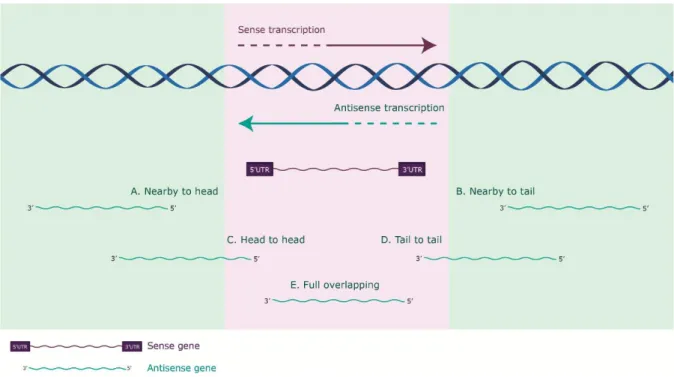
Antisense RNAs can exert their function in a cis- or in a trans-acting manner, whether the interacting sense transcript is transcribed from the same genomic region, or from a distant locus, respectively. Despite low global expression levels of asRNA, a plausible biological role played by cis-acting transcripts can be hypothesized: given that there are two copies of DNA for a given gene in a cell, two antisense lncRNA molecules are theoretically sufficient to interact with the two gene copies and elicit a regulatory effect (Faghihi and Wahlestedt, 2009). Cis-acting antisense transcripts can be further categorized according to their genomic origin, regarding proximity between sense and antisense partners in the
10
genome (Figure 1.4.): nearby to head, when the 5’ end of the sense gene is in proximity to the 5’ end of the antisense gene (commonly with bidirectional promoters); nearby to tail, when the 3’ end of the sense gene is in proximity to the 3’ end of the antisense gene; head-to-head, when the 5’ ends of both sense and antisense genes overlap (divergent); tail-to-tail, when the 3’ ends of both sense and antisense genes overlap (convergent); and fully overlapping, when the sense gene completely overlaps with the antisense one (Villegas and Zaphiropoulos, 2015). Partially or fully overlapping asRNAs share complementarity to the sense expressed transcript and are found to overlap promoters, exons, 5’- and 3’-UTRs, as well as introns.
Basal expression levels of sense transcripts and respective antisense non-coding transcripts may be positively or negatively correlated in different tissues and cell lines (Katayama et al., 2005). Moreover, antisense lncRNAs are functionally very diverse, as they can act as positive or negative modulators of expression of their counterpart sense transcripts (Numata and Kiyosawa, 2012).
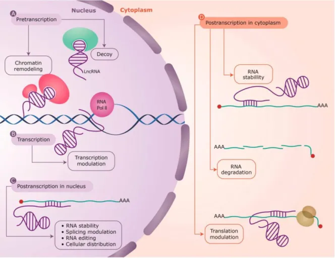
A myriad of mechanisms has been proposed for asRNA-mediated regulatory mechanisms, acting at nearly every level of gene regulation. As such, asRNA-mediated regulatory mechanisms can be divided in three main categories, briefly approached below: pretranscriptional asRNA-mediated regulation, transcriptional asRNA-mediated regulation and posttranscriptional asRNA-mediated regulation (Villegas and Zaphiropoulos, 2015) (Figure 1.5.).
Figure 1.4. Categorization of antisense transcripts according to genomic origin.
Categorization of antisense transcripts according to their genomic origin, regarding proximity between sense (depicted in purple) and antisense non-coding (depicted in green) genes in the genome. (A) Nearby to head, the 5’ end of the sense gene is in proximity to the 5’ end of the antisense gene; (B) Nearby to tail, the 3’ end of the sense gene is in proximity to the 3’ end of the antisense gene; (C) Head-to-head, the 5’ ends of both sense and antisense genes overlap (divergent); (D) Tail-to-tail, the 3’ ends of both sense and antisense genes overlap (convergent); (E) Full overlapping where the sense gene completely overlaps with the antisense gene. Adapted from (Villegas and Zaphiropoulos, 2015).
11
Pretranscriptionally, antisense lncRNAs can act as guides or scaffolds of proteins into specific parts of the genome, or as decoys keeping proteins away from chromatin. Such regulatory mechanisms rest upon the formation of RNA:protein complexes, as lncRNAs – namely antisense lncRNAs – comprise distinct protein-binding domains (Mercer and Mattick, 2013). Specific RNA:DNA interactions can efficiently and selectively recruit proteins to genomic loci. As RNA-interacting proteins are often found to be key regulators of gene transcription – namely epigenetic factors (Mercer and Mattick, 2013) – antisense lnRNAs in the nucleus can act as regulators of their counterpart expression by modulating chromatin structure and bridging epigenetic effectors and regulatory complexes at specific loci (Magistri et al., 2012). Proteins found among such key epigenetic factors are the following: DNA methyltransferases, such as DNA methyltransferase 3A (DNMT3A); members of the Polycomb Repressive Complex PRC2, such as the histone methyltransferase enhancer of zeste homolog 2
Figure 1.5. Antisense lncRNA-mediated regulatory mechanisms found at every level of gene regulation. Representation of antisense lncRNA-mediated regulatory mechanisms. (A) Pretranscriptionally, antisense lncRNAs can act as protein guides, scaffolds or decoys (as depicted), recruiting proteins into specific parts of the genome or holding proteins away from chromatin; (B) Antisense lncRNAs may also be involved in the modulation of an ongoing-transcriptional process, affecting gene expression; (C, D) Posttrancriptionally, antisense lncRNA can affect sense RNA structure (interfering with RNA stability, splicing or RNA editing), or cellular compartmental distribution, either in the nucleus or in the cytoplasm. LncRNAs are depicted in purple, and the interacting protein factors in green and red. Sense RNAs are shown as green lines and the base pair interactions highlighted by short purple lines. RNA polymerase II (RNA pol II) is depicted in pink, genomic DNA is depicted as a blue helix and a translating ribosome on the mRNA is depicted in yellow. Adapted from (Villegas and Zaphiropoulos, 2015).
12
(EZH2), which elicits histone H3 lysine 27 trimethylation (H3K27me3); or G9a/GLP methyltransferases, targeting histone H3 lysine 9 (H3K9).
The X-inactivation centre illustrates how an intricate network of lncRNAs regulates gene expression pretranscriptionally, being pivotal in such a fundamental process as the inactivation of one X-chromosome in an early development stage. Such process involves asymmetric expression of the lncRNAs X-inactive specifc transcript (Xist) and reverse of Xist (Tsix) in a pair of X-chromosomes. At
the onset of X-inactivation, Xist accumulates on one of two Xs, working as a functional lncRNA molecule
that recruits the PRC2 towards one of the female X-chromosomes, in cis, establishing and spreading the H3K27me3 repressive chromatin mark, leading to heterochromatinization and inactivation of the chromosome. In the other X-chromosome, the antisense lncRNA Tsix is transcribed, negatively regulates Xist by recruiting the DNA methyltransferase DNMT3A in cis. This induces DNA methylation at the Xist promoter and protects heterochromatinization of the X-chromosome by Xist, allowing such chromosome to remain active (Sun et al., 2006).
Even though the genomic arrangement of sense:asRNA transcription suggests a more plausible regulatory role in a cis-acting manner, trans-regulatory mechanisms have also been identified and described for antisense lncRNAs. The functional mechanism of the lncRNA HOX Antisense Intergenic RNA (HOTAIR) exemplifies a trans-acting asRNA-mediated pretranscriptional regulatory role played by an antisense ncRNA. HOTAIR is transcribed from the Homeobox Protein C (HOXC) locus, one of the identified human Homeobox (HOX) loci, crucial in the morphogenesis process of development. HOTAIR represses transcription in trans across 40 kilobases of the Homeobox Protein D (HOXD) locus, by interacting with PRC2 and establishing the H3K27me3 chromatin mark at HOXD locus. Thus, this antisense lncRNA demarcates chromosomal domains of gene silencing at a distance (Rinn et al., 2007). Antisense lncRNAs may also be involved in the modulation of an ongoing-transcriptional process, affecting gene expression. According to this mechanism, the act of transcription in the antisense direction, but not the antisense RNA molecule per se, modulates transcription of the sense gene. Transcription in the antisense direction is suggested to exert alterations in gene expression in the event of transcriptional collision (Faghihi and Wahlestedt, 2009). An in silico study showed that, in humans and in mice, the expression levels of cis-acting asRNAs decreases as the length of the overlapping region increases (Osato et al., 2007). Such observation may suggest a putative clash of RNA polymerases, to the point that the probability of collision increases as the length of the overlapping region increases.
Posttranscriptionally, functions of antisense lncRNAs can be exerted through RNA:RNA interactions, affecting sense RNA structure or cellular compartmentalization, either in the nucleus or the cytoplasm. Given the sequence complementarity found among antisense and sense partner transcripts, RNA duplexes tend to be formed. This can have different posttranscriptional outcomes, all of which modulate sense RNA expression: interfering with splicing, RNA editing, stability, subcellular distribution, transport, nuclear retention or even modulating translation of the sense RNA transcripts (Faghihi and Wahlestedt, 2009).
An example of antisense lncRNA-mediated posttrancriptional modulation of sense RNA, namely through regulation of RNA stability, is the PTEN pseudogene-encoded antisense RNA β (PTENpg1
13
asRNA β). This antisense lncRNA is transcribed from the reverse strand of the locus from which the lncRNA PTEN pseudogene sense (PTENpg1 sense) is transcribed. These transcripts interact through an RNA:RNA pairing interaction, maintaining stable levels of PTENpg1 sense in the cytoplasm. Ultimately, such interaction affects the stability of Phosphatase and Tensin Homolog (PTEN) mRNA through microRNA (miRNA) sponge activity, by sequestering PTEN-targeting miRNAs away from PTEN protein-coding transcript. This results in an increased amount of PTEN protein (Johnsson et al., 2013).
1.2.3. Biological Settings of Long Non-Coding RNAs
As the extensive diversity found at the level of cell phenotypes lays upon intricate and complex networks of regulation of gene expression, most of the mammalian genome and indeed that of all eukaryotes is expressed in a cell- and tissue-specific manner. While non-protein-coding sequences increasingly dominate the genomes of multicellular organisms as their complexity increases, the number of protein-coding genes remains relatively static. (Amaral and Mattick, 2008). Given that, there is growing evidence that the observed transcription of non-coding sequences, namely of lncRNAs, is involved in the regulation of fundamental cell biological processes associated with differentiation and development. Such processes include maintenance of telomeric structure (Silanes et al., 2010), alternative splicing (Tripathi et al., 2010), retinal, erythroid and breast development (Young et al., 2005; Hu et al., 2011; Askarian-Amiri et al., 2011), epidermal differentiation (Kretz et al., 2013), among many others.
Besides the described involvement of lncRNAs in a multitude of physiological mechanisms, these RNA molecules are also found in association with dysregulation of cellular events tied to pathological conditions, namely to disorders that stand amongst leading causes of death in western societies, including cancer (further discussed below), cardiovascular diseases, such as coronary artery disease, and diabetes (Broadbent et al., 2008).
It is increasingly evident that many of the genomic mutations in cancer reside inside regions that do not encode proteins, namely regions that are transcribed into, or directly regulate the expression of, ncRNAs. The recent application of next-generation sequencing to a growing number of cancer transcriptomes has revealed many lncRNAs whose aberrant expression is associated with specific cancer types (Yan et al., 2015). Among the few that have been functionally characterized, several have been linked to malignant transformation (Huarte, 2015). Given that, participation of lncRNAs in the regulation of cellular pathways whose disruption is associated with cancer ensures a link between lncRNAs and tumorigenesis. Such pathways relate to chromosome maintenance, transcriptional regulation, mRNA and protein control, apoptosis and senescence (Khorkova et al., 2015).
An example of a lncRNA with a vastly characterized oncogenic function is the well-studied Metastasis Associated Lung Adenocarcinoma Transcript 1 (MALAT1). This lncRNA has been found to promote cell proliferation and metastasis, and its overexpression has been linked to lung adenocarcinoma, breast, pancreatic, colon, prostate and hepatocellular carcinomas. A single-nucleotide polymorphism in MALAT1 gene has been associated with hepatocellular carcinoma (Huarte, 2015).
14
1.2.3.1. Long Non-Coding RNAs in Melanoma
Despite the limited amount of studies focused on the characterization of functional roles of lncRNAs, there is increasing evidence for the involvement of lncRNAs in cancer progression and a role for several lncRNAs in development and progression of melanoma has been suggested.
Examples of melanoma-related lncRNAs associated with cellular proliferation, migration and apoptosis are presented below, focusing on the lncRNAs BANCR, GAS5 and SAMMSON.
Activating mutations in the BRAF gene, namely mutations that give rise to expression of active mutant BRAFV600E protein, have previously been implicated in development of CMM – as discussed in
subsection 1.1.3.1. Transcriptome analysis of BRAFV600E-mutant human melanomas revealed that induced expression of BRAFV600E regulates expression of ~100 lncRNAs. One of the transcripts most
highly induced by oncogenic BRAF, which is recurrently overexpressed in melanoma, is BRAF-regulated lncRNA 1 (BANCR). This lncRNA is known to regulate a set of genes involved in cell migration and is required for full migratory capacity of melanoma cells (Flockhart et al., 2012). Additionally, BANCR was demonstrated to activate ERK1/2 and CRAF in vitro and in vivo, thus being implicated in regulation of MAPK/ERK pathway, which led to proliferation of melanoma cells. Its expression was also directly correlated with tumour stage (Li et al., 2014). Altogether, a novel lncRNA-mediated regulatory mechanism of melanoma proliferation has been proposed.
The Growth Arrest-Specific Transcript 5 (GAS5) is a lncRNA whose expression has been shown to be downregulated in melanoma cell lines. It has been demonstrated that suppression of GAS5 leads to higher ability of melanoma cells to migrate. Additionally, induced overexpression in such cells reduced migratory ability, partially as a consequence of decreased expression and activity of matrix mettaloproteinase 2 protein (MMP) – a protein required for breakdown of extracellular matrix and cell migration (Chen et al., 2016). Ultimately, GAS5 may function as a tumour suppressor associated with melanoma.
Survival-Associated Mitochondrial Melanoma-Specific Oncogenic Non-Coding RNA (SAMMSON) is located downstream of the melanoma-specific oncogene Melanogenesis-Associated Transcription Factor (MITF) and is co-amplified in around 10% of all melanoma cases. Mechanistically, the lncRNA SAMMSON acts in trans as a decoy, by targeting p32 – a master regulator of mitochondrial homeostasis and metabolism associated with cell apoptosis. This increases mitochondrial targeting and pro-oncogenic function of p32. While expression of SAMMSON was detectable in over 90% of human primary melanoma and metastasis, no expression is observed in normal healthy tissue. Knockdown experiments established a role for this lncRNA in melanoma cell viability and growth, irrespective of mutational status of melanoma cells. Additionally, suppression of SAMMSON transcript led to enhanced cell sensitivity towards MAPK-targeting therapeutics (Leucci et al., 2016; Richtig et al., 2017).
1.2.4. Therapeutic Aspects of Long Non-Coding RNAs
The study of ncRNAs, namely lncRNAs, has become increasingly attractive as a starting point for identification of new therapeutic targets and development of new pharmacological compounds, as further efforts are concentrated in the characterization of such RNA molecules. This rests upon observed biological features of lncRNAs, such as the identified functional roles related to the regulation of
15
expression of a myriad of protein-coding and other non-protein coding genes, in a multitude of biological settings, including many pathological contexts.
Regarding the effects of lncRNAs in gene expression regulation, namely in gene repression, one of the main advantages of lncRNAs as therapeutic targets is that these molecules may allow the indirect manipulation of proteins previously considerable as “undruggable”. Such proteins are commonly required to be specifically manipulated and/or activated/upregulated in order to achieve beneficial outcomes in disease management/treatment. Resorting to novel strategies to specifically suppress expression of lncRNAs – easily achieved through oligonucleotide-based drugs –, consequent activation/upregulation of desired genes may be attained. Additionally, blockage of interacting motifs in the structure of lncRNA molecules, as protein-interacting sites, may also be employed to achieve an identical outcome. Moreover, the elevated target-specificity of lncRNAs makes these molecules highly suitable for modulating the expression of one gene or a small group of related genes (Khorkova et al., 2015).
Furthermore, the detection of lncRNAs in biological fluids and their vast involvement in pathological settings makes such molecules as ideal candidates for the development of diagnostic assays, namely as prognosis and/or predictive biomarkers (Khorkova et al., 2015). Additionally, expression profiling of human tumours based on the expression of ncRNAs, namely lncRNAs, has identified signatures associated with diagnosis, staging, progression, prognosis, and response to treatment (Kim and Reitmair, 2013). As an example, the ncRNA HOTAIR (whose functional molecular mechanism was briefly described in subsection 1.2.2.3 of this thesis) was shown to be involved in cancer metastasis, with an up to 2000-fold increased transcription being observed in breast cancer cells over normal breast tissue. As such, the detection of this lncRNA may present a potential application as a marker of prognosis in patients with primary breast cancer (Gupta et al., 2010; Kim and Reitmair, 2013).
1.3. C/EBPβ and C/EBPβ-AS 1.3.1. C/EBPβ
The CCAAT/enhancer-binding proteins (C/EBPs) are a family of exclusively eukaryotic transcription factors, that bind as dimers to sequence-specific, double-stranded DNA. Such proteins contain a highly conserved C-terminal basic leucine zipper domain (bZIP domain) – involved in dimerization and sequence-specific DNA binding – and a less conserved N-terminal domain – comprising three short motifs, referred to as activation domains, which interact with transcriptional coactivators and components of the basal transcription machinery (Ramji and Foka, 2002). Dimerization is a prerequisite for DNA binding of C/EBPs and because the bZIP domain is conserved, all the C/EBPs are capable of forming intrafamilial homodimers or heterodimers with each other (Vinson et al., 1989). Additionally, heterodimerization among C/EBP family members can result in a myriad of regulatory effects on gene expression (Zahnow, 2009). All C/EBP dimers bind to the same DNA consensus sequence: RTTGCGYAAY, where A, T, G and C stand for the nucleotides adenine, thymine, guanine and cytosine, respectively, R stands for nucleotides adenine or guanine, and Y for nucleotides cytosine or thymine (Osada et al., 1996).
CCAAT/enhancer-binding protein β (C/EBPβ) is a member of the C/EBP family of proteins and is encoded by an intronless gene – C/EBPβ – located in human chromosome 20 at position q13.13,