Decreases Endoplasmic Reticulum Stress
Maya Styner1*, Mark B. Meyer2, Kornelia Galior1, Natasha Case1, Zhihui Xie1, Buer Sen1, William R. Thompson1, John Wesley Pike2, Janet Rubin1
1Department of Medicine, University of North Carolina, Chapel Hill, North Carolina, United States of America,2Department of Biochemistry, University of Wisconsin-Madison, Wisconsin-Madison, Wisconsin, United States of America
Abstract
Exercise prevents marrow mesenchymal stem cell (MSC) adipogenesis, reversing trends that accompany aging and osteoporosis. Mechanical input, the in-vitro analogue to exercise, limits PPARcexpression and adipogenesis in MSC. We considered whether C/EBPbmight be mechanoresponsive as it is upstream to PPARc, and also is known to upregulate endoplasmic reticulum (ER) stress. MSC (C3H10T1/2 pluripotent cells as well as mouse marrow-derived MSC) were cultured in adipogenic media and a daily mechanical strain regimen was applied. We demonstrate herein that mechanical strain represses C/EBPbmRNA (0.6-fold60.07, p,0.05) and protein (0.4-fold60.1, p,0.01) in MSC. SiRNA silencing ofb-catenin prevented mechanical repression of C/EBPb. C/EBPb overexpression did not override strain’s inhibition of adipogenesis, which suggests that mechanical control of C/EBPbis not the primary site at which adipogenesis is regulated. Mechanical inhibition of C/EBPb, however, might be critical for further processes that regulate MSC health. Indeed, overexpression of C/ EBPbin MSC induced ER stress evidenced by a dose-dependent increase in the pro-apoptotic CHOP (protein 4-fold60.5, p,0.05) and a threshold reduction in the chaperone BiP (protein 0.6-fold60.1, p = 0.2; mRNA 0.3-fold60.1, p,0.01). ChIP-seq demonstrated a significant association between C/EBPband bothCHOPandBiPgenes. The strain regimen, in addition
to decreasing C/EBPbmRNA (0.5-fold60.09, p,0.05), expanded ER capacity as measured by an increase in BiP mRNA (2-fold 60.2, p,0.05) and protein. Finally, ER stress induced by tunicamycin was ameliorated by mechanical strain as demonstrated by decreased C/EBPb, increased BiP and decreased CHOP protein expression. Thus, C/EBPbis a mechanically responsive transcription factor and its repression should counter increases in marrow fat as well as improve skeletal resistance to ER stress.
Citation:Styner M, Meyer MB, Galior K, Case N, Xie Z, et al. (2012) Mechanical Strain Downregulates C/EBPbin MSC and Decreases Endoplasmic Reticulum Stress. PLoS ONE 7(12): e51613. doi:10.1371/journal.pone.0051613
Editor:Xing-Ming Shi, Georgia Health Sciences University, United States of America
ReceivedJuly 24, 2012;AcceptedNovember 2, 2012;PublishedDecember 12, 2012
Copyright:ß2012 Styner et al. This is an open-access article distributed under the terms of the Creative Commons Attribution License, which permits unrestricted use, distribution, and reproduction in any medium, provided the original author and source are credited.
Funding:This work was supported by the following National Institutes of Health awards: 5K12HD001441, 1K08AR062097, AR056655 and AR042360, DK072281. The funders had no role in study design, data collection and analysis, decision to publish, or preparation of the manuscript.
Competing Interests:The authors have declared that no competing interests exist. * E-mail: [email protected]
Introduction
Exercise increases skeletal strength through mechanical effects that improve bone mineral content and architecture [1–3]. The positive effect of exercise on the skeleton depends, at least partially, on the ability of mechanical input to regulate output of osteoblasts from progenitor mesenchymal stem cells (MSC). Decreased adipocytes and increased pre-osteoblasts have been demonstrated in the marrow of running rats [4] and climbing mice [5], indicating that MSCs are targeted by mechanical input. MSC adipogenesis, recapitulatedin-vitro, is highly sensitive to mechan-ical loading [6]. We have shown that mechanmechan-ical input applied to MSC slows adipogenesis in a process marked by downregulation of PPARcas well as activation ofb-catenin [6–8].
Several studies demonstrate that negative regulators of adipo-genesis exert their effects via a critical transcription factor upstream of PPARc, C/EBPb[9–13]. Nutrients, hormones, and genetic factors induce C/EBPb, which upregulates both PPARc and its dimeric transcription partner, C/EBPa [14,15]_EN-REF_23. As mechanical input leads to reduced expression of PPARc [6], we hypothesized that mechanical signals might regulate C/EBPb.
novel mechanisms for the beneficial effects of exercise on skeletal health.
Methods
Ethics Statement
All animal work was conducted according to relevant national and international guidelines. Wild Type C57/BL6 mice were used for the isolation of marrow-derived MSC (mdMSC). This animal work was approved by the University of North Carolina Animal Care and Use Committee (IACUC). The mice were sacrificed via CO2 and University of North Carolina-approved physical disruption method. Appropriate steps were taken to ameliorate suffering in accordance with our institutional IACUC.
Reagents
Culture medium, antibiotics, trypsin-EDTA, reverse transcrip-tase, siRNA, MgCl2, 106PCR buffer, and Taq polymerase were obtained from Invitrogen (Carlsbad, CA). Fetal bovine serum was from Atlanta Biologicals (Atlanta, GA). Insulin, indomethacin, dexamethasone, and SB415286 were from Sigma-Aldrich (St. Louis, MO). LipoD293 and PepMute were from SignaGen (Ijamsville, MD).
Cell Culture
C3H10T1/2 pluripotent stem cells or mouse marrow-derived MSC (mdMSC) were maintained in growth medium (a-MEM along with 10% FBS and 100mg/ml penicillin/streptomycin). Cells were plated in six-well plates at a density of 16105except as otherwise noted one day prior to initiation of experiments. Adipogenic medium including 0. 1mM dexamethasone, 50mM indomethacin, and 5mg/ml insulin was added on day zero. Key experiments in the C3H10T1/2 cells were replicated in a marrow derived MSC line generated from C57/BL6 wild-type mice using the procedure of [27,28]. These cells readily undergo differenti-ation into osteogenic, adipogenic or alternative lineages using standard differentiation media [28]. We have termed these cells ‘‘marrow derived MSC’’ (mdMSC) in the text. Early adipogenesis is measurable in mdMSC a day earlier than in the C3HT101/2 cells under the same conditions and thus the timing of adipogenesis experiments was adjusted to reflect this as noted in figure legends.
Mechanical Input
Daily strain regimens (2% magnitude, 0.17 Hz, 3600 cycles/ day or 300 cycles twice/day) previously shown to repress MSC adipogenesis [6,29] were applied using the Flexcell FX-4000 system (Flexcell International, Hillsborough, NC). For adipogen-esis experiments, strain was applied each day in culture as indicated on figure legends.
RNA Interference
MSC were transfected at 50% confluence with siRNA (20 nM; Invitrogen) in DMEM-high glucose media for 6 hours using PepMute siRNA reagent per the manufacturer’s protocol. Protein isolation was performed 72 hours after transfection. C/EBPb siRNA sequence was as follows: 59 -UUG-GCC-ACU-UCC-AUG-GGU-CUA-AAGG-39.
Protein Analysis
Whole cell lysates were prepared and western blotting performed as previously described in [30]. Antibodies for immunoblotting include: active b-catenin (clone 8E7, Upstate,
Temecula, CA), totalb-catenin (BD, Bedford, MA), adiponectin, b-tubulin, BiP, and CHOP (Santa Cruz Biotechnology, CA), aP2 (ProSci, Inc., Poway, CA), C/EBPb, phospho-C/EBPb(Thr235), phospho-GSK3b (serine-9), PPARc (Cell Signaling, Danvers, MA), and GSK3b (Chemicon, Billerica, MA). Densitometry was determined using NIH ImageJ, 1.37v.
Real Time PCR
Total RNA was isolated and one microgram was reverse transcribed and analyzed via real time PCR as previously described [30]. Ten microliters of cDNA from each experimental condition were pooled and diluted 1:10, 1:100, 1:1,000 and 1:10,000 to generate a 5-point standard curve. A non-template control was added to each PCR reaction. PPARc2, adiponectin, and 18S primers were as in [6,31] and C/EBPbprimers were as in [32]. Standards and samples were run in duplicate. PCR products were normalized to 18S amplicons.
Plasmids and Transfection
The pcDNA3.1 (2) mouse C/EBPbplasmid was obtained from Addgene (plasmid 12557, deposited by Peter Johnson). 2.56105 cells were seeded in 6-well plates in triplicate. On day zero, cells were transfected with pcDNA3.1 (2)C/EBPb at indicated concentrations versus pcDNA3.1 expression vector using the LipoD293 reagent per manufacturer protocol at 3ml per 1mg of DNA.Salmon sperm DNA was added to equalize microgram of DNA transfected per experimental condition (Figure 3B–C). Transfection reagent was applied at the same concentration for each experimental condition.
Chromatin Immunoprecipitation
MC3T3 cells as well as mdMSC were treated for 3 hours with vehicle or 50 mM LiCl followed by chromatin immunoprecipita-tion performed as described previously [33–36]. Briefly, Isolated DNA [or DNA acquired before precipitation (input)] was subjected to further preparation for ChIP-seq analysis and results were additionally confirmed by quantitative real-time PCR (qPCR). Antibodies tob-catenin (H-102, sc-7199; C-18, sc-1496; E-5, sc-7963) were purchased from Santa Cruz Biotechnology, Inc. (Santa Cruz, CA). TCF-4 (clone 6H5–3) antibody was purchased from Millipore Corp. (Billerica, MA). ChIP-seq was performed as previously described in [37].
Statistical Analysis
Results are expressed as mean6SEM. Statistical significance was evaluated by t-test, one-way ANOVA with a Tukey’s post hoc test, or two-way ANOVA with a Bonferroni posttest (GraphPad Prism 5.04). All experiments were replicated at least twice to assure reproducibility. Statistical significance is indicated on graphs and figure legends.
Results
C/EBPbis a Mechanical Target in MSC
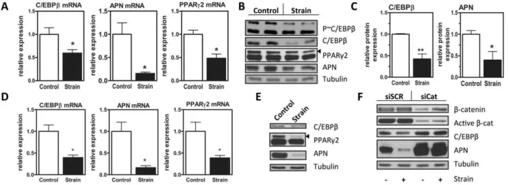
repression of C/EBPb mRNA we measured decreases in expression of fat markers adiponectin (p,0.05) and PPARc (p,0.05). Mechanical inhibition of C/EBPb mRNA predicted reductions of C/EBPbprotein as well as PPARc2 and adiponectin (figure 1B). Densitometry confirmed that mechanical repression of C/EBPband adiponectin proteins were significant (figure 1C, p,0.01 and p,0.05, respectively). As C/EBPb is activated by phosphorylation at a consensus ERK/GSK3b site, we next explored whether mechanical strain alters C/EBPb activation. The reduction in phospho-C/EBPb, the active form of this transcription factor, correlated with reduced expression of total C/ EBPband was not observed to be greater than the effect on total C/EBPb expression (figure 1B), suggesting that mechanical repression of C/EBPbis at a transcriptional level.
Key findings in C3H10T1/2 cells were reproduced in mouse marrow-derived MSC (mdMSC). Similar to the mechanical effect in C3H10T1/2 cells, C/EBPb mRNA and protein were downregulated by mechanical input in mdMSC at day 3 of adipogenic differentiation (figure 1D, 2E). As well, PPARcand adiponectin mRNA and protein were decreased in the presence of mechanical stimulation.
b-catenin contributes significantly to strain repression of adipogenesis [7]. We asked ifb-catenin participated in mechanical regulation of C/EBPb. SiRNA targeting b-catenin or a control siRNA sequence was transfected into cultures prior to induction of adipogenesis. In cultures treated with control siRNA, mechanical strain had expected effects to repress adipogenesis, shown here by reduced expression of adiponectin (figure 1F,left lanes). As in figure 1A, 1B, mechanical strain repressed C/EBPb protein expression (figure 1F). Knockdown of b-catenin (figure 1F, right lanes) accelerated adipogenesis as shown by increased expression of adiponectin. b-catenin knockdown interfered not only with mechanical repression of adiponectin, but also prevented the mechanical inhibition of C/EBPb. Thus, C/EBPb is mechanically sensitive and predicts that multiple C/EBPbtargets, such as PPARcexpression and the ER stress response should be susceptible to mechanical input.
C/EBPbOverexpression does not Rescue Adipogenesis from Mechanical Inhibition
Having established C/EBPbas a mechanical target, we sought to determine if C/EBPbwas necessary for mechanical repression of adipogenesis. Adipogenic differentiation of MSC was marked by an early expression peak of C/EBPbmRNA at day 2 (figure 2A) and protein at day 3 (figure 2B). The rise in C/EBPbwas noted concomitantly with the expression of PPARcas well as PPARc target genes adiponectin and aP2. Consistent with prior studies in 3T3-L1 pre-adipocytes [38], knockdown of C/EBPbwith siRNA prevented adipogenesis of multipotential MSC (figure 2C). To investigate whether mechanical repression of C/EBPbwas critical for mechanical inhibition of adipogenesis, we studied adipogenesis during C/EBPb overexpression (figure 2D). As C/EBPb does not induce adipogenesis alone, adipogenic medium was added to cultures transfected with a C/EBPb expression or empty vector. C/EBPboverexpression enhanced adipogenesis (figure 2D).
MSC were treated with a daily mechanical regimen for 3 days during adipogenic culture. The mechanical regimen effectively inhibited adipogenesis, as measured by decreases in adiponectin and PPARc2 mRNA as well as adiponectin protein, despite overexpression of C/EBPb(figure 2E, F). We additionally asked if pharmacologicalb-catenin activation could overcome C/EBPb-mediated adipogenesis. SB415286, a GSK3binhibitor, was added to cultures undergoing adipogenesis, causing a reduction in in adipogenic differentiation as measured by PPARcand aP2 protein (figure 2G). The effect of SB41528 to repress adipogenesis, similar to that of mechanical strain, was maintained in the presence of C/EBPboverexpression(figure 2G, right lanes). As such, mechanical repression of adipogenesis was not rescued by C/EBPb. Despite this result, the combination of our data showing that C/EBPb is not, alone, enough to stimulate adipogenesis of MSC, and our confirmation that adipogenesis requires C/EBPb expression, supports that that repression of C/EBPb must contribute to the anti-adipogenic effects of mechanical stimulation. In addition to the importance of our findings for adipogenic differentiation, we explored other potentially salutary downstream effects of C/EBPbdownregulation.
Figure 1. C/EBPbis a mechanical target in MSC.A, mRNA of C3H10T1/2 cells in adipogenic media subjected to strain, analyzed via real time PCR for C/EBPb, adiponectin (APN), and PPARc2, n = 3, *p,0.05. B, Immunoblot of day 3 C3H10T1/2 cells in adipogenic media subjected to strain. C, Densitometry from (B) n = 4, * p,0.05, ** p,0.01. D, mRNA of mdMSC in adipogenic media treated with strain, analyzed via real time PCR for C/EBPb, adiponectin (APN), and PPARc2, n = 3,*p,0.05. E, Representative immunoblot of day 2 mdMSC cells in adipogenic media subjected to strain, n = 3. F, C3H10T1/2 cells transiently transfected with siRNA tob-catenin. 24 hr later, induction of adipogenesis was initiated with adipogenic media. Strain was applied and immunoblot performed forb-catenin, activeb-catenin, as well as C/EBPb, and adiponectin (APN).
C/EBPb is Involved in ER Stress in MSC
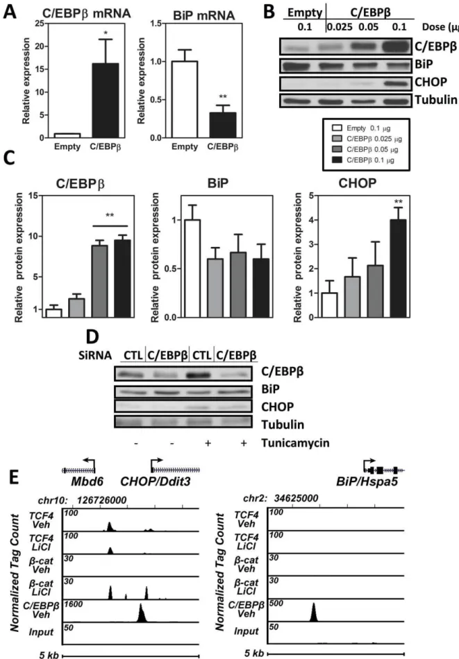
As C/EBPbhas been shown to trigger endoplasmic reticulum stress in pancreaticb-cells and fibroblasts [16,17], we considered whether C/EBPbmight play a similar role in the MSC unfolded protein response. We first examined the effect of C/EBPb overexpression on components of the ER stress response. C/ EBPb overexpression downregulated the ER chaperone BiP mRNA (figure 3A; p,0.01). Concomitant with the reduction in BiP, C/EBPb overexpression upregulated the pro-apoptotic CHOP protein in a C/EBPb-dose-dependent manner (figure 3B; p,0.01) as confirmed by densitometry (figure 3C). Moreover, ablation of C/EBPbby siRNA decreases expression of CHOP and increases expression of BiP (figure 3D). As BiP confers improved capacity to resist ER stress [39], a reduction of BiP in the presence of C/EBPboverexpression is consistent with a reduction in the cells ability to withstand ER stress. C/EBPb upregulation of the pro-apoptotic CHOP-as well as the diminution of CHOP with siRNA to C/EBPb- additionally implicates C/ EBPbin exacerbation of ER stress.
We then asked whether C/EBPb might directly regulate expression of these two components of the ER stress response-BiP and CHOP. Indeed, ChIP-seq analysis demonstrated that C/ EBPb is significantly associated with bothBiP and CHOP genes (figure 3E). As mechanical regulation of C/EBPb required b-catenin, we queried ifb-catenin might associate with eitherBiPor CHOP genes in a ChIP-seq analysis. Neither b-catenin nor its associated transcription factor TCF4 showed significant associa-tion with the upstream promoter ofBiP. There was an association ofb-catenin and TCF4 withCHOP; however this association was significantly lower than that of C/EBPbwith theCHOPgene. This suggests that although b-catenin is important for repression of
adipogenesis, C/EBPbregulatory role in ER stress is unlikely to involve direct effects ofb-catenin on these responders. Thus, C/ EBPb might affect ER stress by direct association of the transcription factor with the CHOP and BiP genes, regulating their expression levels in MSC.
Mechanical Strain Increases ER Capacity in MSC
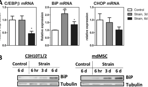
The C/EBPb effect to decrease BiP and increase CHOP suggests that it impairs the MSC’s ability to resist ER stress. Indeed, in pancreatic b-cells, ablation of C/EBPb has been demonstrated to improve ER capacity [16]. We were thus interested ascertain if mechanical repression of C/EBPb would similarly improve ER capacity in MSC. We found that a daily mechanical strain regimen increased BiP mRNA compared to non-strained controls at both 3 and 6 days of a daily mechanical regimen (figure 4A; p,0.01 and p,0.05). Consistent with work demonstrating that endurance exercise ameliorates ER stress in muscle [40], increases in BiP protein were found in both C3H10T1/2 and mdMSC after an in-vitro endurance regimen with increased benefit at 6 days as compared to either 3 days or 6 hours (figure 4B). CHOP mRNA in these cultures showed a trend toward reduction at 6 days, although CHOP protein was not measurable in unstressed cultures. Thus, mechanical strain in the setting of adipogenesis (figure 1) downregulates C/EBPb after 3 days as compared to 6 days required to downregulate C/EBPbin non-adipogenic conditions (figure 4A). This difference is likely due to early adipogenic C/EBPbinduction (see figure 2A) that allows strain effects to be demonstrable at an earlier time point. Consistent with an expansion of the ER as marked by increased BiP, CHOP mRNA expression was decreased in cultures which were treated daily with mechanical strain.
Figure 2. C/EBPboverexpression does not rescue adipogenesis from mechanical inhibition.A, C3H10T1/2 cells cultured in adipogenic media and analyzed on indicated days by real time PCR for C/EBPb, PPARc, and adiponectin (APN) n = 3, **p,0.01 relative to day zero B, C3H10T1/2 cells cultured in adipogenic media and blotted for C/EBPb, markers of adipogenesis. C, Immunoblot of C3H10T1/2 cells transfected with siRNA to C/ EBPb. 24 hours after transfection, adipogenic medium was added for 72 hours. D, C3H10T1/2 cells were transfected with 1mg of overexpression
vector for C/EBPbor control empty vector. 24 later adipogenic media was added and cells were analyzed at 48 hours by immunoblot for C/EBPb, adiponectin (APN) or aP2. E, C3H10T1/2 cells overexpressing C/EBPbas in A were subjected to strain for 3 days and analyzed by real-time PCR for C/ EBPb, PPARc2 and adiponectin mRNA expression. F, experiment as described in (E) analyzed by immunoblot. G, C3H10T1/2 cells were transiently transected with a C/EBPboverexpression vector. 24 hours later adipogenic media+/2SB41528 was added and maintained for 3 days prior to protein analysis for markers of adipogenesis and C/EBPb.
Figure 3. C/EBPbis involved in ER stress in MSC.A, C3H10T1/2 cells were transfected with 1mg of C/EBPbversus empty vector. 48 hours after
Mechanical Strain Ameliorates Tunicamycin-induced ER Stress in MSC
We next tested whether mechanical induction of BiP could afford protection from ER stress. Endoplasmic reticulum stress was induced with tunicamycin, which blocks synthesis of N-linked glycoproteins, leading to an accumulation of protein in the ER lumen. Tunicamycin dose-dependently increased the expression of C/EBPb, as well as that of CHOP and BiP in MSC (figure 5A). Additionally tunicamycin induced alternative splicing of XBP1 in MSC (figure 5B) consistent with activation of a second arm of the unfolded protein response [26]. Mechanical strain applied for 6 hours, 3 days or 6 days effectively ameliorated tunicamycin-induced ER stress as evidenced by decreased C/EBPbprotein as well as increased BiP and decreased CHOP (figure 5C). Increasing the days of mechanical treatment prior to tunicamycin treatment increased this beneficial response. C/EBPb transmits deleterious effects of ER stress [16] and enables adipogenesis. In Figure 5C, the small rise in C/EBPbmeasured in strained cells conflicts with the significant repression of C/EBPb expression shown in figure 2 and 4. It is likely that mechanical effects on expression of this gene are context dependent: we have reproducibly measured, here, the ability of mechanical strain to repress stimulated rises in C/EBPb. Thus, repetitive mechanical input, akin to endurance exercise, improved MSC ability to resist a significant ER stressor.
Discussion
Exercise is critical to musculoskeletal health. Mechanical stimulation biases the bone marrow MSC away from the fat lineage, permitting stem cell entry into other higher order tissues [6,7,28,31]. When daily mechanical loads are decreased – as in paralysis or in microgravity of spaceflight - osteopenia rapidly ensues [41,42]. We have now shown that mechanical input limits expression of the C/EBPb, a transcription factor central for processes associated with reduced skeletal health: adipogenesis and endoplasmic reticulum stress [16,24,25,38].
C/EBPb regulates adipogenesis at multiple levels. C/EBPb shares significant target gene-binding homology with C/EBPa as well as PPARc, and the loss of any one of these factors interferes with the adipogenic program [43]. Recent work suggests that a majority of adipocyte genes are not regulated by PPARcalone but require simultaneous actions of C/EBPaand C/EBPbrather than individual actions in discrete sequential steps [43]. As such, mechanical control of C/EBPb can be expected to contribute to repression of early steps of adipogenesis. The failure of C/EBPb overexpression to prevent mechanical inhibition of adipogenesis suggests that its contribution is less than other adipogenic factors that are subject to mechanical control. For example, mechanically activatedb-catenin downregulates PPARc activity [44], and b-catenin is activated by mechanical stimuli [45]. Another potential mechanism is the known effect of mechanical input to stimulate MAPK, which can phosphorylate and repress PPARc-directed transcription [46]. As such mechanical effects, including those due to preservation and activation ofb-catenin, act at multiple sites. indicated doses and 48 hours after transfection, protein analysis was performed. A representative blot is shown, n = 3. C, Densitometry for (B). D, Immunoblot of C3H10T1/2 cells transfected with siRNA to C/EBPbor control siRNA shown at 72 hours after transfection. Tunicamycin at 1mg/ml was
added for the final 6 hours. E, ChIP-seq analysis of C/EBPb,b-catenin, TCF4 binding at the mouseCHOPandBiPgene loci. MC3T3 cells were treated for 3 hours with vehicle or 50 mM LiCl prior to ChIP-seq assay. The genomic interval on the indicated mouse chromosome and the location of theCHOP orBiPtranscription unit including the direction of transcription (arrow) is shown at the top. Tracks indicate tag densities (normalized to 107reads) for vehicle or LiCl treatedb-catenin or TCF4 binding. Note the scale for peak height is different for each track to highlight peak activities.
doi:10.1371/journal.pone.0051613.g003
Figure 4. Mechanical strain increases ER capacity in MSC.A,MSC seeded on the same day were treated with mechanical strain for indicated time prior to mRNA (n = 3 * P,0.05, ** P,0.01) (A) or protein analysis (B) performed with C3H10T1/2 cells and mdMSC, as indicated. Control cultures were seeded on the same day as strained cultures and housed in similar conditions for 6 days prior to analysis.
This also suggests that the mechanical regulation of C/EBPbmay be important for non-adipogenic functions of this widely expressed transcription factor.
That C/EBPb is subject to mechanical control further widens the net of cellular events subject to biophysical control. In addition to its role in adipogenesis, this transcription enhancer plays a role in immune responses [47,48], cancer [49], hepatocyte differenti-ation [50], and has notably been shown to upregulate ER stress [16]. BiP buffers the cell from ER stress by providing docking sites for ATF6, IRE1 and PERK [26]. When these three ER transducers are released, downstream effectors traffic from the ER to the nucleus, triggering apoptosis via a rise in CHOP out of proportion to BiP [51–53]. Increased expression of C/EBPb can accelerate negative aspects of the unfolded protein response [16,17] activating CHOP and pro-apoptotic caspase-3. In MSCs, we have now shown that overexpression of C/EBPb increases CHOP expression, and decreases BiP, thus decreasing cellular capacity for handling of ER stress.
Besides increasing transcription of CHOP, C/EBPb serves as the principal dimerization partner for the pro-apoptotic CHOP [17]. As such, C/EBPboverexpression in pancreatic bcells was shown to increase these pro-apoptotic proteins while decreasing the beneficial chaperone BiP [16]. Conversely, ablation of C/ EBPbresulted in an increased ER capacity, reflected by increased BiP. Accordingly, C/EBPb deficient MSC show resistance to
tunicamycin-induced apoptosis [16]. In line with these studies, we found that mechanical repression of C/EBPb correlates with reduced ER stress in MSC, suggesting that mechanical input may have salutary effects that are transmitted through a C/EPBb control point.
Accumulating evidence suggests that exercise serves to lower ER stress by preserving and enhancing ER capacity [19,20]. For example, exercise downregulates C/EBPbin cardiomyocytes and ablation of C/EBPb in trained mice improves resistance to a heart-specific pathological stress [54]. Our data is the first to suggest that for MSCs, mechanical inhibition of C/EBPb promotes MSC cell health through increasing resistance to stress. Furthermore, we have identified that C/EBPb is an important mechanical target in MSC, and is likely to be in other tissue where C/EBPbcontrols cell differentiation and function. Thus, C/EBPb is a mechanically responsive transcription factor and its biophys-ical control advances our understanding of how exercise is beneficial to MSC/skeletal health.
Author Contributions
Conceived and designed the experiments: MS JR. Performed the experiments: MBM KG. Analyzed the data: MS MBM KG. Contributed reagents/materials/analysis tools: NC ZX BS WRT JWP. Wrote the paper: MS MBM KG JWP JR.
References
1. Ozcivici E, Luu YK, Adler B, Qin YX, Rubin J, et al. (2010) Mechanical signals as anabolic agents in bone. Nat Rev Rheumatol 6: 50–59.
2. Dalsky GP, Stocke KS, Ehsani AA, Slatopolsky E, Lee WC, et al. (1988) Weight-bearing exercise training and lumbar bone mineral content in postmenopausal women. Ann Intern Med 108: 824–828.
3. Leichter I, Simkin A, Margulies JY, Bivas A, Steinberg R, et al. (1989) Gain in mass density of bone following strenuous physical activity. J Orthop Res 7: 86– 90.
4. David V, Martin A, Lafage-Proust MH, Malaval L, Peyroche S, et al. (2007) Mechanical loading down-regulates peroxisome proliferator-activated receptor gamma in bone marrow stromal cells and favors osteoblastogenesis at the expense of adipogenesis. Endocrinology 148: 2553–2562.
5. Menuki K, Mori T, Sakai A, Sakuma M, Okimoto N, et al. (2008) Climbing exercise enhances osteoblast differentiation and inhibits adipogenic differentia-tion with high expression of PTH/PTHrP receptor in bone marrow cells. Bone 43: 613–620.
6. Sen B, Xie Z, Case N, Ma M, Rubin C, et al. (2008) Mechanical strain inhibits adipogenesis in mesenchymal stem cells by stimulating a durable beta-catenin signal. Endocrinology 149: 6065–6075.
7. Sen B, Styner M, Xie Z, Case N, Rubin CT, et al. (2009) Mechanical loading regulates NFATc1 and beta-catenin signaling through a GSK3beta control node. Journal of Biological Chemistry 284: 34607–34617.
8. Sen B, Xie Z, Case N, Styner M, Rubin CT, et al. (2011) Mechanical signal influence on mesenchymal stem cell fate is enhanced by incorporation of refractory periods into the loading regimen. Journal of Biomechanics 44: 593– 599.
9. Batchvarova N, Wang XZ, Ron D (1995) Inhibition of adipogenesis by the stress-induced protein CHOP (Gadd153). EMBO Journal 14: 4654–4661.
10. Bezy O, Elabd C, Cochet O, Petersen RK, Kristiansen K, et al. (2005) Delta-interacting protein A, a new inhibitory partner of CCAAT/enhancer-binding protein beta, implicated in adipocyte differentiation. Journal of Biological Chemistry 280: 11432–11438.
11. Rochford JJ, Semple RK, Laudes M, Boyle KB, Christodoulides C, et al. (2004) ETO/MTG8 is an inhibitor of C/EBPbeta activity and a regulator of early adipogenesis. Molecular and Cellular Biology 24: 9863–9872.
12. Shi X, Shi W, Li Q, Song B, Wan M, et al. (2003) A glucocorticoid-induced leucine-zipper protein, GILZ, inhibits adipogenesis of mesenchymal cells. EMBO Rep 4: 374–380.
13. Tong Q, Dalgin G, Xu H, Ting CN, Leiden JM, et al. (2000) Function of GATA transcription factors in preadipocyte-adipocyte transition. Science 290: 134–138. 14. Farmer SR (2006) Transcriptional control of adipocyte formation. Cell Metab 4:
263–273.
15. Tang QQ, Otto TC, Lane MD (2003) Mitotic clonal expansion: a synchronous process required for adipogenesis. Proc Natl Acad Sci U S A 100: 44–49. 16. Matsuda T, Kido Y, Asahara S-i, Kaisho T, Tanaka T, et al. (2010) Ablation of
C/EBPballeviates ER stress and pancreaticbcell failure through the GRP78 chaperone in mice. J Clin Invest 120: 115–126.
17. Zinszner H, Kuroda M, Wang X, Batchvarova N, Lightfoot RT, et al. (1998) CHOP is implicated in programmed cell death in response to impaired function of the endoplasmic reticulum. Genes and Development 12: 982–995. 18. Ron D, Walter P (2007) Signal integration in the endoplasmic reticulum
unfolded protein response. Nat Rev Mol Cell Biol 8: 519–529.
19. Kim Y, Park M, Boghossian S, York DA (2010) Three weeks voluntary running wheel exercise increases endoplasmic reticulum stress in the brain of mice. Brain Res 1317: 13–23.
Figure 5. Mechanical strain ameliorates tunicamycin-induced ER stress in MSC.A, mdMSC were cultured in the presence of tunicamycin (TM) at indicated doses for 6 hours and analyzed via immunoblot for C/EBPb, BiP and CHOP. B, as in (A) mdMSC cultured in the presence of tunicamycin 1mg/ml for 6 hours and analyzed via conventional PCR for XBP1. C, C3H10T1/2 cells were seeded on the same day and treated with
mechanical strain for the indicated number of days. Tunicamycin (TM) was added for the final 6 hours prior to protein analysis (representative blot, n = 3).
20. Hirasawa H, Jiang C, Zhang P, Yang FC, Yokota H (2010) Mechanical stimulation suppresses phosphorylation of eIF2alpha and PERK-mediated responses to stress to the endoplasmic reticulum. FEBS Lett 584: 745–752. 21. Pavalko FM, Gerard RL, Ponik SM, Gallagher PJ, Jin Y, et al. (2003) Fluid
shear stress inhibits TNF-alpha-induced apoptosis in osteoblasts: a role for fluid shear stress-induced activation of PI3-kinase and inhibition of caspase-3. J Cell Physiol 194: 194–205.
22. Blanco-Gelaz MA, Suarez-Alvarez B, Ligero G, Sanchez L, Vidal-Castineira JR, et al. (2010) Endoplasmic reticulum stress signals in defined human embryonic stem cell lines and culture conditions. Stem Cell Rev 6: 462–472.
23. Cho YM, Jang YS, Jang YM, Chung SM, Kim HS, et al. (2009) Induction of unfolded protein response during neuronal induction of rat bone marrow stromal cells and mouse embryonic stem cells. Experimental and Molecular Medicine 41: 440–452.
24. Zhang P, McGrath B, Li S, Frank A, Zambito F, et al. (2002) The PERK eukaryotic initiation factor 2 alpha kinase is required for the development of the skeletal system, postnatal growth, and the function and viability of the pancreas. Mol Cell Biol 22: 3864–3874.
25. Liu J, Hoppman N, O’Connell JR, Wang H, Streeten EA, et al. (2011) A functional haplotype in EIF2AK3, an ER stress sensor, is associated with lower bone mineral density. Journal of Bone and Mineral Research.
26. Todd DJ, Lee AH, Glimcher LH (2008) The endoplasmic reticulum stress response in immunity and autoimmunity. Nat Rev Immunol 8: 663–674. 27. Peister A, Mellad JA, Larson BL, Hall BM, Gibson LF, et al. (2004) Adult stem
cells from bone marrow (MSCs) isolated from different strains of inbred mice vary in surface epitopes, rates of proliferation, and differentiation potential. Blood 103: 1662–1668.
28. Case N, Xie Z, Sen B, Styner M, Zou M, et al. (2010) Mechanical activation of beta-catenin regulates phenotype in adult murine marrow-derived mesenchymal stem cells. Journal of Orthopaedic Research 28: 1531–1538.
29. Sen B, Xie Z, Case N, Styner M, Rubin CT, et al. (2010) Mechanical signal influence on mesenchymal stem cell fate is enhanced by incorporation of refractory periods into the loading regimen. J Biomech.
30. Styner M, Sen B, Xie Z, Case N, Rubin J (2010) Indomethacin promotes adipogenesis of mesenchymal stem cells through a cyclooxygenase independent mechanism. J Cell Biochem.
31. Case N, Ma M, Sen B, Xie Z, Gross TS, et al. (2008) Beta-catenin levels influence rapid mechanical responses in osteoblasts. Journal of Biological Chemistry 283: 29196–29205.
32. Styner M, Sen B, Xie Z, Case N, Rubin J (2010) Indomethacin promotes adipogenesis of mesenchymal stem cells through a cyclooxygenase independent mechanism. Journal of Cellular Biochemistry 111: 1042–1050.
33. Kim S, Yamazaki M, Zella LA, Shevde NK, Pike JW (2006) Activation of receptor activator of NF-kappaB ligand gene expression by 1,25-dihydroxyvi-tamin D3 is mediated through multiple long-range enhancers. Molecular and Cellular Biology 26: 6469–6486.
34. Kim S, Shevde NK, Pike JW (2005) 1,25-Dihydroxyvitamin D3 stimulates cyclic vitamin D receptor/retinoid X receptor DNA-binding, co-activator recruitment, and histone acetylation in intact osteoblasts. Journal of Bone and Mineral Research 20: 305–317.
35. Fretz JA, Zella LA, Kim S, Shevde NK, Pike JW (2006) 1,25-Dihydroxyvitamin D3 regulates the expression of low-density lipoprotein receptor-related protein 5 via deoxyribonucleic acid sequence elements located downstream of the start site of transcription. Molecular Endocrinology 20: 2215–2230.
36. Zella LA, Kim S, Shevde NK, Pike JW (2006) Enhancers located within two introns of the vitamin D receptor gene mediate transcriptional autoregulation by 1,25-dihydroxyvitamin D3. Molecular Endocrinology 20: 1231–1247. 37. Meyer MB, Goetsch PD, Pike JW (2012) VDR/RXR and TCF4/beta-Catenin
Cistromes in Colonic Cells of Colorectal Tumor Origin: Impact on c-FOS and c-MYC Gene Expression. Molecular Endocrinology 26: 37–51.
38. Hamm JK, Park BH, Farmer SR (2001) A role for C/EBPbeta in regulating peroxisome proliferator-activated receptor gamma activity during adipogenesis in 3T3-L1 preadipocytes. J Biol Chem 276: 18464–18471.
39. Kammoun HL, Chabanon H, Hainault I, Luquet S, Magnan C, et al. (2009) GRP78 expression inhibits insulin and ER stress-induced SREBP-1c activation and reduces hepatic steatosis in mice. Journal of Clinical Investigation 119: 1201–1215.
40. Wu J, Ruas JL, Estall JL, Rasbach KA, Choi JH, et al. (2011) The unfolded protein response mediates adaptation to exercise in skeletal muscle through a PGC-1alpha/ATF6alpha complex. Cell Metab 13: 160–169.
41. Minaire P, Edouard C, Arlot M, Meunier PJ (1984) Marrow changes in paraplegic patients. Calcif Tissue Int 36: 338–340.
42. Lang T, LeBlanc A, Evans H, Lu Y, Genant H, et al. (2004) Cortical and trabecular bone mineral loss from the spine and hip in long-duration spaceflight. J Bone Miner Res 19: 1006–1012.
43. Lefterova MI, Zhang Y, Steger DJ, Schupp M, Schug J, et al. (2008) PPARgamma and C/EBP factors orchestrate adipocyte biology via adjacent binding on a genome-wide scale. Genes Dev 22: 2941–2952.
44. Liu J, Wang H, Zuo Y, Farmer SR (2006) Functional interaction between peroxisome proliferator-activated receptor gamma and beta-catenin. Mol Cell Biol 26: 5827–5837.
45. Thompson WR, Rubin CT, Rubin J (2012) Mechanical regulation of signaling pathways in bone. Gene.
46. Hu E, Kim JB, Sarraf P, Spiegelman BM (1996) Inhibition of adipogenesis through MAP kinase-mediated phosphorylation of PPARgamma. Science 274: 2100–2103.
47. Xu G, Zhang Y, Zhang L, Roberts AI, Shi Y (2009) C/EBPbeta mediates synergistic upregulation of gene expression by interferon-gamma and tumor necrosis factor-alpha in bone marrow-derived mesenchymal stem cells. Stem Cells 27: 942–948.
48. Akira S, Isshiki H, Sugita T, Tanabe O, Kinoshita S, et al. (1990) A nuclear factor for IL-6 expression (NF-IL6) is a member of a C/EBP family. EMBO J 9: 1897–1906.
49. Carro MS, Lim WK, Alvarez MJ, Bollo RJ, Zhao X, et al. (2010) The transcriptional network for mesenchymal transformation of brain tumours. Nature 463: 318–325.
50. Descombes P, Chojkier M, Lichtsteiner S, Falvey E, Schibler U (1990) LAP, a novel member of the C/EBP gene family, encodes a liver-enriched transcrip-tional activator protein. Genes Dev 4: 1541–1551.
51. Ron D, Walter P (2007) Signal integration in the endoplasmic reticulum unfolded protein response. Nat Rev Mol Cell Biol 8: 519–529.
52. Schroder M, Kaufman RJ (2005) ER stress and the unfolded protein response. Mutat Res 569: 29–63.
53. Xu C, Bailly-Maitre B, Reed JC (2005) Endoplasmic reticulum stress: cell life and death decisions. J Clin Invest 115: 2656–2664.