Acute liver failure: An up-to-date approach
☆
,
☆☆
Filipe S. Cardoso, MD MSc
a,⁎
, Paulo Marcelino, MD, PhD
a, Luís Bagulho, MD
a,
Constantine J. Karvellas, MD, SM
ba
Intensive Care Unit, Curry Cabral Hospital, Central Lisbon Hospital Center, Lisbon, Portugal
b
Divisions of Gastroenterology (Liver Unit) and Critical Care, University of Alberta Hospital, Edmonton, Canada
a b s t r a c t
a r t i c l e i n f o
Acute liver failure is a rare but potentially devastating disease. Throughout the last few decades, acute liver failure outcomes have been improving in the context of the optimized overall management. This positive trend has been associated with the earlier recognition of this condition, the improvement of the intensive care unit management, and the developments in emergent liver transplantation. Accordingly, we aimed to review the current diagnostic and therapeutic approach to this syndrome, especially in the intensive care unit setting.
© 2017 Elsevier Inc. All rights reserved.
Keywords: Hepatitis Liver failure Critical care Contents 1. Introduction . . . 25
2. Definition and epidemiology . . . 26
3. Pathophysiology and clinical manifestations . . . 26
4. General management . . . 26
4.1. General therapeutic approach . . . 27
4.1.1. Hemodynamics . . . 27
4.1.2. Hepatic encephalopathy . . . 27
4.1.3. Coagulopathy . . . 27
4.1.4. Infection . . . 27
4.2. Etiology-specific therapeutic approach . . . 27
4.2.1. Acetaminophen . . . 27
4.2.2. Non-acetaminophen . . . 28
5. Intensive care management . . . 28
5.1. Acute lung injury . . . 28
5.2. Hemodynamics . . . 28
5.3. Acute kidney injury . . . 28
5.4. Cerebral edema and intracranial hypertension . . . 28
5.5. Extracorporeal liver support systems . . . 28
6. Liver transplantation . . . 29
7. Conclusions . . . 29
References . . . 29
1. Introduction
Acute liver failure (ALF) is a rare condition characterized by new and rapidly evolving hepatic dysfunction associated with neurologic dysfunc-tion and coagulopathy. It is more frequent in young individuals and its etiologies vary geographically, with impact on both clinical course and outcomes. Throughout the last decades, ALF outcomes have been improv-ing in the context of the optimized overall management. However, its
☆ Conflicts of interest: None. ☆☆ Funding: None.
⁎ Corresponding author at: Polyvalent Intensive Care Unit, Curry Cabral Hospital, Central Lisbon Hospital Center, Rua da Beneficência, n.° 8, 1069-166 Lisbon, Portugal. Tel.: +351 21 792 4200; fax: +351 21 792 4392.
E-mail address:fi[email protected](F.S. Cardoso).
http://dx.doi.org/10.1016/j.jcrc.2017.01.003
0883-9441/© 2017 Elsevier Inc. All rights reserved.
Contents lists available atScienceDirect
Journal of Critical Care
j o u r n a l h o m e p a g e :w w w . j c c j o u r n a l . o r gpresent morbidity and mortality remain high in patients fulfilling poor prognostic criteria and without emergent liver transplantation (LT). 2. Definition and epidemiology
Acute liver failure definition has evolved throughout the time and presently includes the following features: international normalized ratio (INR) at least 1.5, neurologic dysfunction with any degree of hepat-ic encephalopathy (HE), no preexisting cirrhosis, and disease course of 26 weeks or less[1]. Exceptions to this definition may be patients with acute presentations of Wilson disease, autoimmune hepatitis, or vertically transmitted hepatitis B if by the time of the new hepatic insult, they also have underlying cirrhosis known for 26 weeks or less.
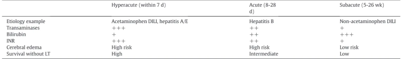
Acute liver failure has been subdivided into 3 phenotypes based on the time between jaundice and HE onset (Table 1): hyperacute (within 7 days), acute (8-28 days), or subacute (5-26 weeks)[2]. However, the clinical utility of this classification has not been entirely clarified be-cause its impact on outcomes seems to be mostly associated with un-derlying etiologies[3].
Acute liver failure incidence has been reported as fewer than 10 cases per million people per year in developed countries[4]. Its etiolo-gies vary worldwide: whereas in Eastern developing countries, viruses (mainly hepatitis A, B, or E) may account for up to 95% of all ALF cases, the Western developed countries have reported more heterogeneous causes of ALF[5]. In the United Kingdom and the United States, acet-aminophen overdose has been the leading cause of ALF[3,6]. In other parts of Europe (eg, Germany and Portugal), the most common causes of ALF have been non-acetaminophen drug–induced liver injury, seroneg-ative (indeterminate) liver injury, and viruses (mostly hepatitis B)[7,8]. Acute liver failure etiologies are summarized inTable 2 [1,3,4,9].
Acute liver failure outcomes have been improving throughout the decades with overall hospital survival increasing from 17% in 1973 to
1978 to 62% in 2004 to 2008 in a single center from the United Kingdom
[10]. In a multicenter registry from the United States in 1998 to 2010, ALF 2-year survival has been reported as 92% for liver transplant (LT) recip-ients, 90% for acetaminophen overdose spontaneous survivors, and 76% for non-acetaminophen spontaneous survivors[11]. This positive trend has been attributed to the earlier recognition of this condition, the improve-ment of the intensive care unit (ICU) manageimprove-ment, and the developimprove-ments in emergent LT[10]. Overall, the most common causes of death in ALF have been multiorgan failure (18%), liver failure (17%), and sepsis (12%)[11]. 3. Pathophysiology and clinical manifestations
In ALF, the liver insult results in extensive death of hepatocytes with activation of the innate immune system responses (Kupffer cells and circulating monocytes) causing a large production of inflammatory me-diators. The“spill-over” of these inflammatory mediators into the circu-lation ultimately leads to the systemic disturbances and clinical manifestations of ALF[12]. An overwhelming systemic inflammatory response syndrome (SIRS) is associated with the several organ failures that may ensue.
In parallel to the proinflammatory response, a compensatory anti-inflammatory response develops[13]. Although within the injured liver, the production of anti-inflammatory mediators serves to dampen proin-flammatory responses, limit the extent of tissue injury, and promote liver regeneration, their release into the systemic circulation is associated with a predisposition to infection[14]. At this stage, circulating monocytes may become functionally impaired and less able to respond to infectious stimuli, a state often referred as immune paresis[15,16]. Consequently, sepsis and multiorgan failure are frequently the cause of death in ALF. A summary of clinical manifestations related to the organ failures that may ensue in the context of ALF is presented inTable 3.
4. General management
The initial management of acute liver injury (hepatitis) or ALF is sup-portive with the objective to optimize conditions for the liver to
Table 1 ALF phenotypes
Hyperacute (within 7 d) Acute (8-28 d)
Subacute (5-26 wk)
Etiology example Acetaminophen DILI, hepatitis A/E Hepatitis B Non-acetaminophen DILI
Transaminases +++ ++ +
Bilirubin + ++ +++
INR +++ ++ +
Cerebral edema High risk High risk Low risk
Survival without LT High Intermediate Low
DILI indicates drug-induced liver injury.
Table 2 ALF etiologies Viruses Hepatitis A, B, D, or E viruses Cytomegalovirus Epstein-Barr virus Herpes simplex virus Varicella zoster virus Parvovirus
Drug-induced liver injury Acetaminophen
Non-acetaminophen (eg, isoniazid, phenytoin, valproate, propylthiouracil, nitrofurantoin)
Recreational drugs (eg, cocaine, MDMA) Autoimmune hepatitis
Ischemic/congestive hepatitis Budd-Chiari syndrome Wilson disease Amanita phalloides
Pregnancy (eg, acute fatty liver of pregnancy, HELLP syndrome) Heat stroke
Malignant infiltration Seronegative (indeterminate)
MDMA indicates 3,4-methylenedioxy-N-methylamphetamine.
Table 3 ALF organ failures
Organ failure Pathophysiology
Liver Hyperlactatemia: decreased lactate clearance Hyperammonemia: decreased ammonia clearance Coagulopathy: decreased synthesis of procoagulant and anticoagulant factors
Hypoglycemia: decreased gluconeogenesis
Portal hypertension: may develop especially in subacute disease Brain HE: circulating inflammatory mediators and hyperammonemia
Cerebral edema: inflammatory mediators from microglial cells and glutamine accumulation in astrocytes
Cardiovascular Hypotension or shock especially if sepsis superimposes Lungs Acute lung injury or ARDS: SIRS, sepsis, and/orfluid overload Kidneys AKI: SIRS, sepsis, and/or hypovolemia
Pancreas Acute pancreatitis: SIRS and/or drug toxicity (eg, acetaminophen) Multi-organ
failure
regenerate and prevent and treat as early as possible complications[17]. Although most patients with acute liver injury may be managed in a regular ward, patients with ALF should be referred to the ICU, ideally one in a center capable of providing emergent LT, as soon as possible as they may deteriorate quickly[1].
The initial diagnostic approach should be directed at detecting and assessing the severity of hepatitis, HE, coagulopathy, and other organ fail-ures. Clinical history taking, if possible, may be important to understand the time between jaundice and HE onset, etiology factors (eg, alcohol in-take, viral infection, drugs, mushroom, or tisane ingestion), and suspected or known previous liver disease. Physical examination allows to detect signs of organ failures and should include a mental status evaluation (West-Haven criteria for overt HE and, if feasible, a neuropsychological test for covert HE) and a search for stigmata of chronic liver disease. A list of useful initial laboratory tests is displayed inTable 4. Abdominal im-aging with ultrasonography (with Doppler studies) or computed tomo-graphic (CT) scan (with intravenous contrast if safe) helps to exclude features of chronic liver disease and biliary, pancreatic, or other intra-abdominal pathology that could be the origin or contribute to the liver failure. Liver biopsy (transcutaneous or transjugular) should be consid-ered whenever the etiology remains unclear and histopathology informa-tion may help to change management decisions, including LT assessment.
4.1. General therapeutic approach 4.1.1. Hemodynamics
Patients with ALF are often volume depleted at presentation and re-quirefluid resuscitation. As in many other hypovolemic syndromes, crys-talloids should be the initialfluids of choice, with normal saline (sodium chloride 0.9%) being the most frequently in use[17]. Nevertheless, it may be considered the use of a chloride-restrictive solution (eg,
Plasma-Lyte 148, Hartmann's solution) as it has been shown in patients with acute illness that it may decrease the risk of metabolic acidosis and acute kidney injury (AKI)[18,19]. In a non-ICU setting, some of the sim-plest parameters can be used as targets for the initialfluid replenishment (while avoidingfluid overload), for example, mean arterial pressure at least 60 to 65 mm Hg, urine output at least 0.5 mL kg−1h−1, and lactate 2 mmol/L or less.
On the contrary, patients with congestive hepatitis (eg, heart failure) may befluid overloaded and diuretics may help to improve liver function. 4.1.2. Hepatic encephalopathy
The correction of all factors that may contribute to the acute confu-sion (eg, dehydration, infection, serum ions derangements) is important for HE treatment. With this objective, sedative medications should be avoided, except in situations deemed necessary (eg, agitation, periendotracheal intubation for progressive HE). To exclude intracranial pathology, a CT scan needs to be considered. Regular oral or enteral feeding should be maintained whenever feasible[20]. Lactulose may add to these patients' transplant-free survival, but careful should be taken with possible abdominal distension[21].L-Ornithine-L-aspartate
has not shown benefit to neither HE grade nor overall survival[22]. 4.1.3. Coagulopathy
In ALF, overall hemostasis as measured by thromboelastography has been shown to be normal by several compensatory mechanisms, even in patients with markedly elevated INR[23]. In the absence of active bleeding or invasive procedure, it is not advisable to correct the INR with fresh-frozen plasma, because clinically significant blood loss is rare and correction obscures trends in the INR, an important marker of prognosis[1]. Nevertheless, vitamin K deficiency may be present in a minority of patients with ALF; thus, vitamin K supplementation may be used at least once[1,24].
In ALF, bleeding due tofibrinolysis has been found to be limited due to high levels of plasminogen activator inhibitor-1[25]. In the absence of ac-tive bleeding or invasive procedure, the platelet count should be main-tained at least above the appropriate spontaneous bleeding threshold. 4.1.4. Infection
Acute liver failure risk of immune paresis increases susceptibility to infection which may preclude emergent LT. Therefore, surveillance for infection (including chest radiography and periodic cultures of sputum, urine, and blood) should be undertaken, while maintaining a low threshold for starting antimicrobial therapy[1]. Prophylactic antimicro-bials have not been proven to improve 21-day survival in ALF[26]. Nev-ertheless, the development of grade III-IV HE or SIRS has been associated with infection and worse outcomes[27,28].
4.2. Etiology-specific therapeutic approach 4.2.1. Acetaminophen
N-acetylcysteine (NAC) has been largely used as an antidote for acet-aminophen intoxication. Its mechanism of action has to do mainly with glutathione replenishment, a crucial molecule for acetaminophen de-toxification in the liver[29,30]. In ALF, NAC use has been shown to
Table 4
Initial laboratory tests for acute liver injury or ALF Assessment Test
Severity (serum) Arterial blood gas Arterial lactate Arterial ammonia
Hemoglobin, leukocytes, platelets INR, aPTT,fibrinogen, factor V
AST, ALT, bilirubin, albumin, alkaline phosphatase, LDH, amylase Creatinine, urea, sodium, chloride, potassium, calcium, magnesium, phosphorus, creatine kinase
Etiology (serum or urine)
HAV IgM, HBsAg, HBc IgM, anti-HCV, anti-HEV, CMV IgM, EBV IgM, HSV IgM, VZV IgM, anti-HIV
Ceruloplasmin, copper
Anti–nuclear antibody, anti–smooth muscle antibody, immunoglobulins
Acetaminophen, toxicology screen Blood type
Pregnancy test (females)
aPTT indicates activated partial thromboplastin time; AST, asparte aminotransferase; ALT, alanine aminotransferase; LDH, lactate dehydrogenase; HAV, hepatitis A virus; HBs, hepa-titis B virus surface antigen; HBc, hepahepa-titis B virus core antibody; HCV, hepahepa-titis C virus; HEV, hepatitis E virus; CMV, cytomegalovirus; EBV, Epstein-Barr virus; HSV, herpes sim-plex virus; VZV, varicella zoster virus; HIV, human immunodeficiency virus.
Table 5
NAC protocols in ALF Route and dosage[1,30]
Oral or enteral Loading dose of 140 mg/kg in glucose 5%, followed by 70 mg/kg in glucose 5% every 4 h × 17 doses (overall 72 h) Intravenous Loading dose of 150 mg/kg in glucose 5% for 15 min, followed by 50 mg/kg in glucose 5% for 4 h, and followed
by 100 mg/kg in glucose 5% for 16 h (overall 20 h) Length of therapy[1,30]
Controversy exists but at least a 72-h period irrespective of route has been suggested. A decision to stop based on consistent clinical improvement of patients has also been suggested.
More frequent adverse effects[1,30]
significantly improve overall survival with a low rate of adverse effects
[31]. Although reported more effective within 48 hours of acetamino-phen ingestion, NAC is currently being started in many centers when acetaminophen intoxication is suspected (eg, reported history of acet-aminophen intake outside dose normal range or transaminases≥3500 IU/L, especially in an alcohol intake or fasting context), irrespective of acetaminophen dose, time since ingestion, or acetaminophen serum level because it is often difficult to establish when was the initial drug in-take or if that was a one-time ingestion or a staggered overdose[1,32]. N-acetylcysteine administration protocols are presented inTable 5. 4.2.2. Non-acetaminophen
In non–acetaminophen-related ALF, namely in drug-induced liver injury or hepatitis B, NAC seems to play a therapeutic role also as it has been shown to improve transplant-free survival when given during early HE stages (grade I-II)[33,34]. These results may be related to other effects that have been attributed to NAC, for example, improving hemo-dynamics and oxygen use and decreasing the risk of cerebral edema[26, 35]. The mechanism of such effects has not been cleared, but it may in-volve reducing the production of proinflammatory cytokines (eg, IL-17), scavenging of free radicals, or changes in the hepatic bloodflow[35-37]. Several ALF etiologies may require specific therapies, for example: (1) consider antiviral therapy for hepatitis B virus, cytomegalovirus, herpes simplex virus, or varicella zoster virus–related ALF; (2) consider steroid therapy for autoimmune hepatitis–related ALF; (3) consider penicillin G or silibinin in Amanita phalloides–related ALF; and (4) con-sider emergent delivery for pregnancy-related ALF (eg, acute fatty liver of pregnancy or hemolysis, elevated liver enzymes, and low plate-lets [HELLP] syndrome)[1].
5. Intensive care management
Acute liver failure may lead to several organ failures; therefore, ICU ad-mission should be considered as early as possible, especially when HE is being difficult to control in a regular ward or coagulopathy is progressing. In this context, supporting organ failures follows the rules of general ICU patients but with some specificities, which deserve to be emphasized. 5.1. Acute lung injury
Acute lung injury may occur in patients with ALF, more often at a later stage of their clinical course, namely with liver regeneration or sepsis[4]. Although patients with acute respiratory distress syndrome (ARDS) may require greater number of ventilator days, the requirement for vasopressors or renal replacement therapy (RRT), ICU stay, and mor-tality have been described as similar to patients without ARDS[38].
Management of ARDS in this context, albeit following general inten-sive care rules, may be more difficult because increasing positive end-expiratory pressure may exacerbate cerebral edema or liver congestion
[39]. Nevertheless,finding the optimum positive end-expiratory pres-sure for each patient may help to improve oxygenation while minimiz-ing such adverse consequences[40,41].
5.2. Hemodynamics
Patients with ALF typically present with a hyperdynamic circulation characterized by high cardiac output and low peripheral vascular resis-tance, a pattern resembling that of sepsis. For patients who continue to have hypotension despite volume repletion, noradrenaline is the pre-ferred vasopressor, with or without adjunctive use of vasopressin or its analogs[42].
Echocardiography or invasive hemodynamic monitoring (eg, Pulse Contour Continuous Cardiac Output, Swan-Ganz catheter) should be used to assess cardiac function and help titrate decisions on the doses offluids and/or vasopressors. Elevated troponin levels have been associ-ated with an increase in grade III-IV HE and overall mortality[43].
Nevertheless, it is unclear whether that represents subclinical myocar-dial ischemia or simply reflects metabolic stress[44].
Relative adrenal insufficiency may be present and the use of steroids has been associated with a decrease in noradrenaline dose and overall mortality[45].
5.3. Acute kidney injury
Acute kidney injury may develop in up to 70% of patients with ALF and has been associated with worse overall survival[46]. Hyperphosphatemia develops with AKI if there is impaired liver regeneration and has been as-sociated with poorer outcomes[47]. Acute kidney injury is often multifac-torial and more common with certain etiologies: ischemic hepatitis, acute fatty liver of pregnancy, HELLP syndrome, heat stroke, hepatitis A virus, or drug-induced liver injury due to acetaminophen, phenytoin, trimethoprim-sulfamethoxazole, or macrolides[48].
In this context, classic indications for RRT initiation also apply, includ-ing severe acidosis, hyperkalemia, anuria, and/orfluid overload[49]. Fur-ther indications may include the following: removal of toxic substances (eg, acetaminophen, ammonia), difficult to treat hyponatremia, or diffi-cult to treat hyperthermia[50,51]. Continuous modes of RRT are pre-ferred over intermittent ones because the latter have been associated with increased risk of hemodynamic instability and cerebral edema[52]. 5.4. Cerebral edema and intracranial hypertension
Cerebral edema incidence in patients with ALF has been decreasing throughout the last decades[10]. This positive trend may reflect im-provements in the preventive care and use of emergent LT[17].
In ALF, astrocyte swelling may result in cytotoxic brain edema which may culminate in tonsillar herniation and death. Liver-brain proin flamma-tory signaling mechanisms involve the transduction of systemically de-rived cytokines as well as the gliotoxic effects of ammonia and lactate[53]. Frequent neurologic examination (including pupils) and the use of transcranial Doppler are simple strategies to monitor for signs of cere-bral edema and intracranial pressure. On the contrary, CTfindings com-patible with intracranial hypertension often present too late in the course of this condition. Invasive monitoring of intracranial pressure has not been shown to improve these patients’ hospital survival[54]. Nevertheless, it is still not clear if patients with the highest risk for cere-bral edema and intracranial hypertension (eg, ammoniaN150 µmol/l, vasopressors requirement, or RRT requirement) may benefit from such monitoring capacity[55].
The approach to cerebral edema and intracranial hypertension con-sists of the following: (1) head of the bed greater than 30°; (2) minimize patient stimulation; (3) sedation and invasive mechanical ventilation; (4) treat fever (although active hypothermia has not been proven to pre-vent cerebral edema and intracranial hypertension)[56,57]; (5) treat sei-zures (although prophylaxis has unclear value)[1]; (6) aim for a mean arterial pressure of at least 75 mm Hg withfluids and/or vasopressors, with the goal being to maintain an intracranial pressure less than 25 mm Hg and a cerebral perfusion pressure greater than 50 mm Hg; (7) consider using RRT to promote more effective ammonia clearance
[51]; (8) aim for a serum sodium of 145 to 155 mmol/L with hypertonic saline (3%-30% infusion) for prophylaxis in patients with grade III-IV HE
[58]; (9) consider using mannitol (0.5-1 g/kg bolus) to transiently reduce intracranial pressure when there is established intracranial hypertension (repeat if serum osmolalityb320 mOsm/L)[59]; and (10) consider using hyperventilation (aiming for a PCO2 25-30 mm Hg) in cases of established intracranial hypertension despite optimized treatment to try to delay the progression to tonsillar herniation[60].
5.5. Extracorporeal liver support systems
The rationale for using an extracorporeal liver support (ECLS) system in ALF is to help maintain homeostasis while the liver regenerates (eg,
ischemic hepatitis or acetaminophen-related ALF) or until an organ is available for transplantation (all etiologies). An ideal ECLS system would be one capable of assisting on 3 major liver functions: removal of toxins, biosynthesis, and immune modulation[61]. However, none of the arti fi-cial (depuration through membranes) or bioartificial (hepatocytes) sys-tems available performs efficiently all of these functions[62,63].
Two artificial ECLS systems have been studied in ALF with random-ized controlled trials: molecular adsorbent recirculating system (MARS) and high-volume plasmapheresis (HVP). With MARS, blood is dialyzed across an albumin-impregnated high-flux dialysis membrane (pores with 50 kDa) and against a dialysate with albumin. Subsequent-ly, 2 sequential adsorbent columns containing activated charcoal and anion exchange resin remove most of the water-soluble and albumin-bound toxins. In a recent study, MARS has not been proven to improve 6-month survival in ALF[64]. However, a confounder may have been the median listing-to-transplant time which was only 16 hours, with 75% of enrolled patients undergoing LT within 24 hours.
With HVP, patients' plasma is replaced by fresh-frozen plasma with the objective of removing plasma cytokines and adhesion molecules, re-placing plasma factors, and potentially modulating the immune system. In a recent study, HVP has been shown to significantly improve hospital survival, especially for patients with contraindications for LT[65]. Nev-ertheless, a limitation to account for may have been the 12-year inclu-sion period. With both MARS and HVP, adverse effects have been reported as similar to standard therapy[64,65].
Several bioartificial ECLS systems have been clinically tested, for ex-ample: HepatAssist (Arbios, formerly Circe, Waltham, Mass), ECLS de-vice (ELAD; Vital Therapies, San Diego, Calif), modular ECLS system (MELS; Charité, Berlin, Germany), bioartificial liver support system (BLSS; Excorp Medical, Minneapolis, Minn), and Amsterdam Medical Center bioartificial liver (AMCBAL; AMC, Amsterdam, the Netherlands)
[1]. A meta-analysis has shown that these systems failed to improve sur-vival in ALF[66]. Nevertheless, more well-designed clinical trials in humans are likely needed to fully evaluate these systems efficacy and safety[67].
6. Liver transplantation
Liver transplantation is the only definitive treatment for patients with ALF. Overall survival after LT has been reported to be lower for pa-tients with ALF in comparison to papa-tients with cirrhosis until 1 year after transplant, but it tends to be similar from then on[68]. In fact, most deaths after LT for patients with ALF occur from infection during the first 3 postoperative months[4]. Nevertheless, survival after LT for ALF has been improving throughout the last decades, with 21-day survival reaching 96% for the period of 2006 to 2013[69].
Cadaveric donor LT has been the norm in ALF, but living-donor trans-plant has been performed in some large-volume centers with accept-able outcomes. Despite this, controversy remains in regard to ethical issues, namely the risk of living-donor coercion and morbidity or
mortality[70]. Another strategy to prevent waitlist mortality has been auxiliary transplantation (partial graft as temporary support for native liver regeneration), which has shown reasonable outcomes too[71].
In ALF, stratifying patients based on their risk of death without LT is crucial to prioritize them for transplantation. King's College criteria have been used worldwide for this purpose (Table 6)[72]. A recent meta-analysis has revealed its prognostic ability in comparison with the MELD score: for acetaminophen-related ALF, sensitivities were 58% and 80%, respectively, and specificities were 89% and 53%, respectively; for non-acetaminophen etiologies, sensitivities were 58% and 76%, re-spectively, and specificities were 74% and 73%, respectively[73]. There-fore, King's College criteria remain highly specific for acetaminophen-related ALF, but less accurate for non-acetaminophen etiologies. Another useful tool for prognostication of death without LT in patients with hepatitis B–related ALF and grade III-IV HE may be the Clichy criteria. According to this tool, a serum factor V level lower than 20% in patients 30 years or younger or lower than 30% in any other patients prognosti-cates mortality with a positive predictive value of 82% and a negative predictive value of 98%[74]. However, it has been shown that the Clichy criteria perform worse than the King's College criteria overall[75].
Risk stratification in ALF may be improved further if taking into ac-count not only the probability of death without LT but also the probabil-ity of death after transplantation. Studies have suggested that some characteristics were associated with an increased risk of death after LT: male recipient, recipient older than 45 years, recipient on vasopres-sors, ABO-incompatible transplant, and high-risk graft use[76,77].
7. Conclusions
Acute liver failure diagnostic and therapeutic strategies have evolved throughout the time and that has been associated with im-proved outcomes. New advances in basic and clinical research may po-tentiate even more such outcomes. Despite this, these patients' early referral to an LT center, ICU timely treatment, and a comprehensive multidisciplinary strategy in risk stratification and selection for LT will continue to be fundamental steps for a successful approach.
References
[1] Lee W, Larson AM, Stravitz RT. AASLD position paper: the management of acute liver failure: update 2011. Available at: http://www.aasld.org/sites/default/files/guide-line_documents/141022_Position_ALF_4UFb.pdf
[2]O'Grady JG, Schalm SW, Williams R. Acute liver failure: redefining the syndromes. Lancet 1993;342(8866):273–5.
[3]Ostapowicz G, Fontana RJ, Schiødt FV, Larson A, Davern TJ, Han SH, et al. Results of a prospective study of acute liver failure at 17 tertiary care centers in the United States. Ann Intern Med 2002;137(12):947–54.
[4]Bernal W, Wendon J. Acute liver failure. N Engl J Med 2013;369:2525–34.
[5]Acharya SK, Batra Y, Hazari S, Choudhury V, Panda SK, Dattagupta S. Etiopathogenesis of acute hepatic failure: Eastern versus Western countries. J Gastroenterol Hepatol 2002;17(Suppl. 3):S268–73.
[6]Gulmez SE, Larrey D, Pageaux GP, Bernuau J, Bissoli F, Horsmans Y, et al. Liver trans-plant associated with paracetamol overdose: results from the seven-country SALT study. Br J Clin Pharmacol 2015;80(3):599–606.
[7]Hadem J, Tacke F, Bruns T, Langgartner J, Strnad P, Denk GU, et al. Etiologies and out-comes of acute liver failure in Germany. Clin Gastroenterol Hepatol 2012;10(6): 664–9.
[8]Areia M, Romãozinho JM, Ferreira M, Amaro P, Leitão MC. Fulminant hepatic failure: a Portuguese experience. Eur J Gastroenterol Hepatol 2007;19(8):665–9.
[9]Bernal W, Auzinger G, Dhawan A, Wendon J. Acute liver failure. Lancet 2010; 376(9736):190–201.
[10]Bernal W, Hyyrylainen A, Gera A, Audimoolam VK, McPhail MJ, Auzinger G, et al. Les-sons from look-back in acute liver failure? A single centre experience of 3300 pa-tients. J Hepatol 2013;59(1):74–80.
[11]Fontana RJ, Ellerbe C, Durkalski VE, Rangnekar A, Reddy RK, Stravitz T, et al. Two-year outcomes in initial survivors with acute liver failure: results from a prospective, multicentre study. Liver Int 2015;35(2):370–80.
[12]Possamai LA, Thursz MR, Wendon JA, Antoniades CG. Modulation of monocyte/mac-rophage function: a therapeutic strategy in the treatment of acute liver failure. J Hepatol 2014;61(2):439–45.
[13]Antoniades C, Berry P, Wendon J, Vergani D. The importance of immune dysfunction in determining outcome in acute liver failure. J Hepatol 2008;49:845–61.
Table 6
King's College criteria in ALF
Acetaminophen-related ALF Non–acetaminophen-related ALF (A) Single criterion (A) Single criterion
- pHb7.30 or lactate N3.0 mmol/L after adequatefluid resuscitation
- INRN6.5 (B) 3 criteria (B) 3 of 5 criteria
- Grade III-IV (West-Haven) HE - Ageb10 or N40 y
- INRN6.5 - Time from jaundice to comaN7 d - CreatinineN3.4 mg/dL - INRN3.5
- BilirubinN17 mg/dL
- Unfavorable etiology: drug-induced liver injury, Wilson disease, or seronegative liver injury
[14]Antoniades CG, Quaglia A, Taams LS, Mitry RR, Hussain M, Abeles R, et al. Source and characterization of hepatic macrophages in acetaminophen-induced acute liver fail-ure in humans. Hepatology 2012;56:735–46.
[15]Antoniades CG, Khamri W, Abeles RD, Taams LS, Triantafyllou E, Possamai LA, et al. Secretory leukocyte protease inhibitor: a pivotal mediator of anti-inflammatory re-sponses in acetaminophen induced acute liver failure. Hepatology 2014;59:1564–76.
[16]Antoniades CG, Berry PA, Davies ET, Hussain M, Bernal W, Vergani D, et al. Reduced monocyte HLA-DR expression: a novel biomarker of disease severity and outcome in acetaminophen-induced acute liver failure. Hepatology 2006;44:34–43.
[17]Bernal W, Lee WM, Wendon J, Larsen FS, Williams R. Acute liver failure: a curable disease by 2024? J Hepatol 2015;62(1 Suppl.):S112–20.
[18]Yunos MN, Bellomo R, Hegarty C, Story D, Ho L, Bailey M. Association between a chloride-liberal vs chloride-restrictive intravenousfluid administration strategy and kidney injury in critically ill adults. JAMA 2012;308(15):1566–72.
[19]Raghunathan K, Murray PT, Beattie WS, Lobo DN, Myburgh J, Sladen R, et al. Choice offluid in acute illness: what should be given? An international consensus. Br J Anaesth 2014;113(5):772–83.
[20]Plauth M, Cabré E, Riggio O, Assis-Camilo M, Pirlich M, Kondrup J, et al. ESPEN guide-lines on enteral nutrition: liver disease. Clin Nutr 2006;25(2):285–94.
[21]Alba L, Hay JE, Angulo P, Lee WM. Lactulose therapy in acute liver failure. J Hepatol 2002;36:33.
[22]Acharya SK, Bhatia V, Sreenivas V, Khanal S, Panda SK. Efficacy ofL-ornithineL -aspar-tate in acute liver failure: a double-blind, randomized, placebo-controlled study. Gastroenterology 2009;136(7):2159–68.
[23]Stravitz RT, Lisman T, Luketic VA, Sterling RK, Puri P, Fuchs M, et al. Minimal effects of acute liver injury/acute liver failure on hemostasis as assessed by thromboelastography. J Hepatol 2012;56(1):129–36.
[24]Pereira SP, Rowbotham D, Fitt S, Shearer MJ, Wendon J, Williams R. Pharmacokinet-ics and efficacy of oral versus intravenous mixed-micellar phylloquinone (vitamin K1) in severe acute liver disease. J Hepatol 2005;42:365–70.
[25]Pernambuco JR, Langley PG, Hughes RD, Izumi S, Williams R. Activation of the fibrino-lytic system in patients with fulminant liver failure. Hepatology 1993;18(6):1350–6.
[26]Karvellas CJ, Cavazos J, Battenhouse H, Durkalski V, Balko J, Sanders C, et al. Effects of an-timicrobial prophylaxis and blood stream infections in patients with acute liver failure: a retrospective cohort study. Clin Gastroenterol Hepatol 2014;12(11):1942–9.
[27]Vaquero J, Polson J, Chung C, Helenowski I, Schiodt FV, Reisch J, et al. Infection and the progression of encephalopathy in acute liver failure. Gastroenterology 2003;125:755–64.
[28]Rolando N, Wade J, Davalos M, Wendon J, Philpott-Howard J, Williams R. The systemic inflammatory response syndrome in acute liver failure. Hepatology 2000;32:734–9.
[29]Saito C, Zwingmann C, Jaeschke H. Novel mechanisms of protection against acet-aminophen hepatotoxicity in mice by glutathione and N-acetylcysteine. Hepatology 2010;51(1):246–54.
[30]Heard KJ. Acetylcysteine for acetaminophen poisoning. N Engl J Med 2008;359:285–92.
[31]Keays R, Harrison PM, Wendon JA, Forbes A, Gove C, Alexander GJ, et al. Intravenous acetylcysteine in paracetamol induced fulminant hepatic failure: a prospective con-trolled trial. BMJ 1991;303(6809):1026–9.
[32]Zimmerman HJ, Maddrey WC. Acetaminophen (paracetamol) hepatotoxicity with regular intake of alcohol: analysis of instances of therapeutic misadventure. Hepatology 1995;22(3):767–73.
[33]Singh S, Hynan LS, Lee WM, Acute Liver Failure Study Group. Improvements in he-patic serological biomarkers are associated with clinical benefit of intravenous N-acetylcysteine in early stage non-acetaminophen acute liver failure. Dig Dis Sci 2013;58(5):1397–402.
[34]Lee WM, Hynan LS, Rossaro L, Fontana RJ, Stravitz RT, Larson AM, et al. Intravenous N-acetylcysteine improves transplant-free survival in early stage non-acetaminophen acute liver failure. Gastroenterology 2009;137(3):856–64.
[35]Harrison PM, Wendon JA, Gimson AES, Alexander GJM, Williams R. Improvement by acetylcysteine of hemodynamics and oxygen transport in fulminant hepatic failure. N Engl J Med 1991;324:1852–7.
[36]Stravitz RT, Sanyal AJ, Reisch J, Bajaj JS, Mirshahi F, Cheng J, et al. Effects of N-acetylcysteine on cytokines in non-acetaminophen acute liver failure: potential mech-anism of improvement in transplant-free survival. Liver Int 2013;33(9):1324–31.
[37]Jones AL. Mechanism of action and value of N-acetylcysteine in the treatment of early and late acetaminophen poisoning: a critical review. J Toxicol Clin Toxicol 1998;36:277–85.
[38]Audimoolam VK, McPhail MJ, Wendon JA, Willars C, Bernal W, Desal SR, et al. Lung injury and its prognostic significance in acute liver failure. Crit Care Med 2014; 42(3):592–600.
[39]Sass DA, Shakil AO. Fulminant hepatic failure. Liver Transpl 2005;11(6):594–605.
[40]Suter PM, Fairley B, Isenberg MD. Optimum end-expiratory airway pressure in pa-tients with acute pulmonary failure. N Engl J Med 1975;292(6):284–9.
[41]Mascia L, Grasso S, Fiore T, Bruno F, Berardino M, Ducati A. Cerebro-pulmonary inter-actions during the application of low levels of positive end-expiratory pressure. In-tensive Care Med 2005;31(3):373–9.
[42]Eefsen M, Dethloff T, Frederiksen H-J, Hauerberg J, Hansen BA, Larsen FS. Compari-son of terlipressin and noradrenalin on cerebral perfusion, intracranial pressure and cerebral extracellular concentrations of lactate and pyruvate in patients with acute liver failure in need of inotropic support. J Hepatol 2007;47:381–6.
[43]Parekh NK, Hynan LS, De Lemos J, Lee WM, Acute Liver Failure Study Group. Elevated troponin I levels in acute liver failure: is myocardial injury an integral part of acute liver failure? Hepatology 2007;45(6):1489–95.
[44]Audimoolam VK, McPhail MJ, Sherwood R, Willars C, Bernal W, Wendon JA, et al. Elevated troponin I and its prognostic significance in acute liver failure. Crit Care 2012;16(6):R228.
[45]Marik PE, Gayowski T, Starzl TE, Hepatic Cortisol Research and Adrenal Pathophysiology Study Group. The hepatoadrenal syndrome: a common yet unrec-ognized clinical condition. Crit Care Med 2005;33(6):1254–9.
[46]Tujios SR, Hynan LS, Vazquez MA, Larson AM, Seremba E, Sanders CM, et al. Risk fac-tors and outcomes of acute kidney injury in patients with acute liver failure. Clin Gastroenterol Hepatol 2015;13(2):352–9.
[47]Schmidt LE, Dalhoff K. Serum phosphate is an early predictor of outcome in severe acetaminophen-induced hepatotoxicity. Hepatology 2002;36:659–65.
[48]Urrunaga NH, Magder LS, Weir MR, Rockey DC, Mindikoglu AL. Prevalence, severity, and impact of renal dysfunction in acute liver failure on the US liver transplant waiting list. Dig Dis Sci 2016;61(1):309–16.
[49]Bagshaw SM, Wald R. Acute kidney injury: timing of renal replacement therapy in AKI. Nat Rev Nephrol 2016;12(8):445–6.
[50]Gosselin S, Juurlink DN, Kielstein JT, Ghannoum M, Lavergne V, Nolin TD, et al. Extra-corporeal treatment for acetaminophen poisoning: recommendations from the EXTRIP workgroup. Clin Toxicol (Phila) 2014;52(8):856–67.
[51]Slack AJ, Auzinger G, Willars C, Dew T, Musto R, Corsilli D, et al. Ammonia clearance with haemofiltration in adults with liver disease. Liver Int 2014;34(1):42–8.
[52]Davenport A, Will EJ, Davidson AM. Improved cardiovascular stability during contin-uous modes of renal replacement therapy in critically ill patients with acute hepatic and renal failure. Crit Care Med 1993;21:328–38.
[53]Butterworth RF. Pathogenesis of hepatic encephalopathy and brain edema in acute liver failure. J Clin Exp Hepatol 2015;5:S96–S103.
[54]Karvellas CJ, Fix OK, Battenhouse H, Durkalski V, Sanders C, Lee WM, et al. Outcomes and complications of intracranial pressure monitoring in acute liver failure: a retro-spective cohort study. Crit Care Med 2014;42(5):1157–67.
[55]Bernal W, Hall C, Karvellas CJ, Auzinger G, Sizer E, Wendon J. Arterial ammonia and clinical risk factors forEncephalopathy and intracranial hypertension in acute liver failure. Hepatology 2007;46(6):1844–52.
[56]Bernal W, Murphy N, Brown S, Whitehouse T, Bjerring PN, Hauerberg J, et al. A multicentre randomized controlled trial of moderate hypothermia to prevent intracra-nial hypertension in acute liver failure. J Hepatol 2016 [pii: S0168–8278(16)30030–7].
[57]Karvellas CJ, Todd Stravitz R, Battenhouse H, Lee WM, Schilsky ML, US Acute Liver Failure Study Group. Therapeutic hypothermia in acute liver failure: a multicenter retrospective cohort analysis. Liver Transpl 2015;21(1):4–12.
[58]Murphy N, Auzinger G, Bernal W, Wendon J. The effect of hypertonic sodium chloride on intracranial pressure in patients with acute liver failure. Hepatology 2002;39:464–70.
[59]Canalese J, Gimson AE, Davis C, Mellon PJ, Davis M, Williams R. Controlled trial of dexamethasone and mannitol for the cerebral oedema of fulminant hepatic failure. Gut 1982;23(7):625–9.
[60]Ede RJ, Gimson AE, Bihari D, Williams R. Controlled hyperventilation in the preven-tion of cerebral oedema in fulminant hepatic failure. J Hepatol 1986;2:43–51.
[61]Karvellas CJ, Subramanian RM. Current evidence for extracorporeal liver support systems in acute liver failure and acute-on-chronic liver failure. Crit Care Clin 2016;32(3):439–51.
[62]Khuroo MS, Khuroo MS, Farahat KL. Molecular adsorbent recirculating system for acute and acute-on-chronic liver failure: a meta-analysis. Liver Transpl 2004; 10(9):1099–106.
[63]Karvellas CJ, Gibney N, Kutsogiannis D, Wendon J, Bain VG. Bench-to-bedside re-view: current evidence for extracorporeal albumin dialysis systems in liver failure. Crit Care 2007;11(3):215.
[64]Saliba F, Camus C, Durand F, Mathurin P, Letierce A, Delafosse B, et al. Albumin dial-ysis with a noncell artificial liver support device in patients with acute liver failure: a randomized, controlled trial. Ann Intern Med 2013;159(8):522–31.
[65]Larsen FS, Schmidt LE, Bernsmeier C, Rasmussen A, Isoniemi H, Patel VC, et al. High-volume plasma exchange in patients with acute liver failure: an open randomised controlled trial. J Hepatol 2016;64(1):69–78.
[66]Kjaergard LL, Liu J, Als-Nielsen B, Gluud C. Artificial and bioartificial support systems for acute and acute-on-chronic liver failure: a systematic review. JAMA 2003; 289(2):217–22.
[67]Lee KC, Stadlbauer V, Jalan R. Extracorporeal liver support devices for listed patients. Liver Transpl 2016;22(6):839–48.
[68]Berg CL, Steffick DE, Edwards EB, Heimbach JK, Magee JC, Washburn WK, et al. Liver and intestine transplantation in the United States 1998-2007. Am J Transplant 2009; 9(4 Pt 2):907–31.
[69]Reuben A, Tillman H, Fontana RJ, Davern T, McGuire B, Stravitz RT, et al. Outcomes in adults with acute liver failure between 1998 and 2013: an observational cohort study. Ann Intern Med 2016;164(11):724–32.
[70]Brown Jr RS, Russo MW, Lai M, Shiffman ML, Richardson MC, Everhart JE, et al. A sur-vey of liver transplantation from living adult donors in the United States. N Engl J Med 2003;348(9):818–25.
[71]van Hoek B, de Boer J, Boudjema K, Williams R, Corsmit O, Terpstra OT. Auxiliary ver-sus orthotopic liver transplantation for acute liver failure. EURALT Study Group. European Auxiliary Liver Transplant Registry. J Hepatol 1999;30:699–705.
[72]O'Grady JG, Alexander GJM, Hayllar KM, Williams R. Early indicators of prognosis in fulminant hepatic failure. Gastroenterology 1989;97:439–55.
[73]McPhail MJ, Farne H, Senvar N, Wendon JA, Bernal W. Ability of King's College criteria and model for end-stage liver disease scores to predict mortality of patients with acute liver failure: a meta-analysis. Clin Gastroenterol Hepatol 2016;14(4):516–25.
[74]Bismuth SD, Gugenheim J, Castaing D, Bernuau J, Rueff B, et al. Emergency liver transplantation for fulminant hepatitis. Ann Intern Med 1987;107:337–41.
[75]Pauwels A, Mostefa-Kara N, Florent C, Lévy VG. Emergency liver transplantation for acute liver failure. Evaluation of London and Clichy criteria. J Hepatol 1993;17(1):124–7.
[76]Bernal W, Cross TJ, Auzinger G, Sizer E, Heneghan MA, Bowles M, et al. Outcome after wait-listing for emergency liver transplantation in acute liver failure: a single centre experience. J Hepatol 2009;50(2):306–13.
[77]Germani G, Theocharidou E, Adam R, Karam V, Wendon J, O'Grady J, et al. Liver transplantation for acute liver failure in Europe: outcomes over 20 years from the ELTR database. J Hepatol 2012;57(2):288–96.