Maria do Pilar Castillo Portela
Licenciatura em BioquímicaStructural and functional
characterization of the periplasmic
cytochrome PpcA from
Geobacter metallireducens
Dissertação para obtenção do Grau de Mestre em Bioquímica
Orientador: Prof. Doutor Carlos A. Salgueiro, Professor
Associado com Agregação, Faculdade de Ciências e
Tecnologia, Universidade NOVA de Lisboa
Júri:
Presidente: Prof. Doutor José Ricardo Ramos Franco Tavares Arguente: Prof. Doutora Maria Alice Santos Pereira
Vogal: Prof. Doutor Carlos Alberto Gomes Salgueiro Setembro 2019
Universidade Nova de Lisboa
Maria do Pilar Castillo Portela
Licenciatura em Bioquímica
Structural and functional
characterization of the periplasmic
cytochrome PpcA from
Geobacter metallireducens
Dissertação para obtenção do Grau de Mestre em Bioquímica
Orientador: Prof. Doutor Carlos A. Salgueiro, Professor Associado com
Agregação, Faculdade de Ciências e Tecnologia, Universidade NOVA de
Lisboa
Structural and functional
characterization of the periplasmic
cytochrome PpcA from
Geobacter metallireducens
“Copyright”
Maria do Pilar Castillo Portela Faculdade de Ciências e Tecnologia Universidade NOVA de Lisboa
Os capítulos 4 e 5 foram parcialmente reproduzidos de artigos publicados, sob permissão dos editores originais e sujeito às restrições de cópia impostas pelos mesmos.
A Faculdade de Ciências e Tecnologia e a Universidade NOVA de Lisboa têm o direito, perpétuo e sem limites geográficos, de arquivar e publicar esta dissertação através de exemplares impressos reproduzidos em papel ou de forma digital, ou por qualquer outro meio conhecido ou que venha a ser inventado, e de a divulgar através de repositórios científicos e de admitir a sua cópia e distribuição com objetivos educacionais ou de investigação, não comerciais, desde que seja dado crédito ao autor e editor.
Structural and functional
characterization of the periplasmic
cytochrome PpcA from
Geobacter metallireducens
The results in Chapter 4 were published on the Biomolecular NMR Assignments journal1 and
were presented as a flash presentation in the Modern Methods of Structure Elucidation Course 20192. Part of the results obtained in Chapter 5 were published on The Journal of Physical
Chemistry B3.
1P.C. Portela, J.M. Dantas, C.A. Salgueiro, Backbone, side chain and heme resonance assignment
of the triheme cytochrome PpcA from Geobacter metallireducens in the oxidized state. Biomolecular NMR Assignments (2019).
2P.C. Portela, J.M. Dantas, C.A. Salgueiro, Looking for the cofactors: NMR characterization of the
triheme cytochrome PpcA from Geobacter metallireducens in the oxidized state, 2019.
2 J.M. Dantas, P.C. Portela, A.P. Fernandes, Y.Y. Londer, X. Yang, N.E.C. Duke, M. Schiffer, P.R.
Pokkuluri, C.A. Salgueiro, Structural and functional relevance of the conserved residue V13 in the triheme cytochrome PpcA from Geobacter sulfurreducens, The Journal of Physical Chemistry B, 123 (2019) 3050-3060.
The work developed in this thesis was supported by Fundação para a Ciência e Tecnologia (FCT) through the grants: PTDC/BBB-BQB/3554/2014 and PTDC/BIA-BQM/31981/2017. This work was also supported by the Applied Molecular Biosciences Unit – UCIBIO which is financed by national funds from FCT/MCTES (UID/Multi/04378/2019). The NMR spectrometers at FCT NOVA are part of Rede Nacional de RMN (PTNMR), supported by FCT-MCTES (ROTEIRO/0031/2013 - PINFRA/22161/2016) co-funded by FEDER through COMPETE 2020, POCI, and PORL and FCT through PIDDAC.
Agradecimentos
A presença de muitas pessoas, tanto dentro como fora do laboratório, foi essencial para a concretização deste trabalho e aqui deixo o meu agradecimento a todas elas.
Gostaria de agradecer ao Professor Doutor Carlos Salgueiro pela maneira irrepreensível com que desempenhou o seu papel de orientador. Agradeço toda a dedicação, paciência, trabalho de equipa e ensinamentos transmitidos que permitiram que eu crescesse como profissional. A par disto, as sempre sensatas palavras, especialmente em alturas mais difíceis, e a sua disponibilidade para me ouvir fizeram sempre com que a célebre frase de Confúcio se aplicasse “Escolhe um trabalho de que gostes, e não terás que trabalhar nem um dia na tua vida”.
À Doutora Marta Silva pela ajuda crucial nas titulações UV-Visível na câmara e nos crescimentos dos mutantes, pelo entusiasmo pelo nosso trabalho bem como pelas grandes conversas (especialmente em dias de titulação!).
À Doutora Leonor Morgado por estar sempre disponível para as minhas dúvidas, pelos sempre proveitosos conselhos bem como pelas conversas diárias.
Aos colegas de laboratório, Mestres Liliana Teixeira, Marisa Ferreira e Tomás Fernandes por tornarem o Lab. 611 um local de trabalho tão divertido quanto de aprendizagem e por sempre estarem disponíveis para me ajudar. Gostaria de agradecer de forma especial à Liliana pelos conhecimentos de PCR transmitidos, essenciais para a concretização dos mutantes desta tese. À Doutora Joana Dantas pelo apoio em todos os assignments e cálculos que levaram à estrutura do PpcA, bem como por toda a ajuda em muitas outras dúvidas que foram surgindo.
Aos meus Pais, por fazerem com que seja quem sou. Por todo o carinho, por toda a compreensão nas muitas horas de trabalho fora de casa e pelo ânimo durante a escrita desta tese. Espero que com este documento possam ver como gosto daquilo que faço! Aos meus irmãos, por todos os risos e brincadeiras. Pelas certeiras piadas do Luís e pelos calorosos abraços da Victória. A todos os meus avós e restante família por me apoiarem diariamente. Um agradecimento especial aos meus Abuelos por la pequeña gran casa Agarimo con su hermoso jardín, fundamental na escrita desta tese.
Ao Paulo, por estares sempre perto desde sempre! Pela tua paciência, carinho, compreensão e apoio incondicional todos os dias tanto nos bons como nos maus momentos. Por tudo o resto que é impossível de materializar em palavras mas que é “essencial mas invisível aos olhos”. Por tornares teus também os meus sonhos!
Abstract
The bacterium Geobacter metallireducens (Gm) is capable of transferring electrons to the cell’s exterior and thus reduce extracellular electron acceptors. Because of this, Gm has been used for electricity harvesting upon its association with electrodes and for bioremediation of contaminated waters with Cr(VI) and U(VI), for example, which serve as extracellular acceptors. The triheme c-type cytochrome PpcA from Gm is abundant in the periplasm and crucial for bridging the electron transfer between the cytoplasm and the cell’s exterior. It shares 80% of identity with the well-characterized PpcA from Geobacter sulfurreducens (Gs) but the functional properties, namely the reduction potential and the hemes’ order of oxidation, are markedly different. In this work, we have used nuclear magnetic resonance spectroscopy to structurally characterize PpcA from Gm and to probe pH-linked conformational changes in the reduced and oxidized states. The structural role of the highly conserved residue Val13 in the PpcA family of Gm and Gs was probed
by replacing it with alanine, isoleucine, serine and threonine. Replacement of Phe6 and Trp45 in
PpcA from Gm with the correspondent amino acids in PpcA from Gs – leucine and methionine – has been achieved to probe their influence in the reduction potential. The obtained results suggest that the structure in the reduced and oxidized states is conserved and similar to that of PpcA from Gs, with localized differences in the polypeptide segments near hemes I and III. Val13
has been shown to be essential for the maintenance of a single heme core conformation. The substitution of Phe6 and Trp45 yielded opposite effects on the cytochrome’s reduction potential
values, suggesting that these residues play different roles in the modulation of this property. These observations emphasize the preponderant role of key residues in the structure of PpcA from Gm and in the fine-tuning of its reduction potential values.
Keywords: Geobacter, extracellular electron transfer, multiheme cytochrome, nuclear magnetic resonance, protein structure
Resumo
A bactéria Geobacter metallireducens (Gm) tem a capacidade de transferir eletrões para o seu exterior e reduzir aceitadores extracelulares. Assim, é utilizada na produção de corrente elétrica quando associada a elétrodos e na biorremediação de águas contaminadas com compostos tóxicos/radioativos, como Cr(VI) ou U(VI), que atuam como aceitadores de eletrões. O citocromo c tri-hémico PpcA de Gm é extremamente abundante no periplasma e assegura a correta transferência de eletrões entre os componentes associados à membrana celular interna e externa. Embora possua 80% de identidade com o citocromo PpcA de Geobacter sulfurreducens (Gs) as propriedades funcionais de ambos – nomeadamente o potencial de redução e a ordem de oxidação dos hemos – são claramente distintas. Neste trabalho, utilizou-se a espetroscopia de ressonância magnética nuclear para caracterizar estruturalmente e estudar mudanças conformacionais associadas à variação do pH no citocromo PpcA de Gm no estado reduzido e oxidado. Avaliou-se o papel estrutural do resíduo Val13, conservado na família PpcA de Gm e Gs, através da sua
substituição por uma alanina, isoleucina, serina ou treonina. Também se substituíram os resíduos Phe6 e Trp45 pelos equivalentes no PpcA de Gs – leucina e metionina – e avaliou-se o impacto
destas substituições no potencial de redução. Verificou-se que a estrutura do PpcA de Gm não se altera significativamente consoante o estado de oxidação e que é semelhante à do PpcA de Gs apresentando, contudo, diferenças junto dos hemos I e III. Concluiu-se que a Val13 é essencial
para manter uma conformação única dos três hemos em solução e que os aminoácidos Phe6 e
Trp45 desempenham papéis opostos na modulação do potencial de redução. Os resultados obtidos
ilustram a importância de certos resíduos para a estrutura e modulação das propriedades funcionais do PpcA de Gm.
Palavras-chave: Geobacter, transferência eletrónica extracelular, citocromo multi-hémico, ressonância magnética nuclear, estrutura de proteínas
Table of Contents
1 Introduction ... 1
1.1 The Geobacter genus ... 3
1.2 Geobacter metallireducens and Geobacter sulfurreducens ... 4
1.3 The Role of Cytochromes c in Extracellular Electron Transfer Mechanisms ... 4
1.3.1 Characteristics of c-type Cytochromes ... 5
1.3.2 The Extracellular Electron Transfer Chain in Geobacter ... 6
1.4 The PpcA Family of Triheme Cytochromes ... 7
1.4.1 Structural Features of Triheme Cytochromes ... 9
1.4.2 Functional Characterization of Triheme Cytochromes ... 10
1.5 Objectives and Thesis Outline ... 13
1.6 References ... 14
2 Materials and Methods ... 17
2.1 Expression Vectors and Site-directed Mutagenesis ... 19
2.2 Protein Expression and Purification ... 20
2.2.1 Transformation of E. coli (BL21(DE3)) Cells ... 21
2.2.2 Natural Abundance Protein Overexpression ... 21
2.2.3 Isotopically Labeled Protein Overexpression ... 21
2.2.4 Protein Isolation and Purification ... 22
2.2.5 Protein Purity Evaluation and Quantification ... 22
2.3 Redox Titrations Followed by UV-Visible Spectroscopy ... 23
2.4 Nuclear Magnetic Resonance Studies ... 23
2.4.1 NMR Fundamentals ... 24
2.4.2 NMR Experiments in the Reduced State ... 28
2.4.3 NMR Experiments in the Oxidized State ... 29
2.4.4 pH Titrations in the Reduced and Oxidized State ... 30
2.4.5 Assignment Methodologies ... 30
2.4.5.1 Backbone and Side Chain Resonance Assignment ... 30
2.4.5.2 Heme Substituents Assignment ... 32
2.4.5.3 Solution Structure Determination in the Reduced State ... 34
2.4.6 Prediction of Secondary Structural Elements with TALOS+ ... 34
3 Structural and Functional Studies of PpcA in the Reduced State ... 37
3.1 Results and Discussion ... 40
3.1.1 Solution Structure Determination ... 40
3.1.2 Analysis of PpcA Structure ... 42
3.1.3 Pinpointing the Differences Between PpcA from Gm and PpcA from Gs ... 43
3.1.4 pH-linked Redox Conformational Changes ... 46
3.2 Conclusions ... 51
3.3 References ... 52
4 Structural and Functional Studies of PpcA in the Oxidized State ... 55
4.1 Results and Discussion ... 58
4.1.1 Heme, Backbone and Side Chain NMR Resonance Assignments ... 58
4.1.2 Probing of pH-linked Redox Conformational Changes ... 63
4.2 Conclusions ... 66
4.3 References ... 67
5 Role of Phe6, Val13 and Trp45 Residues in PpcA ... 69
5.1 Results and Discussion ... 72
5.1.1 Structural Role of Val13 ... 72
5.1.2 Structural and Functional Role of Phe6 and Trp45 ... 76
5.1.2.1 Cloning, Overexpression and Purification of the Mutants ... 76
5.1.2.2 Structural Impact of the Mutations ... 80
5.1.2.3 Functional Impact of the Mutations ... 84
5.2 Conclusions ... 87
5.3 References ... 88
6 Final Conclusions and Future Work ... 89
A Appendix ... 93
A.1 Supplementary Figures ... 95
A.2 Supplementary Tables ...101
A.3 Deduction ...119
List of Figures
1 Introduction
Figure 1.1 - Schematic representation of a c-type heme and the correspondent polypeptide binding motif ... 6 Figure 1.2 - Model of the extracellular electron transfer pathway in Geobacter sulfurreducens ... 7 Figure 1.3 - Alignment of the amino acid sequences of the periplasmic triheme cytochromes
from Geobacter metallireducens and Geobacter sulfurreducens ... 8 Figure 1.4 - Electronic properties of a low-spin iron attached to the heme group and
UV-Visible spectroscopic properties of low-spin c-type cytochromes ... 9 Figure 1.5 - Structures of PpcA family members from Geobacter sulfurreducens ... 10 Figure 1.6 - Electronic distribution network for a triheme cytochrome and the respective
1D 1H NMR spectra in the reduced and oxidized states ... 11
Figure 1.7 - Preferential pathway for electron/proton transfer in PpcA from
Geobacter sulfurreducens and Geobacter metallireducens at pH 7 .. ... 13 2 Materials and Methods
Figure 2.1 – Overview of the NZYMutagenesis kit protocol ... 19 Figure 2.2 – Illustration of 3D NMR spectral features that permit the specific assignment of
alpha and beta carbons ... 31 Figure 2.3 - Diagram of a heme c numbered according to the IUPAC-IUB nomenclature ... 32 3 Structural and Functional Studies of PpcA in the Reduced State
Figure 3.1 - Sequential NOE connectivities involving HN, Hα and Hβ protons observed in
the 2D 1H,1H-NOESY spectrum of PpcA in the reduced state ... 40
Figure 3.2 - Number of NOE restraints per residue used for the calculation of the current
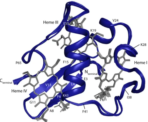
structure of PpcA from Gm ... 41 Figure 3.3 - Overlay of the 20 lowest-energy NMR solution structures of PpcA in the
reduced state ... 42 Figure 3.4 - Average pairwise backbone and heavy atom RMSD values per residue of the
family of 20 conformers for the solution structure of PpcA ... 43 Figure 3.5 - Comparison of secondary structural motifs of PpcA given by TALOS+ and
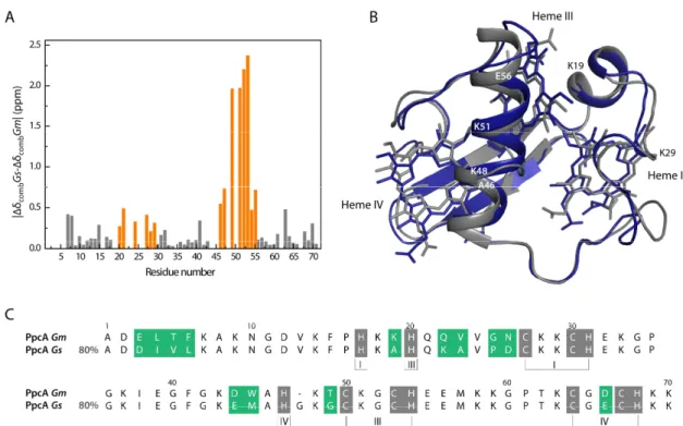
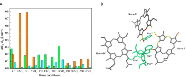
from structure calculations of this work ... 43 Figure 3.6 – Structural differences between PpcA cytochromes from Gm and Gs . ... 44 Figure 3.7 – Location of the heme’s substituents with higher chemical shift differences
between PpcA from Gm and PpcA from Gs . ... 45 Figure 3.8 - Comparison of pH-linked conformational changes in PpcA from Gm and
PpcA from Gs ... 47 Figure 3.9 – Spatial disposition of residues with significant chemical shift variation on
the pH titration, located in the vicinity of heme IV ... 48 Figure 3.10 - Spatial disposition of residues with significant chemical shift variation on
Figure 4.2 - Inter-heme NOE connectivities observed for PpcA in the oxidized state ... 59
Figure 4.3 - 2D 1H,15N-HSQC spectrum of PpcA in the oxidized state ... 60
Figure 4.4 - TALOS+ secondary structural motifs prediction for PpcA in the oxidized state ... 61
Figure 4.5 - Mapping of structural differences between PpcA from Gm and Gs in the oxidized state ... 62
Figure 4.6 - Comparison of pH-linked conformational changes in PpcA from Gm and PpcA from Gs ... 64
5 Role of Phe6, Val13 and Trp45 Residues in PpcA Figure 5.1 - Spatial location of the residue Val13 in the solution structure of PpcA from Gs ... 72
Figure 5.2 - Selected regions of 2D 1H,1H-TOCSY and 2D 1H,1H-NOESY NMR spectra to illustrate the presence of different conformations in solution . ... 74
Figure 5.3 - Impact of the mutations V13A, V13I, V13S and V13T on the heme core architecture ... 75
Figure 5.4 – Agarose gel electrophoresis of the PCR products for the assessment of the best PCR conditions ... 77
Figure 5.5 - Purification of 15N labeled PpcA F6L ... 78
Figure 5.6 - SDS-PAGE gels of the purification process of the mutants ... 79
Figure 5.7 - Spatial location of the Phe6 and Trp45 residues in the solution structure of PpcA from Gm in the reduced state ... 80
Figure 5.8 - Comparison between the 1H,15N-HSQC spectrum of fully reduced F6L and W45M mutants with the spectrum of wild-type PpcA ... 81
Figure 5.9 - Selected regions of 2D 1H,1H-TOCSY and 2D 1H,1H-NOESY NMR spectra to illustrate the observed differences in the hemes' substituents ... 82
Figure 5.10 - Impact of the F6L and W45M mutations on the heme core architecture ... 83
Figure 5.11 - UV-Visible spectra of PpcA F6L in the fully oxidized and reduced states ... 84
Figure 5.12 - Redox titrations followed by UV-Visible spectroscopy for PpcA F6L and W45M mutants at pH 8, 15 °C ... 85
A Appendix Figure A1.1 - pCSGmet2902 plasmid ... 95
Figure A1.2 - Overview of the NZYMiniprep protocol used for the isolation of plasmid DNA ... 95
Figure A1.3 - Protein molecular weight markers used in this work for the SDS-PAGE gels . ... 95
Figure A1.4 - DNA ladder used in this work for the 0.8% agarose gels ... 96
Figure A1.5 - Inter-heme NOE connectivities for PpcA from Gm in the reduced state ... 96
Figure A1.6 - Purification of natural abundance PpcA F6L ... 97
Figure A1.7 - Purification of natural abundance PpcA W45M ... 98
Figure A1.8 - Purification of 15N labeled PpcA W45M ... 99
List of Tables
1 Introduction
Table 1.1 - Heme reduction potentials and pairwise interactions of the fully reduced and protonated forms of PpcA, PpcB, PpcD, PpcE of Gs and PpcA of Gm at 15 ºC,
250 mM ionic strength and pH 7 ... 12
2 Materials and Methods Table 2.1 - PCR mix components used for site-directed mutagenesis of PpcA ... 20
Table 2.2 - PCR program used for site-directed mutagenesis of PpcA ... 20
Table 2.3 - 2D NMR experiments used in this work ... 26
Table 2.4 - 3D NMR experiments used in this work ... 27
Table 2.5 - NMR acquisition parameters for each experiment in the reduced state ... 28
Table 2.6 - NMR acquisition parameters for each experiment in the oxidized state ... 29
3 Structural and Functional Studies of PpcA in the Reduced State Table 3.1 - Summary of restraint violations and quality analysis of the current family of solution structures of PpcA in the reduced state ... 41
Table 3.2 - Thermodynamic parameters of the fully reduced and protonated forms of PpcA from Gm and PpcA from Gs at 15 ºC, 250 mM of ionic strength, pH 7 ... 46
5 Role of Phe6, Val13 and Trp45 Residues in PpcA Table 5.1 - Protein yields of PpcA F6L and W45M mutants expressed in natural abundance and with 15N-isotopic labeling ... 79
Table 5.2 - Apparent and macroscopic reduction potentials for PpcA mutants F6L and W45M, wild-type PpcA and PpcA from Gs at pH 8, 15 ºC ... 85
A Appendix Table A2.1 - Genotype of E. coli DH5α cells ...101
Table A2.2 - Genotype of E. coli BL21(DE3) cells ...101
Table A2.3 - Composition of the 15% SDS-PAGE gel ...102
Table A2.4 - 1H and 13C chemical shifts of the heme substituents of PpcA in the oxidized state, pH 5.5, 25 ºC ...102
Table A2.5 - Backbone and side chain chemical shifts of oxidized PpcA, pH 5.5, 25 ºC ...103
Table A2.6 - 1H chemical shifts of the heme substituents of PpcA V13 mutants in the reduced state, pH 8 and 15 ºC ...112
Table A2.7 - Backbone chemical shifts of reduced PpcA F6L, pH 7.1, 25 ºC . ...114
Table A2.8 - Backbone chemical shifts of reduced PpcA W45M, pH 7.1, 25 ºC ...116
Table A2.9 - 1H chemical shifts of the heme substituents of PpcA F6L and PpcA W45M in the reduced state, pH 7.1, 25 ºC ...118
Abbreviations, Symbols and Constants
1D One dimensional
2D Two dimensional
2xYT 2x yeast extract – tryptone
3D Three dimensional
AMP Ampicillin
ATP Adenosine triphosphate
B0 Magnetic Field
bp Base pair
BLAST Basic Local Alignment Search Tool BMRB Biological Magnetic Resonance Bank
Ccm Cytochrome c maturation
CLO Chloramphenicol
E. coli Escherichia coli
Eapp/E0 Macroscopic apparent midpoint reduction potential
Da Dalton
DIET Direct Interspecies Electron Transfer DMSO Dimethyl sulfoxide
EDTA Ethylenediaminetetraacetic acid FID Free Induction Decay
Gm Geobacter metallireducens Gs/GSU Geobacter sulfurreducens
H-bond Hydrogen bond
HSQC Heteronuclear Single Quantum Coherence
I Spin quantum number
Imc Inner membrane cytochrome
IPTG Isopropyl β-D-thiogalactoside
IUPAC-IUB International Union of Pure and Applied Chemistry – International Union of Biochemistry
J Coupling constant
LB Luria Bertrani
lov Lower limits for volumes
Mac Membrane associated cytochrome
mI Magnetic quantum number
MFC Microbial Fuel Cell
MQH2 Menaquinol
MQ Menaquinone
NADH Nicotinamide adenine dinucleotide (reduced form)
NHE Normal Hydrogen Electrode
NMR Nuclear Magnetic Resonance NOE Nuclear Overhauser Effect
NOESY Nuclear Overhauser Effect SpectroscopY OD600 Optical Density at 600 nm
Omc Outer membrane cytochrome
P Angular momentum
PX Oxidation fraction x
pijk Microstate with hemes i, j, k oxidized
PCR Polymerase Chain Reaction
PDB Protein Data Bank
pI Isoelectric point
Ppc Periplasmic cytochrome ppm Parts per million
RMSD Root mean square deviation rpm Rotations per minute
SX Oxidation stage x
SDS-PAGE Sodium Dodecyl Sulfate–PolyAcrylamide Gel Electrophoresis
TAE Tris-Acetate EDTA
TEMED Tetramethylethylenediamine TOCSY Total Correlation SpectroscopY Tris Tris(hydroxymethyl)aminomethane upv Upper limits for volumes
UV-Visible Ultraviolet-Visible
WT Wild-Type
δ Chemical Shift
γ Magnetogyric ratio
ε Molar extinction coefficient
μ Magnetic moment
t Evolution time
τm Mixing period
υ0 Larmor frequency
δaverage Delta average
δcomb Combined chemical shift
F Faraday constant (96 485 C mol-1)
h Planck constant (6.63x10⁻³⁴ m2 kg s-1)
kB Boltzmann constant (1.38x10⁻²³ m2 kg s-2 K-1)
Amino Acid Abbreviations Alanine Ala A Arginine Arg R Asparagine Asn N Aspartate Asp D Cysteine Cys C Glutamate Glu E Glutamine Gln Q Glycine Gly G Histidine His H Isoleucine Ile I Leucine Leu L Lysine Lys K Methionine Met M Phenylalanine Phe F Proline Pro P Serine Ser S Threonine Thr T Tryptophan Trp W Tyrosine Tyr Y Valine Val V
_____________________________________________________________________________
Chapter 1
Introduction
1.INTRODUCTION
1 Introduction
Anaerobic environments are prolific in microbial communities that have developed different strategies to obtain energy without using oxygen as terminal electron acceptor in the respiration processes. Dissimilatory metal-reducing organisms, such as the ones belonging to the Geobacter genera, couple cellular growth to the reduction of metals present in the environment [1]. This is only possible due to the development of an extracellular electron transport chain that is able to send the electrons from the cell’s interior to the environment, crossing the cellular membrane. Not only this metabolism has its specificities in order to cope with electron transfer across a lipidic barrier, but also influences the biogeochemistry of the microbe’s environment. Taking advantage of Geobacter physiological hallmarks, these bacteria have been used for bioremediation of contaminated waters and energy production in Microbial Fuel Cells (MFC).
1.1 The Geobacter genus
The Geobacteraceae family belongs to the Desulfuromonadales order in the δ-proteobacteria class and comprises four genera of Gram-negative, nonspore-forming, dissimilative iron-reducing bacteria: Geobacter, Desulfuromonas, Desulfuromusa and Pelobacter [1, 2].
Geobacter species typically colonize aquatic sediments, wetlands, rice paddies, and subsurface environments [3] where the concentration of Fe(III) in sediments frequently exceeds that of other electron acceptors such as oxygen, nitrate and sulfate [4, 5]. Not only this reduction is highly advantageous for the bacteria, but also plays an essential role in the ecosystem’s geochemistry because it releases dissolved Fe(II) and Mn(II) as well as trace metals, metalloids, and phosphate that adsorb onto Fe(III) and Mn(IV) oxides. In fact, the studies that led to the discovery of the first Geobacter species were initially designed to better understand the flux of phosphate from aquatic sediments that contributes to algal blooms [3].
Currently, there are 19 known species of Geobacter isolated from different environments such as aquatic sediments (Geobacter metallireducens), contaminated ditches (Geobacter sulfurreducens), Fe(III)-reducing subsurface sediments (Geobacter bemidjiensis), freshwater sediments (Geobacter lovleyi) or uranium-contaminated subsurface sediments (Geobacter uraniireducens). Geobacter species have attracted much attention since they present a few physiological features that had never been found in living organisms: (i) use of Fe(III), Mn(IV) or humic substances as electron terminal acceptors in respiration processes; (ii) production of extracellular magnetite from microbial Fe(III) reduction; (iii) anaerobic oxidation of aromatic hydrocarbons in pure culture; (iv) microbial reduction of water contaminants such as U(VI), Cr(VI) or Co(III); (v) oxidation of organic compounds to carbon dioxide with an electrode serving as a sole electron acceptor; (vi) potential for interaction with syntrophic partners via Direct Interspecies Electron Transfer (DIET) [3].
1.2 Geobacter metallireducens and Geobacter sulfurreducens
Geobacter metallireducens (Gm) was the first member of the Geobacter species to be isolated in culture from sediments from the Potomac River Estuary, Maryland, in 1987 [5, 6]. Even though Gm was the first member of Geobacter to be described, only in 2009 its genome was fully sequenced, which allowed its genetic manipulation and study of electron transfer processes [7]. Geobacter sulfurreducens (Gs) was isolated in 1994 from surface sediments of a hydrocarbon-contaminated ditch near Norman, Oklahoma [8] and, contrarily to Gm, the development of a genetic system that permitted the manipulation of its genome in 2001 has established this species as the representative of the Geobacter genus [9].
Gm presents the unique ability of oxidizing an enormous variety of organic compounds both aromatic (such as benzaldehyde, phenol, benzene and toluene) and non-aromatic (such as butanol, ethanol and propanol) compared to Gs, which is only able to oxidize acetate, formate, lactate or pyruvate (only with hydrogen as electron donor) [3, 7]. Regarding the electron acceptors, both species are capable of reducing U(VI) and Tc(VII), but only Gm is capable of reducing V(V). This physiological hallmark has been used for the bioremediation of waters contaminated with these compounds [10-12]. The first DIET mechanism was documented in co-cultures of Gm and Gs grown in a medium with ethanol as the electron donor and fumarate as the electron acceptor [13]. DIET was required since Gm could metabolize ethanol but could not use fumarate as an electron acceptor and Gs could reduce fumarate but was not capable of metabolizing ethanol. Gm is also capable of performing DIET with Methanosaeta species in the conversion of waste waters’ organic compounds to methane, an important process used for bioenergy production [14].
Besides the referred applications of Geobacter in the bioremediation of contaminated waters, the bacterial metabolism can be used for production of electrical current from a wide range of organic wastes in MFC devices, a promising form of sustainable energy. However, the present MFC are still far from generating a great amount of electric current [15], and several strategies are being employed for their improvement such as the design of Geobacter strains with enhanced extracellular electron transfer rates [16, 17]. For this, it is necessary to elucidate these mechanisms which are still poorly understood [3].
1.3 The Role of Cytochromes c in Extracellular Electron Transfer Mechanisms
The sequenced genomes of the Geobacter species have presented more than 100 genes encoding for c-type cytochromes with an average of 85% with more than one heme group [18]. The presence of multiple heme groups may confer several advantages: (i) the overlapping of the collective contribution of the individual heme reduction potential ranges extends the protein’s global working reduction potential range; (ii) the presence of more than one heme group1.INTRODUCTION
protonable centers in the protein that modulate the potential according to the solution’s pH (redox-Bohr effect); (iii) the capability of acting as capacitor to maintain the bacteria’s metabolism during periods of time in which the cells are unable to reduce extracellular electron transfer acceptors [19].
In Geobacter the cytochromes are distributed along the inner membrane, periplasm and outer membrane and are responsible for guiding the electron flow from the cytoplasm to the cell’s exterior, constituting an excellent target for the improvement of extracellular electron transfer rates [18].
1.3.1 Characteristics of c-type Cytochromes
The c-type cytochromes are metalloproteins with an essential role in aerobic and anaerobic respiratory processes. The heme group is constituted by a protoporphyrin IX ring with an iron atom in the center, coordinated by the four nitrogen atoms of the ring. The iron (0) is a transition metal with 26 electrons arranged in the electronic configuration 1s22s22p63s23p63d64s2,
where the numbers correspond to the orbitals (s, p and d) and the superscript represents the electron occupancy. An alternative description of the electronic distribution is given by [Ar]3d64s2 which emphasizes the electron occupancy of the higher energy orbitals. In electron
transfer cytochromes, iron generally exists in the ferrous (Fe(II)) and in the ferric (Fe(III)) states with the respective electronic configurations [Ar]3d64s0 and [Ar]3d54s0 [20]. The high and
low-spin states of the iron are consequence of the distribution of the electrons in the five 3d orbitals (two orbitals eg with higher energy and three orbitals t2g with lower energy, in accordance with
the Crystal Field Theory [21]), which in turn are modulated by the nature of the ligands coordinated to the atom. If the d-orbital splitting is smaller than the pairing energy (weak crystal field) the electrons tend to remain mostly unpaired in the orbitals and give rise to a high-spin state. Otherwise, if the d-orbital splitting is larger than the pairing energy (strong crystal field) the electrons tend to pair firstly on the t2g orbitals and give rise to a low-spin state [20].
The heme cofactor is covalently linked to the polypeptide chain via two thioether bonds between the thiol groups of cysteines and the vinyl groups of the heme. As a consequence of this, there is a conserved binding motif sequence of the heme groups: CXXCH, where C represents the amino acid cysteine, X any given amino acid and H the amino acid histidine, which is the proximal ligand (Figure 1.1). The iron atom is always coordinated with five atoms: four nitrogen atoms of the protoporphyrin IX ring and one nitrogen atom of the imidazole ring of the histidine side chain. The sixth coordination position – that corresponds to the distal ligand – can either be transiently vacant or occupied. In cytochromes with enzymatic activity, the sixth coordination is transiently vacant [22, 23] whereas in electron transfer cytochromes it can be occupied by the side chain of a: (i) methionine, which predominates in monoheme cytochromes c, (ii) histidine, particularly in multiheme cytochromes or (iii) asparagine or tyrosine, which occur less frequently. In each case, the axial ligating atom is different: (i) nitrogen, in the case of histidine
or asparagine, (ii) sulfur in the case of methionine, and (iii) oxygen in the case of tyrosine [20, 22, 23].
Figure 1.1 - Schematic representation of a c-type heme and the correspondent polypeptide binding motif. The axial coordination position labeled with A can be free or occupied by the side chain of a, for example, methionine, histidine, asparagine or tyrosine residue. The IUPAC-IUB nomenclature for tetrapyrroles is illustrated in gray [24]. Figure reproduced from [19].
1.3.2 The Extracellular Electron Transfer Chain in Geobacter
Currently, the extracellular electron transfer mechanisms of Gs have been the most studied given the earlier development of a genetic system for proteomic and gene-knockout studies in this microorganism. The current model for electron transfer (Figure 1.2) postulates that electrons originating from the oxidation of organic compounds are transferred to a menaquinone pool via a NADH hydrogenase located in the inner membrane [19]. Depending on the redox potential of the final electron acceptor, the electrons are transferred to either one of two pathways: the CbcL dependent pathway, which operates when the final electron acceptors are at or below redox potentials of -100 mV (vs. the normal hydrogen electrode, NHE) while the ImcH pathway is used when final electron acceptors are above the referred redox potential [25, 26]. The electrons are then transferred from CbcL or ImcH to cytochromes in the periplasm (mainly to the periplasmic cytochrome family PpcA) and from these to porin-cytochrome trans-outer membrane complexes (such as the OmaB-OmbB-OmcB or OmaC-OmbC-OmcC) which convey the electrons to the extracellular electron acceptors [27]. In addition to these electron transfer proteins, Geobacter bacteria possess electrically conductive pili that establish electrical contacts with electrodes and other methanogenic species, and flagella that allow them to move toward solid extracellular electron acceptors [28]. In this year, it was discovered that one of the extracellular cytochromes – OmcS (Outer membrane cytochrome S) –, is capable of polymerizing and forming electrically conductive filaments, illustrating the vast complexity of
1.INTRODUCTION
Figure 1.2 - Model of the extracellular electron transfer pathway in Geobacter sulfurreducens. The proposed electron transfer pathway is represented in white: the electrons from the oxidation of organic compounds are transferred to the menaquinol (MQH2)/menaquinone (MQ) pool at the inner membrane
and from here either to CbcL or ImcH cytochromes. Then, the electrons are transferred to the PpcA family of periplasmic cytochromes (PpcA (PDB code 1OS6), PpcB (PDB code 3BXU), PpcC (PDB code 3H33), PpcD (PDB code 3H4N) and PpcE (PDB code 3H34)) which mediate the electron transfer to outer membrane cytochromes. Pili and the newly discovered polymeric assembly of cytochrome OmcS are also represented and are thought to be involved in long-range electron transfer. MacA (PDB code 4AAL), a membrane-associated diheme cytochrome c which is important for reduction of Fe(III) and U(VI) oxides [30], GSU1996 (PDB code 3OV0), a “nanowire” that may function as a capacitor to enhance the bacterial electron-storage capacity [31], and OmcF (PDB code 3CU4), a monoheme cytochrome which absence negatively affects the Fe(III) citrate reduction and production of electric current [32], are also represented.
In Gm, the extracellular electron transfer mechanisms have been considerably less studied. The existent studies point to the conservation of some electron transfer components, such as the PpcA family of triheme cytochromes and outer membrane cytochromes OmcB and OmcE, but further structural and functional information on these components is scarce [33-36].
1.4 The PpcA Family of Triheme Cytochromes
The PpcA family of c7 triheme cytochromes belongs to class III cytochromes since these proteins
possess multiple covalently attached heme groups with low reduction potentials and bis-histidinyl coordination [20]. The PpcA family is one of the most conserved among Geobacter, with multiple homologs in most of the genomes: five in G. sulfurreducens, five in G. metallireducens, four in G. uraniireducens, three in G. bemidjiensi, and one in G. lovleyi. The PpcA family in Gs is constituted by PpcA, PpcB, PpcC, PpcD, PpcE whereas in Gm the family is constituted by PpcA, PpcB, PpcC, PpcE, PpcF. The PpcD degree of homology is very low between the Gs and Gm families and therefore the designation PpcF was adopted in Gm [33].
In Gs, PpcA, PpcB and PpcC were detected in cultures grown in the presence of Fe(III) citrate and Fe(III) oxide and the deletion of these genes affected the reduction of Fe(III) and U(VI). PpcD is more abundant during growth with Fe(III) oxides and PpcE was only detected in cultures in presence of Fe(III) citrate. The deletion of the genes of PpcD and PpcE imparted the reduction of U(VI) [37]. In Gm, the PpcA family is known to be essential for Fe(III) and U(VI) reduction [38] and the ppcA gene is generally up-regulated in DIET processes of co-cultures of Gm with other organisms such as Gs or Methanosarcina barkeri [39, 40].
The cytochromes from both families possess about 70 amino acids with a molecular mass of approximately 10 kDa and a pI of 9 [33, 34, 41]. The sequence alignment, as well as the identity relatively to PpcA of Gm, are indicated in Figure 1.3.
Figure 1.3 - Alignment of the amino acid sequences of the periplasmic triheme cytochromes from Geobacter metallireducens and Geobacter sulfurreducens. Black and gray boxes color the conserved non-heme attached and non-heme-attached residues, respectively. The residues studied within the scope of this work are highlighted in red. The heme numbering and the respective attached residues are indicated at the bottom of the last cytochrome amino acid sequence. The percentage of sequence identity relative to PpcA from Gm obtained from the basic local alignment search tool (BLAST) [42] is indicated. The alignment was performed with CLUSTAL Ω [43].
Three c-type heme groups are covalently attached to the polypeptide chain as confirmed by the existence of three CXXCH motifs in each sequence. The hemes are axially coordinated by two histidine residues and are low-spin both in the reduced diamagnetic state (Fe(II), S=0) and in the oxidized paramagnetic state (Fe(III), S=1/2) which gives rise to specific features in the UV-Visible spectrum (Figure 1.4).
1.INTRODUCTION
Figure 1.4 - Electronic properties of a low-spin iron attached to the heme group and UV-Visible spectroscopic properties of low-spin c-type cytochromes. (A) Electronic distribution of octahedral Fe(II) and Fe(III) in the low-spin state in which the d-orbital splitting, Δo, is higher than the energy
required to pair electrons in the same orbital. (B) The spectral features are illustrated by the UV-Visible spectra of PpcA from Gm in the fully oxidized (solid line) and fully reduced (dashed line) states. The characteristic bands of the spectra in both redox states are indicated.
1.4.1 Structural Features of Triheme Cytochromes
To date, only the structure of the triheme cytochromes from Gs has been determined (Figure 1.5), showing several conserved motifs: a two-strand β-sheet segment at the N terminal, followed by several α-helixes located in different regions of the protein [44]. The spatial arrangement of the hemes in these cytochromes is conserved, with heme III nearly perpendicular to both hemes I and IV [44]. The hemes’ designation in c7 triheme cytochromes
derives from the superimposition of the hemes with those of the structurally homologous tetraheme cytochromes c3 in which the formers lack heme II and the correspondent polypeptide
Figure 1.5 - Structures of PpcA family members from Geobacter sulfurreducens. PpcA is represented by its solution structure in the reduced state (PDB code 2LDO) and PpcB (PDB code 3BXU), PpcC (PDB code 3H33), PpcD (PDB code 3H4N), PpcE (PDB code 3H34) are represented by their crystal structures. PpcB and PpcD display two molecules in the crystal asymmetric unit (A and B monomers) of which monomer A is represented. The molecules are all in the same orientation. Figure produced with PyMOL [46].
Regarding the cytochromes of the PpcA family from Gm, only cytochromes PpcA and PpcF have been preliminarily characterized. Nuclear Magnetic Resonance (NMR) studies of these proteins in the reduced state have revealed conservation of the heme core of the proteins in comparison with PpcA from Gs. The assignment of the 2D 1H,15N-HSQC (Heteronuclear Single
Quantum Coherence) NMR spectrum of PpcA and its comparison with PpcA from Gs has suggested that the overall folding is maintained in both structures, with important structural differences in segments that include the cleft between hemes I and III [34]. Furthermore, the hemes’ magnetic properties and axial ligands’ orientation have been determined for PpcA from Gm in the oxidized state and have shown that there is conservation of the orientation compared to PpcA from Gs [47].
1.4.2 Functional Characterization of Triheme Cytochromes
In monoheme cytochromes, the redox potential can be obtained directly from the Nernst equation and the Eapp (i.e. the point at which the oxidized and reduced fractions of the protein
are equal) corresponds to the reduction potential of the heme. In the case of multiheme cytochromes, the co-existence of several heme groups that generally possess similar reduction potentials does not allow the direct discrimination of the individual potentials by voltammetry or potentiometric redox titrations. Instead, the Eapp determined by these methods is
1.INTRODUCTION
In a triheme cytochrome, four different redox stages can be defined as a consequence of the three consecutive reversible steps of one-electron transfer that convert the fully reduced state (Stage 0, S0) in the fully oxidized state (Stage 3, S3) (Figure 1.6A). At each stage, several
microstates with the same number of oxidized hemes co-exist and, additionally, for each microstate, the protonable center(s) in the protein that modulate the hemes’ reduction potential (redox-Bohr effect) can be protonated or deprotonated, leading to a minimum of 16 possible microstates. Due to the close spatial disposition of the heme groups within the protein, the reduction potential of one heme is modulated by the oxidation stage of its neighbor (redox interactions) and by the solution pH (redox-Bohr interactions). Therefore, the complete characterization of the redox centers of a multiheme cytochrome comprises the determination of the individual heme reduction potentials, the redox interactions and the properties of the redox-Bohr center(s). Turner and co-workers have developed a strategy that permits the determination of these parameters by combining the information from UV-Visible redox titrations with NMR to determine the individual heme oxidation profiles [48]. This is possible due to the spectral signatures of the NMR spectra of low-spin multiheme cytochromes in the reduced and oxidized states (Figure 1.6B).
Figure 1.6 - Electronic distribution network for a triheme cytochrome and the respective 1D 1H NMR
spectra in the reduced and oxidized states. (A) Electronic distribution scheme for a triheme cytochrome with one proton-linked equilibrium showing the 16 possible microstates. The full and dashed triangles correspond to the protonated and deprotonated microstates, respectively. In each microstate, the heme groups are colored green (heme I), orange (heme III) and blue (heme IV), which can be either reduced (filled symbols) or oxidized (open symbols). The microstates are grouped according to the number of oxidized hemes in four oxidation stages connected by three one-electron redox steps indicated by S0, S1, S2
and S3. P0, P1, P2 and P3 indicate the molar fractions associated to each stage concerning microstates with
zero, one, two or three oxidized hemes respectively. Ex(x = 1-3) represents the macroscopic reduction
potential associated with each stage. p0 and p0H indicate the deprotonated and protonated reduced
microstates, respectively. pijk and pijkH correspond to the deprotonated and protonated microstates,
respectively, with heme(s) i, j, and k oxidized. (B) 1D 1H NMR spectra of the reduced (upper) and
oxidized (lower) forms of PpcA from Gm (25 °C and pH 7). Figure reproduced from [34].
When the interconversion between microstates within the same oxidation stage – intramolecular electron exchange – is fast on the NMR timescale and the interconversion between microstates of different oxidation stages – intermolecular electron exchange – is slow, the individual heme signals in the different oxidation stages can be discriminated. Moreover, the NMR paramagnetic shifts are proportional to the degree of oxidation of a particular heme group and, therefore, can be used to monitor the oxidation of each heme. The heme methyl resonances are the easiest to identify in the NMR spectra and their largest paramagnetic shifts make them the ideal candidates for following the stepwise oxidation of the hemes throughout the redox titration. However, this information only indicates the relative heme reduction potentials and heme redox interactions. Therefore, to determine the absolute potentials, the total reduced protein fractions need to be measured through redox titrations followed by UV-Visible spectroscopy.
The known thermodynamic parameters of the PpcA family from Gs as well as of PpcA from Gm [35, 41] are summarized in Table 1.1.
Table 1.1 - Heme reduction potentials and pairwise interactions (mV) of the fully reduced and protonated forms of PpcA, PpcB, PpcD, PpcE of Gs and PpcA of Gm at 15 ºC, 250 mM ionic strength and pH 7. Reduction potentials are relative to NHE.
Protein Heme reduction potentials Heme-heme redox interactions Redox-Bohr interactions
I III IV I-III I-IV III-IV I-H III-H IV-H
PpcA Gs -154 -138 -125 27 16 41 -32 -31 -58
PpcB Gs -150 -166 -125 17 8 32 -16 -9 -38
PpcD Gs -156 -139 -149 46 3 14 -28 -23 -53
PpcE Gs -167 -175 -116 27 5 22 -12 2 -13
PpcA Gm -80 -70 -113 35 3 37 -2 -23 -49
The heme reduction potentials are negative, differ from each other and cover different functional ranges. In the particular case of PpcA from Gm and PpcA from Gs, the thermodynamic properties are markedly different despite the 80% of sequence identity. Furthermore, at pH 7, the heme oxidation order is IV-I-III for PpcA from Gm and I-IV-III for PpcA from Gs, with these proteins displaying different preferential pathways for electron transfer (Figure 1.7) but maintaining the ability to couple proton to electron transfer. Therefore, it is striking that two proteins with such similarity in the amino acid content present markedly different functional properties.
1.INTRODUCTION
Figure 1.7 - Preferential pathway for electron/proton transfer in PpcA from (A) Geobacter sulfurreducens and (B) Geobacter metallireducens at pH 7. The black arrows indicate the preferential electron transfer pathway involving the relevant microstates. An electron/proton coupling is observed between oxidation stages S1 and S2.
1.5 Objectives and Thesis Outline
The functional differences shown by PpcA from Gm and PpcA from Gs, regardless their high sequence similarity, establish these two proteins as an excellent model to identify the key residues that account for the observed functional differences. However, to attain this objective, it is first necessary to establish a solid characterization of PpcA from Gm in both the reduced and oxidized states.
In this thesis, NMR spectroscopy was used to structurally characterize PpcA from Gm in the reduced and oxidized states and to probe the structural and functional impact of residues Val13,
Phe6 and Trp45 in the modulation of redox properties. Chapter 1 provides a general
contextualization of Geobacter and the role of multiheme cytochromes in extracellular electron transfer mechanisms. Chapter 2 describes the experimental procedures employed in this work and introduces the fundamental principles of NMR spectroscopy. Chapters 3 and 4 describe the structural and functional characterization of PpcA in the reduced and oxidized states, respectively. Site-directed mutagenesis on the above mentioned residues was carried out and the structural and functional characterization of these mutants are described in Chapter 5. Finally, Chapter 6 highlights the main conclusions of this thesis and presents future directions that can be pursued.
1.6 References
[1] M. Madigan, J. Martinko, K. Bender, D. Buckley, D. Stahl, Brock Biology of Microorganisms, 14 ed., Benjamin Cummings, Boston, 2014.
[2] W.F.M. Röling, The family Geobacteraceae, in: E. Rosenberg, E.F. DeLong, S. Lory, E. Stackebrandt, F. Thompson (Eds.) The Prokaryotes: Deltaproteobacteria and Epsilonproteobacteria, Springer Berlin Heidelberg, Berlin, Heidelberg, 2014, pp. 157-172.
[3] D.R. Lovley, T. Ueki, T. Zhang, N.S. Malvankar, P.M. Shrestha, K.A. Flanagan, M. Aklujkar, J.E. Butler, L. Giloteaux, A.-E. Rotaru, D.E. Holmes, A.E. Franks, R. Orellana, C. Risso, K.P. Nevin, Geobacter: The Microbe Electric's Physiology, Ecology, and Practical Applications, in: R.K. Poole (Ed.) Advances in Microbial Physiology, Academic Press, 2011, pp. 1-100.
[4] D.R. Lovely, Microbial reduction of iron, manganese, and other metals, Advances in Agronomy (USA), v. 54 (1995).
[5] D.R. Lovley, E.J. Phillips, Organic matter mineralization with reduction of ferric iron in anaerobic sediments, Applied and Environmental Microbiology, 51 (1986) 683-689.
[6] D.R. Lovley, E.J. Phillips, Novel mode of microbial energy metabolism: organic carbon oxidation coupled to dissimilatory reduction of iron or manganese, Applied and Environmental Microbiology, 54 (1988) 1472-1480.
[7] M. Aklujkar, J. Krushkal, G. DiBartolo, A. Lapidus, M.L. Land, D.R. Lovley, The genome sequence of Geobacter metallireducens: features of metabolism, physiology and regulation common and dissimilar to Geobacter sulfurreducens, BMC Microbiology, 9 (2009) 109.
[8] F. Caccavo, Jr., D.J. Lonergan, D.R. Lovley, M. Davis, J.F. Stolz, M.J. McInerney, Geobacter sulfurreducens sp. nov., a hydrogen- and acetate-oxidizing dissimilatory metal-reducing microorganism, Applied and Environmental Microbiology, 60 (1994) 3752-3759.
[9] M.V. Coppi, C. Leang, S.J. Sandler, D.R. Lovley, Development of a genetic system for Geobacter sulfurreducens, Applied and Environmental Microbiology, 67 (2001) 3180-3187. [10] G. Liu, J. Zhou, C. Chen, J. Wang, R. Jin, H. Lv, Decolorization of azo dyes by Geobacter metallireducens, Applied Microbiology and Biotechnology, 97 (2013) 7935-7942.
[11] I. Ortiz-Bernad, R.T. Anderson, H.A. Vrionis, D.R. Lovley, Vanadium respiration by Geobacter metallireducens: novel strategy for in situ removal of vanadium from groundwater, Applied and Environmental Microbiology, 70 (2004) 3091-3095.
[12] J.R. Lloyd, V.A. Sole, C.V. Van Praagh, D.R. Lovley, Direct and Fe(II)-mediated reduction of technetium by Fe(III)-reducing bacteria, Applied and Environmental Microbiology, 66 (2000) 3743-3749.
[13] Z.M. Summers, H.E. Fogarty, C. Leang, A.E. Franks, N.S. Malvankar, D.R. Lovley, Direct exchange of electrons within aggregates of an evolved syntrophic coculture of anaerobic bacteria, Science, 330 (2010) 1413-1415.
[14] A.-E. Rotaru, P.M. Shrestha, F. Liu, M. Shrestha, D. Shrestha, M. Embree, K. Zengler, C. Wardman, K.P. Nevin, D.R. Lovley, A new model for electron flow during anaerobic digestion: direct interspecies electron transfer to Methanosaeta for the reduction of carbon dioxide to methane, Energy & Environmental Science, 7 (2014) 408-415.
[15] A.E. Franks, K.P. Nevin, Microbial fuel cells, a current review, Energies, 3 (2010) 899-919. [16] M. Izallalen, R. Mahadevan, A. Burgard, B. Postier, R. Didonato Jr, J. Sun, C.H. Schilling, D.R. Lovley, Geobacter sulfurreducens strain engineered for increased rates of respiration, Metabolic engineering, 10 (2008) 267-275.
1.INTRODUCTION
[17] H. Yi, K.P. Nevin, B.-C. Kim, A.E. Franks, A. Klimes, L.M. Tender, D.R. Lovley, Selection of a variant of Geobacter sulfurreducens with enhanced capacity for current production in microbial fuel cells, Biosensors and Bioelectronics, 24 (2009) 3498-3503.
[18] J.E. Butler, N.D. Young, D.R. Lovley, Evolution of electron transfer out of the cell: comparative genomics of six Geobacter genomes, BMC Genomics, 11 (2010) 40.
[19] C.A. Salgueiro, J.M. Dantas, Multiheme Cytochromes, in: Multiheme Cytochromes, Springer Berlin Heidelberg, Berlin, Heidelberg, 2016, pp. 1-39.
[20] G.R. Moore, G.W. Pettigrew, Cytochromes c: evolutionary, structural and physicochemical aspects, Springer Science & Business Media, 2012.
[21] J. Van Vleck, Theory of the variations in paramagnetic anisotropy among different salts of the iron group, Physical Review, 41 (1932) 208.
[22] I. Bertini, G. Cavallaro, A. Rosato, Cytochrome c: occurrence and functions, Chemical Reviews, 106 (2006) 90-115.
[23] L.J. Smith, A. Kahraman, J.M. Thornton, Heme proteins—diversity in structural characteristics, function, and folding, Proteins: structure, function, and bioinformatics, 78 (2010) 2349-2368.
[24] G.P. Moss, Nomenclature of tetrapyrroles, European Journal of Biochemistry, 178 (1988) 277-328.
[25] L. Zacharoff, C.H. Chan, D.R. Bond, Reduction of low potential electron acceptors requires the CbcL inner membrane cytochrome of Geobacter sulfurreducens, Bioelectrochemistry, 107 (2016) 7-13.
[26] C.E. Levar, C.H. Chan, M.G. Mehta-Kolte, D.R. Bond, An inner membrane cytochrome required only for reduction of high redox potential extracellular electron acceptors, Mbio, 5 (2014) 1-9.
[27] Y. Liu, Z. Wang, J. Liu, C. Levar, M.J. Edwards, J.T. Babauta, D.W. Kennedy, Z. Shi, H. Beyenal, D.R. Bond, T.A. Clarke, J.N. Butt, D.J. Richardson, K.M. Rosso, J.M. Zachara, J.K. Fredrickson, L. Shi, A trans-outer membrane porin-cytochrome protein complex for extracellular electron transfer by Geobacter sulfurreducens PCA, Environmental Microbiology Reports, 6 (2014) 776-785.
[28] P.-L. Tremblay, M. Aklujkar, C. Leang, K.P. Nevin, D. Lovley, A genetic system for Geobacter metallireducens: role of the flagellin and pilin in the reduction of Fe(III) oxide, Environmental Microbiology Reports, 4 (2012) 82-88.
[29] F. Wang, Y. Gu, J.P. O’Brien, S.M. Yi, S.E. Yalcin, V. Srikanth, C. Shen, D. Vu, N.L. Ing, A.I. Hochbaum, E.H. Egelman, N.S. Malvankar, Structure of microbial nanowires reveals stacked hemes that transport electrons over micrometers, Cell, 177 (2019) 361-369.e310.
[30] B.-C. Kim, D.R. Lovley, Investigation of direct vs. indirect involvement of the c-type cytochrome MacA in Fe(III) reduction by Geobacter sulfurreducens, FEMS Microbiology Letters, 286 (2008) 39-44.
[31] P.R. Pokkuluri, Y.Y. Londer, N.E.C. Duke, M. Pessanha, X. Yang, V. Orshonsky, L. Orshonsky, J. Erickson, Y. Zagyanskiy, C.A. Salgueiro, M. Schiffer, Structure of a novel dodecaheme cytochrome c from Geobacter sulfurreducens reveals an extended 12 nm protein with interacting hemes, Journal of Structural Biology, 174 (2011) 223-233.
[32] J.M. Dantas, M.A. Silva, D. Pantoja-Uceda, D.L. Turner, M. Bruix, C.A. Salgueiro, Solution structure and dynamics of the outer membrane cytochrome OmcF from Geobacter sulfurreducens, Biochimica et Biophysica Acta - Bioenergetics, 1858 (2017) 733-741.
[33] M.R. Ferreira, J.M. Dantas, C.A. Salgueiro, The triheme cytochrome PpcF from Geobacter metallireducens exhibits distinct redox properties, FEBS Open Bio, 8 (2018) 1897-1910.
[34] P.C. Portela, T.M. Fernandes, J.M. Dantas, M.R. Ferreira, C.A. Salgueiro, Biochemical and functional insights on the triheme cytochrome PpcA from Geobacter metallireducens, Archives of Biochemistry and Biophysics, 644 (2018) 8-16.
[35] T.M. Fernandes, L. Morgado, C.A. Salgueiro, Thermodynamic and functional characterization of the periplasmic triheme cytochrome PpcA from Geobacter metallireducens, Biochemical Journal, 475 (2018) 2861-2875.
[36] J.A. Smith, D.R. Lovley, P.-L. Tremblay, Outer cell surface components essential for Fe(III) oxide reduction by Geobacter metallireducens, Appl. Environ. Microbiol., 79 (2013) 901-907. [37] Y.-H.R. Ding, K.K. Hixson, M.A. Aklujkar, M.S. Lipton, R.D. Smith, D.R. Lovley, T. Mester, Proteome of Geobacter sulfurreducens grown with Fe(III) oxide or Fe(III) citrate as the electron acceptor, Biochimica et Biophysica Acta (BBA) - Proteins and Proteomics, 1784 (2008) 1935-1941.
[38] J.R. Lloyd, C. Leang, A.L.H. Myerson, M.V. Coppi, S. Cuifo, B. Methe, S.J. Sandler, D.R. Lovley, Biochemical and genetic characterization of PpcA, a periplasmic c-type cytochrome in Geobacter sulfurreducens, Biochemical Journal, 369 (2003) 153-161.
[39] P.M. Shrestha, A.-E. Rotaru, Z.M. Summers, M. Shrestha, F. Liu, D.R. Lovley, Transcriptomic and Genetic Analysis of Direct Interspecies Electron Transfer, Applied and Environmental Microbiology, 79 (2013) 2397-2404.
[40] A.-E. Rotaru, P.M. Shrestha, F. Liu, B. Markovaite, S. Chen, K.P. Nevin, D.R. Lovley, Direct Interspecies Electron Transfer between Geobacter metallireducens and Methanosarcina barkeri, Applied and Environmental Microbiology, 80 (2014) 4599-4605.
[41] L. Morgado, M. Bruix, M. Pessanha, Y.Y. Londer, C.A. Salgueiro, Thermodynamic characterization of a triheme cytochrome family from Geobacter sulfurreducens reveals mechanistic and functional diversity, Biophysical Journal, 99 (2010) 293-301.
[42] S.F. Altschul, W. Gish, W. Miller, E.W. Myers, D.J. Lipman, Basic local alignment search tool, Journal of Molecular Biology, 215 (1990) 403-410.
[43] F. Madeira, Y.M. Park, J. Lee, N. Buso, T. Gur, N. Madhusoodanan, P. Basutkar, A.R.N. Tivey, S.C. Potter, R.D. Finn, R. Lopez, The EMBL-EBI search and sequence analysis tools APIs in 2019, Nucleic Acids Research, 47 (2019) W636-W641.
[44] P.R. Pokkuluri, Y.Y. Londer, X. Yang, N.E.C. Duke, J. Erickson, V. Orshonsky, G. Johnson, M. Schiffer, Structural characterization of a family of cytochromes c7 involved in Fe(III)
respiration by Geobacter sulfurreducens, Biochimica et Biophysica Acta (BBA) - Bioenergetics, 1797 (2010) 222-232.
[45] D.L. Turner, H.S. Costa, I.B. Coutinho, J. LeGall, A.V. Xavier, Assignment of the ligand geometry and redox potentials of the trihaem ferricytochrome c3 from Desulfuromonas
acetoxidans, Eur. J. Biochem., 243 (1997) 474-481.
[46] Schrodinger, LLC, The PyMOL Molecular Graphics System, Version 1.8, in, 2015.
[47] T.M. Fernandes, L. Morgado, C.A. Salgueiro, D.L. Turner, Determination of the magnetic properties and orientation of the heme axial ligands of PpcA from Geobacter metallireducens by paramagnetic NMR, Journal of Inorganic Biochemistry, 198 (2019) 110718.
[48] D.L. Turner, C.A. Salgueiro, T. Catarino, J. LeGall, A.V. Xavier, NMR studies of cooperativity in the tetrahaem cytochrome c from Desulfovibrio vulgaris, European Journal of
_____________________________________________________________________________
Chapter 2
Materials and Methods
2.MATERIALS AND METHODS
2 Materials and Methods
2.1 Expression Vectors and Site-directed Mutagenesis
The expression vector containing the ppcA gene from Gm – pCSGmet2902 – was already available in the host laboratory. This plasmid contains a lac promotor, an ompA signal sequence from Escherichia coli (E. coli) and a resistance marker to ampicillin [1].
For the site-directed mutagenesis, the protocol followed a strategy based on the NZYMutagenesis kit (NZYTech), which is indicated in Figure 2.1.
Figure 2.1 - Overview of the NZYMutagenesis kit protocol. Adapted from the kit’s instructions manual.
The primers for F6L and W45M mutants were designed by the QuikChange Primer Design program (Agilent Technologies) and synthesized by Invitrogen. For the F6L mutant, the primers used were 5’-CCG CTG ACG AGC TTA CCT TAA AGG CAA AGA ACG-3’ (reverse primer, 52% GC content) and 5’-CGT TCT TTG CCT TTA AGG TAA GCT CGT CAG CGG-3’ (forward primer, 52% GC content) and for the W45M mutant the primers used were 5’-ATC GAG GGC TTT GGC AAG GAT ATG GCT CAC AAG ACT TG-3’ (reverse primer, 50% GC content) and 5’-CAA GTC TTG TGA GCC ATA TCC TTG CCA AAG CCC TCG AT-3’ (forward primer, 50% GC content). The pCSGmet2902 template was amplified by Polymerase Chain Reaction (PCR) using the primers harboring the desired mutations. Due to the high GC content of the primers, two PCR mix reactions using different buffers (Phusion High Fidelity Buffer or GC Buffer) and dimethyl sulfoxide (DMSO) were tested according to the described in Table 2.1. The PCR program is described in Table 2.2. After selection of the best conditions, the parental DNA was eliminated from the selected PCR products by digestion with DpnI (NZYTech), which is specific for methylated DNA, for 1 h at 37 °C after which the enzyme was inactivated by 20 min incubation at 80 °C. The size of the PCR products was confirmed by 0.8% agarose gel using GeneRuler 1kb DNA ladder (New England BioLabs).
Table 2.1 - PCR mix components used for site-directed mutagenesis of PpcA. DMSO is only used with GC Buffer. The Phusion Buffers, DMSO, nucleotides and Phusion DNA polymerase are all from Thermo Fisher Scientific. 1 U is the amount of enzyme needed to convert in one minute 1 μmole of substrate.
PCR mix components Final concentration Phusion High Fidelity Buffer or GC Buffer 1x
Nucleotides 200 μM
Forward Primer 0.5 μM
Reverse Primer 0.5 μM
Template DNA 0.6 ng/μL
Phusion DNA Polymerase 0.02 U/μL
DMSO 3 %
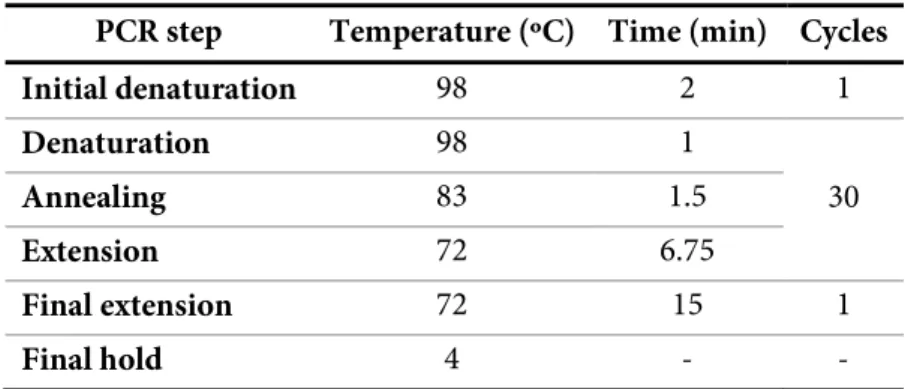
Table 2.2 - PCR program used for site-directed mutagenesis of PpcA.
PCR step Temperature (ºC) Time (min) Cycles
Initial denaturation 98 2 1 Denaturation 98 1 30 Annealing 83 1.5 Extension 72 6.75 Final extension 72 15 1 Final hold 4 - -
E. coli DH5α competent cells were transformed with the PCR products using the Heat Shock Method [2] where cells were incubated 30 minutes on ice, followed by 1-minute incubation at 42 °C and 1-minute incubation on ice again. Then, 500 μL of Luria Bertrani (LB) medium was added and cells were grown aerobically for 1 h at 37 °C, 200 rpm. After the transformation step, the cells were plated on solid LB medium supplemented with 100 μg/mL of ampicillin (AMP) (NZYTech) and incubated overnight at 37 °C. Positive and negative controls were always carried out. Colonies harboring the plasmid with resistance to AMP were inoculated into 5 mL of liquid LB medium supplemented with 100 μg/mL of AMP and were grown overnight aerobically at 37 °C, 200 rpm. The plasmid was isolated from the grown culture using the NZYMiniprep kit (NZYTech) according to the manufacturer’s instructions and the presence of desired mutations was confirmed by DNA sequencing in both strands by STAB VIDA.
2.2 Protein Expression and Purification
co-2.MATERIALS AND METHODS
purified. Wild-type PpcA from Gm both in natural abundance and 13C, 15N-isotopically labeled
were already available in the host lab, as well as PpcA V13 mutants from Gs.
All the descriptions given below apply both to natural abundance and 15N-isotopically labeled
protein overexpression protocols, except for sections 2.2.2 and 2.2.3.
2.2.1 Transformation of E. coli (BL21(DE3)) Cells
Competent E. coli BL21(DE3) cells harboring the plasmid pEC86 (that encodes for cytochrome c maturation gene cluster ccmABCDEFH and with a chloramphenicol (CLO) resistance marker) were transformed with 50 ng of plasmid containing the gene encoding to F6L or W45M mutants following the Heat Shock Method procedure [2] described in section 2.1. Then, cells were incubated with 500 μL of liquid 2x yeast extract – tryptone (2xYT) medium for 1 h at 37 °C, 200 rpm and plated onto solid 2xYT medium supplemented with 100 μg/mL of AMP and 34 μg/mL of CLO (NZYTech). The plates were incubated overnight at 37 °C. Positive and negative controls were also always carried out.
2.2.2 Natural Abundance Protein Overexpression
A colony of transformants was selected and inoculated in 50 mL of liquid 2xYT medium supplemented with 100 μg/mL of AMP, 34 μg/mL of CLO and was incubated aerobically overnight at 30 °C, 200 rpm. On the following day, 10 mL of this culture were transferred to 1 L of liquid 2xYT medium supplemented with the same concentrations of AMP and CLO and the cells were incubated aerobically at 30 °C, 180 rpm until they reached an OD600 between 1.5 and
1.8. Then, protein overexpression was induced with a final concentration of 100 μM of isopropyl β-D-thiogalactoside (IPTG) and cells were incubated overnight at 30 ºC, 160 rpm.
2.2.3 Isotopically Labeled Protein Overexpression
A colony of transformants was inoculated in 5 mL of liquid 2xYT medium supplemented with the same concentration of AMP and CLO as in section 2.2.2 and grown aerobically overnight at 30 ºC, 200 rpm. Then, 500 μL of the culture were transferred to 50 mL of 2xYT liquid medium supplemented with AMP and CLO followed by incubation at 30 °C, 200 rpm until an OD600
between 1.5 and 1.8 was reached. After this, 10 mL of the culture were inoculated in 1 L of 2xYT medium supplemented with AMP and CLO in the usual concentrations and cells were incubated 30 °C, 180 rpm to an OD600 of 1.5–1.8. Cells were then collected by centrifugation
(Avanti J-26 XPI Beckman Coulter) – 6 400 xg, 20 min, 20 °C –, washed twice with 250 mL of salt solution 1xM9 (containing 3 g/L of KH2PO4 (Riedel-de-Haen), 6 g/L of Na2HPO4
(VWRChemicals), 0.5 g/L of NaCl (NZYTech)) and then transferred to minimal medium M9 (in a ratio of 250 mL of minimal medium for each liter of 2xYT medium) containing 2.2 mM


![Figure 2.3 - Diagram of a heme c numbered according to the IUPAC-IUB nomenclature [12]](https://thumb-eu.123doks.com/thumbv2/123dok_br/15787458.1077700/56.892.282.608.340.706/figure-diagram-heme-numbered-according-iupac-iub-nomenclature.webp)

![Table 3.2 - Thermodynamic parameters (in mV) of the fully reduced and protonated forms of PpcA from Gm [6] and PpcA from Gs [20] at 15 ºC, 250 mM of ionic strength, pH 7](https://thumb-eu.123doks.com/thumbv2/123dok_br/15787458.1077700/70.892.120.767.661.780/table-thermodynamic-parameters-fully-reduced-protonated-forms-strength.webp)