Departamento de Fisiologia e Cirurgia Cardiotorácica da
Faculdade de Medicina da Universidade do Porto
Mestrado em Fisiopatologia Cardiovascular
Echocardiographic assessment of the impact of
exercise training at different time points of the
development of experimental pulmonary arterial
hypertension
Nuno Fernando Morais Moreno
AGRADECIMENTOS
Agradeço a todos aqueles que contribuíram para a realização desta tese e me ajudaram no caminho exigente da investigação em Fisiopatologia Cardiovascular.
Ao Professor Doutor Tiago Henriques Coelho, orientador desta tese que me ensinou os conteúdos práticos e teóricos da Fisiopatologia Cardiovascular. Agradeço igualmente pela sua disponibilidade permanente e dedicação durante a minha formação como investigador, pelos seus conselhos e orientação profissional, por ter garantido todas as condições necessárias para o desenvolvimento e realização do projecto científico presente nesta tese, assim como, pela oportunidade e privilégio que me deu em colaborar com a sua equipa de investigação. Desta forma, manifesto a minha sincera gratidão e admiração.
Ao Professor Doutor Daniel Moreira-Gonçalves pelo seu profissionalismo, entusiasmo, rigor, persistência, exigência e dedicação, pela preocupação constante com o decorrer do meu trabalho, por acreditar e incentivar todas as minhas actividades como investigador.
Ao Professor Doutor Adelino Leite-Moreira e a todos os elementos do Serviço de Fisiologia, da Faculdade de Medicina do Porto, o meu profundo reconhecimento pelo apoio que sempre souberam prestar. Ao Professor Doutor André Lourenço pela sua preocupação e disponibilidade. À Daniela Miranda, ao Francisco Vasques Nóvoa, ao Pedro Ferreira e à Dulce Fontoura pelo convívio pessoal e saudável espírito de equipa.
Agradeço à minha orientadora de formação do Internato de Cardiologia Dra. Aurora Andrade e à Directora de Serviço de Cardiologia do Centro Hospitalar do Tâmega e Sousa Dra. Paula Pinto pela simpatia, disponibilidade e colaboracão que sempre souberam prestar.
Por último reservo uma palavra muito especial de agradecimento aos meus pais e à minha namorada por tudo aquilo que sou hoje. Agradeço a confianca que depositaram em mim, o apoio, o carinho e a paciência constantes e toda a coragem transmitida.
RESUMO
Introdução: A hipertensão pulmonar provoca remodelação do ventrículo direito que leva
inexoravelmente à sua insuficiência. Embora sobrecarregue selectivamente o ventrículo direito, a disfunção ventricular esquerda também se manifesta no curso da hipertensão pulmonar. Investigamos se o exercício físico em momentos diferentes pode agir com um modelador, prevenindo a disfunção bi-ventricular num modelo de hipertensão pulmonar induzido pela monocrotalina.
Métodos: Ratos machos Wistar foram aleatoriamente divididos em 4 grupos e submetidos ao
protocolo ecocardiográfico: SED+Cont e SED+MCT (n=5; atividade restringida ao espaço da gaiola e injetados com uma solução salina ou monocrotalina (MCT; 60 mg/kg), n=7, respectivamente) ou a exercício físico crónico num tapete rolante durante (EXafter+MCT, n=6) e após (EXtreat+MCT, n=7) o estabelecimento de sobrecarga de presão sobre o ventrículo direito induzida pela MCT.
Resultados: O exercício preveniu a remodelação do ventrículo direito, com melhoria da função
sistólica (TAPSE, S’ RV e AIV, P<0.05 vs. SED+MCT) e diastólica (IVRT, right atrium, end-diastolic pressure and Tau, P<0.05 vs. SED+MCT) em todos os grupos exercitados. Estes efeitos foram acompanhados por uma melhoria significativa do débito cardíaco (SED+CONT 4.7±0.8 L/min, EXafter+MCT 3.6±0.6 L/min, EXtreat+MCT 2.1±0.5 L/min and SED+MCT 1.7±0.2 L/min) e da tolerância ao exercício.
Conclusões: Estes dados demonstram os efeitos benéficos do exercício físico crónico na função
cardíaca bi-ventricular, num modelo experimental de hipertensão pulmonar, destacando o seu potencial como eventual opção terapêutica adjuntiva.
ABSTRACT
Background: Pulmonary hypertension triggers right ventricle remodeling that inexorably leads to
its failure. Although it selectively overloads the right ventricle, left ventricular dysfunction also manifests in the course of primary PH. We investigated whether exercise training at different time points could act as a modulator, preventing bi-ventricular dysfunction in a monocrotaline (MCT) model of PAH.
Methods: Male Wistar rats were randomly divided into four groups and submitted to
echocardiographic protocol: SED+CONT, SED+MCT (n=5; submitted to normal cage activity and injected with saline or monocrotaline (MCT, 60 mg / kg), n=7, respectively) or to treadmill chronic exercise training during (EXafter+MCT, n=6) and after (EXtreat+MCT, n=7) the establishment of RV pressure overload.
Results: Exercise averted RV remodeling with significant systolic function (TAPSE, S’ RV and
AIV, P<0.05 vs. SED+MCT) and diastolic function (IVRT, right atrium, end-diastolic pressure and Tau, P<0.05 vs. SED+MCT) improvement in all MCT-trained groups. This effects lead to a significant increase in cardiac output (SED+CONT 4.7±0.8 L/min, EXafter+MCT 3.6±0.6 L/min, EXtreat+MCT 2.1±0.5 L/min and SED+MCT 1.7±0.2 L/min) and exercice tolerance in MCT-trained groups.
Conclusions: These data demonstrate the beneficial effects of chronic exercise in bi-ventricular
cardiac function in an experimental model of PAH, emphasizing its potential as a possible adjunctive treatment option.
AGRADECIMENTOS 2 RESUMO 3 ABSTRACT 4 ÍNDICE 5 LISTA DE ABREVIATURAS 6 1) INTRODUCTION 8
1.1 Pulmonary Hypertension Definition and Classification 9
1.2 Pathophysiology 11
1.3 Epidemiology 12
1.4 Therapeutic Approaches 13
1.5 Pulmonary Hypertension and Exercise 16
1.6 Pulmonary Hypertension on Echocardiography 18
1.7 Monocrotaline Model 22
2) OBJECTIVE 24
3) METHODS 25
3.1 Animal procedures 25
3.2 Experimental design 25
3.3 Exercise training protocol 26
3.4 Echocardiographic Assessment 26
3.5 Statistical Analysis 29
4) RESULTS 30
4.1 Effects of exercise on bi-ventricular systolic function 30 4.2 Effects of exercise on bi-ventricular diastolic function 35 4.3 Exercise training improved exercise tolerance in MCT-treated rats 36
5) DISCUSSION 37
LISTA DE ABREVIATURAS
AIV Myocardial Acceleration During Isovolumic Contraction CCB Ca2+ Channel Blockers
CO Cardiac Output
CVC Central Venous Catheter DT Deceleration Time E Early diastolic velocity
E’ Diastolic velocity of mitral annulus by tissue Doppler imaging Ea Arterial elastance
ECHO Echocardiography
EDPVR End-diastolic pressure-volume EF Ejection Fraction
EPC Endothelial Progenitor Cell ERA Endothelin Receptor Antagonists ESPVR End-systolic pressure volume relation ET Ejection Time
HF Heart Failure
iHAP Idiopathic Pulmonary arterial Hypertension IVC Inferior Vena Cava
IVCT Isovolumic Contraction Time IVRT Isovolumic Relaxation Time IVS Interventricular Septum LV Left Ventricle
LVFW Left Ventricular Free Wall LVID Left Ventricular Internal Diameter LVM Left Ventricle Mass
LVOT left ventricle Outflow Tract
mPAP Mean Pulmonary Arterial Pressure MCT Monocrotaline
MI Myocardial Index
PAH Pulmonary Arterial Hypertension PAP Pulmonary Artery Pressure
PASMC Pulmonary Artery Smooth Muscle Cell PASP Pulmonary Arterial Systolic Pressure PCWP Pulmonary Capillary Wedge Pressure PDEi Phosphodiesterase Inhibitors
PDK Pyruvate Dehydrogenase Kinase PH Pulmonary Hypertension
PSAX Parasternal Short-Axis
PVR Pulmonary Vascular Resistance RA Right Atrium
RCT Randomized Controlled Clinical Trials ROS Reactive Oxygen Species
RV Right Ventricle RVF Right Ventricle Failure
S’ RV Tricuspid Annular Plane Maximal Systolic Velocity TCW Time to Clinical Worsening
TAPSE Tricuspid Annular Plane Systolic Excursion TR Tricuspid Regurgitation
1.
INTRODUCTION
Pulmonary hypertension (PH) is a complex syndrome that can be idiopathic, familial, or associated with a wide range of disease processes. It comprehends a heterogeneous group of disorders characterized by complex vascular remodeling which progressively leads to right ventricular (RV) hypertrophy and failure[1]. The development of cardiac dysfunction is the main determinant of mortality [2-3]. Potential mechanisms for adverse cardiac dysfunction leading to RV failure (RVF) include cardiomyocyte remodeling [4-7], neurohumoral activation [8-10], inflammation[11], and oxidative stress[12], among others[13]. Therapies that improve RV function through the modulation of these pathways may be an interesting strategy for PH, as recently proposed [4, 12, 14].
There are strong evidences that aerobic exercise training can prevent or revert left ventricle (LV) maladaptive remodeling in both experimental [15-18] and clinical settings[19-20]. If those benefits can be extended to RVF remains largely unknown. Recent evidence suggests that exercise is safe, improving work capacity and quality of life in patients with stable PH[21-22].
In this study, we intend to perform an echocardiographic (ECHO) evaluation to elucidate whether exercise training performed at different time points, namely, during and after RV chronic pressure overload secondary to experimental PH induced by monocrotaline (MCT) could prevent cardiac dysfunction and remodeling.
1.1 Pulmonary Hypertension Definition and Classification
PH is a progressive disease of the pulmonary vasculature, characterized by elevated pulmonary artery pressure (PAP) and pulmonary vascular resistance (PVR). Pulmonary arterial hypertension is defined by National Institutes of Health (NIH) registry as a sustained elevation of pulmonary arterial pressure to more than 25 mm Hg at rest, with a mean pulmonary-capillary wedge pressure and left ventricular end-diastolic pressure of less than 15 mm Hg, i.e., normal left heart filling pressures [23]. The definition of PH on exercise as a mean PAP of 30 mmHg, as assessed by right heart catheterization (RHC), is no longer supported since recently published data show that healthy individuals can reach much higher values [24]. Thus no definition for PH on exercise as assessed by RHC can be provided at the present time.
The clinical classification of PH has gone through a series of changes since the first version was proposed in 1973. During the fourth World Symposium on PH held in 2008 in Dana Point, California, the consensus agreement of experts worldwide was to maintain the general philosophy and organization of the previous Evian-Venice classifications while amending some specific points to improve clarity and to take into account new information. The current classification divided PH in five categories: i) Pulmonary arterial hypertension (PAH); ii) PH associated with left heart disease; iii) PH associated with lung diseases and/or hypoxemia; iv) PH due to chronic thrombotic and/or embolic disease and v) miscellaneous[25]. The main changes are respective to the group of pulmonary arterial hypertension. The term familial PAH has been replaced by heritable PAH because specific gene mutations have been identified in sporadic cases with no family history. Heritable forms of PAH include clinically sporadic idiopathic PAH (IPAH) with germline mutations and clinical familial cases with or without identified germline mutations [26]. This new category of heritable PAH does not mandate genetic testing in any patient with IPAH or in familial cases of PAH because this would not change the clinical management. The classification of congenital heart disease causing PAH has been updated to include a clinical and an anatomical–pathophysiological version in order to better define each individual patient [27]. Pulmonary arterial hypertension also includes the Associated PAH (APAH) that relates to conditions that can have a similar clinical presentation to that seen in IPAH with identical histological features. APAH accounts for approximately half of the PAH patients followed
such as sickle cell disease, thalassaemia, hereditary spherocytosis, stomatocytosis, and microangiopathic haemolytic anaemia. A particular subgroup remains difficult to classify and has been appointed as group 1’ and includes pulmonary veno-occlusive disease and pulmonary capillary haemangiomatosis. The main problem is that they share some characteristics with IPAH but also demonstrate a number of differences. Given the current evidence, these conditions were enclosed in a distinct category but not completely separated from PAH.
1.2 Pathophysiology
PAH does not have a single cause: a “multi-hit model” is more likely [25]. PAH is inherited in less than 10% of cases[29]. Potential mechanisms of PH include endothelial dysfunction[30], thrombosis[31], increased plasma level of endothelin-1[32], inflammation[33-34], and oxidative stress[35], among others.
It is hypothesized that apoptosis generates apoptosis-resistant endothelial cell phenotypes that cross-talk with PA smooth muscle cell (PASMC) through growth factors such as transforming growth factor- b (TGF-b), that are involved in endothelial cell and fibroblast transdifferentiation and PASMC proliferation [36]. Metalloproteinase activation leads to the disruption of the basement membrane enabling inflammatory cell recruitment and further generation of mitogenic peptides[37]. The main mechanisms involved in inflammation, endothelial progenitor cell (EPC) recruitment, growth factor activity and extracellular matrix remodeling. PAH shares a mitochondrial-metabolic abnormality with cancer, the “Warburg phenotype”, a shift from oxidative phosphorylation to glycolysis (despite adequate O2 supply) that enhances proliferation and
prevents apoptosis. Hyperpolarization of the mitochondrial membrane, reduced production of reactive oxygen species (ROS), normoxic-activation of hypoxia inducible factor-1a (HIF-1), overexpression of pyruvate dehydrogenase kinase (PDK) and decreased expression of O2
-sensitive K+ channels (Kv1.5) have been postulated to underlie changes in mitochondrial O2
sensing[38]. PDK activation suppresses aerobic glucose metabolism and decreased Kv1.5 conductance depolarizes the membrane. Dichloroacetate (DCA), a mitochondrial PDK inhibitor and Kv1.5 channel opener, improved PAH by activating pyruvate dehydrogenase (PDH) and aerobic metabolism, and by restoring membrane potential and ROS production [39].
Adequate knowledge of these pathways may prompt new therapeutic approaches. Modulation of this various pathological pathways may be an interesting strategy for improving RV function. We believe that exercise can be a modulator of this multiple signaling pathways averting cardiac dysfunction.
1.3 Epidemiology
The incidence and prevalence of PAH was estimated to be 2.4 - 7.6 cases/million/year and 15 - 26 cases/million, respectively, in large population studies [28, 40]. Worldwide prevalence is hard to appraise, but it is surely underdiagnosed [41]. Apart from iPAH no precise estimates of incidence or prevalence are available. Nevertheless, non-PAH PH is increasingly recognized as a major health burden. Not only up to 60% of patients with severe LV systolic dysfunction but also 70% of those with heart failure (HF) and normal ejection fraction (EF) [42] develop PH[43]. Moreover, PH afflicts 70% of patients with rheumatic heart disease.[44] Many patients develop chronic thromboembolic PH (CTEPH) after pulmonary thromboembolism (PTE) or PH during the progression of chronic obstructive pulmonary disease (COPD).
Prevalence ranges from 35 to 90% according to stage [45], mostly limited to systolic PAP (SPAP) values of 25 - 35 mmHg, though severe PH is not uncommon in advanced COPD [46].
Some patients develop disproportionate PH and warrant particular attention [45], but even modest PH has a strong impact on quality of life and survival. Right HF, its most severe complication, is responsible for 10 - 30% of admissions due to decompensate HF.[47]
Presently COPD is already responsible for 84% of cor pulmonale cases and, due to smoking, will be the 3rd cause of death by 2020[46].
1.4 Therapeutics approach
In the past few years, treatment of PAH has undergone a large evolution, leading to a significant improvement in patients’ symptomatic status and a slower rate of clinical deterioration.
Initial therapeutic, and largely supportive, approaches to the treatment of PAH are anticoagulation, diuretics, O2 therapy and digoxin. Observational studies suggested improved
survival after anticoagulation in patients with iPAH, therefore most experts recommend anticoagulation (titrated to international normalized ratio of 1.5 - 2.5). This is only advised for severe non-iPAH.[1]
Diuretics are used to manage HF symptoms. O2 therapy in hypoxemia is employed strictly to
avoid vasoconstriction. Based on a short-term effect study, digoxin may be used in patients with low CO particularly with supraventricular arrhythmias. During the last two decades substantial RCT and pharmacological research have yielded several new and more effective alternatives to treat PH. Most of the studies are small scaled and short-termed, not suitable for survival analysis, but a recent meta-analysis found an overall benefit in mortality. Nevertheless, most therapies reduce mPAP by only 10 - 20%, with the exception of strong responders to CCB. Despite all the advancement, many patients still remain symptomatic, with a suboptimal QOL and warrant combined therapy or even invasive or surgical procedures.
The main pharmacological classes are Ca2+ channel blockers (CCB), Prostanoids, Endothelin receptor antagonists (ERA) and Phosphodiesterase inhibitors (PDEi).
Rich et al. [48]have shown a marked improvement in survival rates, for patients with iPAH and a positive vasoreactivity test, after long-term high-dose CCB therapy. Long acting nifedipine, diltiazem, or amlodipine are more commonly used. If there is no recovery to functional classes I or II patients are deemed as non-responders and should discontinue CCB. True responders are rare in non-iPAH[49]. Indiscriminate use is not recommended, due to systemic vasodilation and negative inotropic effects.
Regarding prostanoid therapy there are presently several commercially available prostanoid formulations. Intravenous (iv) epoprostenol was the first shown to improve functional class, hemodynamics and survival in a 12 week follow-up period in patients with iPAH of classes III and
IV [50]. These beneficial effects were reproduced in long-term observational comparisons with historical controls [51].
Moreover, epoprostenol was also evaluated in CTD associated PAH and other forms of non idiopathic PAH with favourable outcomes. Presently, because of the complex administration and cumbersome follow-up, epoprostenol use is mainly confined to highly experienced centers. Patients must keep a central venous catheter (CVC) and handle drug preparation and infusion. Dosing must be carefully titrated. Most patients do well with an initial in-hospital dose of 2 ng/kg/min and a dose range between 25 and 40 ng.kg-1.min-1. Unfortunately, substantial side-effects have been reported, namely flushing, headache, and sudden death after abrupt discontinuation, as well as risk of infection related to CVC. Treprostinil, a longer half-life prostanoid, amenable to administration by subcutaneous (sc) route, circumventing the need for CVC, showed minor beneficial effects in patients with functional classes II – IV of idiopathic, CTD and (CHD) associated PAH. The United States (US) Food and Drug Administration (FDA) approved it for functional classes II - IV also by iv route, when the sc route is not tolerated due to pain or erythema. On another attempt to facilitate administration, iloprost was developed for inhalation by aerosol device. After a 12-weeks administration, iloprost improved the 6 minute walking test and functional class in multicenter RCT enrolling patients with PAH of different aetiologies.
ERA are another important pharmacological class in the treatment of PH. After a small magnitude RCT had shown improvement in the 6MWT, mPAP and CO with the non-selective ERA bosentan[52], a larger scale study conducted in patients with idiopathic or CTD associated PAH, reproduced these findings and reported improvement in time to clinical worsening (TCW), a secondary endpoint defined as a composite of mortality, hospitalization, discontinuation due to lack of recovery or need for epoprostenol or atrial septostomy. Anemia can occur and the US FDA therefore recommends liver function test and haematocrit surveillance [1]. Long-term evaluation as a first-line drug in functional class III patients also revealed good results, although many patients demanded prostanoids [53]. In fact, improved survival was only demonstrated by comparison with historical data from epoprostenol treated iPAH WHO class III patients, and unfortunately the two cohorts were not comparable [54]. By now, bosentan has also been tested in CHD, HIV-associated PAH and CTEPH with favourable results. Moreover, it has been successfully used in a large sample of mildly symptomatic, class II, multiple cause-PAH patients improving haemodynamics and TCW [55]. Ambrisentan, a selective ETA ERA, improved the
6MWT and TCW, which was reproducible in long-term studies [56]. It is approved by the FDA since 2007 for PAH patients in functional classes II and III.
PDEi potentiate the effects of cGMP generated by NO activation of guanylate cyclase. NO and NO donors have been extensively used as a rescue therapy to mitigate mPAP in the perioperative period and in the critically ill patient, particularly in children. Sildenafill, 20, 40, or 80 mg t.i.d. has confirmed favourable results on exercise capacity, symptoms, and haemodynamics [57]. Other PDEi like tadanafil has shown favourable results on exercise capacity, symptoms, haemodynamics, and time to clinical worsening at the largest dose [58]. The side effect profile was similar to that of sildenafil.
The standard of care in many PAH centres is combination therapy although long-term safety and efficacy have not yet been amply explored. Numerous case series have suggested that various drug combinations appear to be safe and effective [59-60].
Balloon atrial septostomy can be a late therapy. The creation of an inter-atrial right-to-left shunt can decompress the right heart chambers, and increase LV preload and CO [61-62].In addition, this improves systemic O2 transport despite arterial O2 desaturationand decreases sympathetic
hyperactivity.
The advent of disease-specific therapy for severe PAH has reduced patient referral for lung transplant programs. Both heart–lung and double-lung transplantation have been performed for PAH, although the threshold of unrecoverable RV systolic dysfunction and/or LV diastolic dysfunction is unknown. The overall 5-year survival following transplantation for PAH is 45–50%, with evidence of continued good quality of life[63].
1.5 Pulmonary Hypertension and exercise
In the 1970s, exercise training was discouraged for HF patients due to concerns of worsening symptoms and detriment to the disease process itself. Early observations in the 1980s documented improvements in exercise function for patients with HF with a low rate of complications. These observations were followed by a series of studies that demonstrated that exercise training was safe, improved exercise tolerance and functional class, provided cardiovascular improvements, reduced lactate production, improved ventilatory reserve, and increased leg blood flow during progressive exercise [64-66]. Many studies confirm now the beneficial effects of careful exercise training programs in stable HF patients at several levels, namely improvement of quality of life [67-69], functional and metabolic abnormalities of the skeletal muscle [67-68], endothelial function [69-70], cardiac function [69, 71-72], reversal of left ventricular remodelling [69, 73], and reduced hospitalization rate [72]. It has been shown that exercise training improves survival and LV remodeling in animal models and clinical settings[15]. This is explained in part by its action on some of the pathways that lead to PH promoting coronary angiogenesis, attenuating LV concentricity and inhibiting myocardial fibrosis[16].
Whether similar benefits can be extended to RVF remains largely unknown. Signaling pathways activated during PAH are positively modulated by exercise training. Healthy persons who exercise on a regular basis show a reduction in systemic inflammation (mitogen-stimulated inflammatory cytokine production, skeletal muscle inflammatory protein content, adipokine production, and serum levels of CRP) [74-75]. In addition, it seems that exercise training is able to reduce the local expression of TNF-alpha, IL-1-beta, IL-6, and iNOS in the skeletal muscle of chronic HF patients [76]. Regarding neurohumoral activation, exercise training was shown to reduce the circulating levels of Ang-II, aldosterone, vasopressin and natriuretic peptides in HF patients [69, 77]. This positive effect was also observed in relation to endothelial function, with regular exercise promoting an increase in nitric oxide bioavailability and number of endothelial progenitor cells[70],as well as decreasing oxidative stress and protein oxidation[69]. Finally, exercise training was shown to interfere with maladaptive hypertrophic signaling, suppressing adverse remodeling of the heart [78].
As in the history of LV we are gradually leaving the classical believe that physical activity or training exert a negative impact on PAH patients. Current guidelines [79] state that patients with
PH should be encouraged to be active within symptom limits. Mild breathlessness is acceptable but patients should avoid excessive physical activity that leads to distressing symptoms. Clinical studies have demonstrated its safety and that it may improve work capacity, quality of life and further prognostic relevant parameters. [22] And so there is growing evidence that these patients should undertake supervised exercise rehabilitation. In laboratory studies, functional improvement after training was found to be beneficial in stable PH due to enhanced RV myocardial capillarization [34]. These improvements were assessed by magnetic resonance imaging showing that it may improve pulmonary perfusion and blood flow in patients with PAH[80].
We intend to evaluate the effects of exercise training in a MCT rat model with RV chronic pressure overload and perceive differences in different time points. We believe that training may have the unique potential to represent a unifying therapy, acting in multiple ways, and operating as an upstream modulator of the multiple signaling pathways involved in RV dysfunction in the context of PAH.
1.6 Pulmonary hypertension on echocardiography
ECHO is an important modality in the noninvasive assessment of PH and has been used to provide functional and morphologic cardiac sequelae of PH, and identification of possible cardiac causes. This imaging method has also been used as useful technique for diagnosing and evaluating cardiovascular disease in small animals. Values obtained from clinically normal animals but also in PH models have been reported in a variety of animals [81-84].
Evaluation of RV structure and function is still a challenge. Although there have been significant improvements in cardiac imaging, many factors contribute to the challenges of RV assessment. These include the complex geometry of the RV, the limited definition of RV endocardial surface caused by the heavily trabeculated myocardium, the retrosternal position of the RV, which can limit echocardiographic imaging windows and the marked load dependence of indices of RV function [85]. Despite these difficulties it has an important role indentifying cardiac disease that has resulted in elevation in PAP. Examples include detection of shunt lesions such as atrial septal defect, mitral stenosis, or severe left ventricular systolic or diastolic dysfunction. Current guidelines recommend ECHO to estimate PAP and to assess for right atrial enlargement, right ventricular enlargement, pericardial effusion, left ventricular systolic or nonsystolic dysfunction, left atrial or ventricular enlargement, and valvular disease as part of the initial evaluation of a patient suspected of having PH. Although limitations to its use in PH and PAH exist, several aspects of ECHO are particularly helpful in the assessment and long-term management of patients with PH.[86]
The diagnosis of PH depends on direct measurement of the mean PAP by right heart catheterization. ECHO can provide an estimate of the pulmonary artery systolic pressure (PASP). Echocardiographically derived peak and mean PAP have been reported to correlate well with invasive measurements in humans [87-88]. Although ECHO cannot be used to establish the diagnosis of PH, because sequelae of PH on the right heart are similar irrespective of the etiology, it is a useful screening test. Regardless, some studies argue that PH due to left heart disease can be determined with accuracy be ECHO. Early diastolic velocity (E)/ tissue Doppler imaging measured the diastolic velocity of mitral annulus on the lateral (E'L) with a E/E'L < 6 can predict 100% PCWP ≤ 15 mm Hg and E/E'L > 15 can predict PCWP > 15 mm Hg with specificity of 98.5%[89].
The most commonly used modality to estimate pulmonary arterial systolic pressure (PASP) is Doppler ECHO. PASP can be determined by measuring the peak systolic pressure gradient from the right ventricle to the right atrium. This is calculated using the modified Bernoulli equation: 4v2, where v is the maximum velocity of the tricuspid valve regurgitant (TR) jet, measured by continuous wave Doppler. TR velocity reliably permits estimation of RVSP with the addition of right atrium (RA) pressure, assuming no significant right ventricle outflow tract obstruction. It is recommended to use the RA pressure estimated from IVC and its collapsibility, rather than arbitrarily assigning a fixed RA pressure. According to recent the American Guidelines for the Echocardiographic Assessment of the Right Heart in Adults [90] RA pressure should be estimated based on inferior vena cava (IVC) diameter ≤2.1 cm that collapses >50% with a sniff suggests a normal RA pressure of 3 mm Hg (range, 0-5 mm Hg), whereas an IVC diameter > 2.1 cm that collapses <50% with a sniff suggests a high RA pressure of 15 mm Hg (range, 10-20 mm Hg). In indeterminate cases in which the IVC diameter and collapse do not fit this paradigm, an intermediate value of 8 mm Hg (range, 5-10 mm Hg) may be used. Unfortunately, there are some limitations and this cannot be transposed to ventilator-dependent patients and also we should be aware that the prevalence of tricuspid regurgitation in patients with a PASP greater than 35 is only 80%, although this increases to greater than 95% in patients with a PASP greater than 50 mmHg[91]. In the rat model up to 84% of rats with pulmonary hypertension after an injection of monocrotaline have TR, at day 35 [84]. The interpretation of PASP estimates should include assessment of the individual patient; population based studies have demonstrated that the resting physiologic range of PASP depends on age, sex, and body mass index and may exceed 35 mmHg in the elderly and obese[92]. Furthermore, highly-trained athletes may generate PASP of 60 mmHg with exertion, related to increased pulmonary blood flow and left atrial pressure[93]. Other ECHO methods have been used to estimate mean PAP in patients with PH. Pulmonary artery acceleration time (PAAT), defined as the interval from the onset to maximal velocity of forward flow in the right ventricular outflow tract, can be used to estimate mean PAP. Acceleration time correlates with invasively derived PAP especially in mild to moderate PAH and has prognostic value as well[94]. The regional right ventricle isovolumetric relaxation time corrected for heart rate (IVRTc, defined as the interval between pulmonary valve closure and tricuspid valve opening) from tissue velocity recordings of the RV myocardial wall motion at the tricuspid
annulus, correlates strongly with invasively-measured PASP. However, this relationship loses its significance with increasing RV dysfunction and thus has limited utility[95].
Morphologic assessment of the RV can be difficult because of its structure and geometric configuration. Because of the complex shape of the right ventricle, triangular from the frontal aspect and crescentic from the apex, it is necessary to image the right ventricle from several projections, each characterized by specific anatomic landmarks. Right ventricular hypertrophy, generally defined as an end-diastolic thickness of the free wall greater than 5 mm, usually reflects chronic increased right ventricular afterload[96].
In PH, the movement of the interventricular septum reflects right ventricular function, pressure, and volume state. Because of the relationship between the RV and LV during the cardiac cycle (ventricular interdependence), inferences from the assessment of the movement of the interventricular septum can be drawn. In the parasternal short-axis (PSAX) view, the LV assumes a progressively more D-shaped cavity as the ventricular septum flattens and progressively loses its convexity with respect to the center of the RV cavity during diastole[97]. This relationship between the left ventricle and right ventricle can be quantified on the basis of the ratio between the LV anteroposterior dimension and the septolateral dimension. This ‘‘eccentricity index’’ is abnormal and suggests RV overload when this ratio is >1.0. Although patients with relatively isolated RV volume overload have the most marked shift of the ventricular septum away from the center of the right ventricle at diastole (with relatively more normal septal geometry at end-systole), patients with relatively isolated RV pressure overload have leftward septal shift away from the center of the right ventricle at both end-systole and end-diastole, with the most marked deformation at endsystole [90, 98].
Evaluation of RV function on ECHO has traditionally been challenging given the geometry and poor endocardial definition of the RV. It is also difficult in part because of the interplay between intrinsic myocardial performance and right ventricular loading conditions. Measures that have been studied to estimate RV function include dP/dT, RV ejection fraction, RV fractional area change, tricuspid annular plane systolic excursion (TAPSE), tricuspid annular plane maximal systolic velocity (S’ RV) and more recently myocardial acceleration during isovolumic contraction (AIV).
Neither dP/dT, RV or ejection fraction are recommended for routine uses. dP/dT because of the lack of data in normal human subjects and EF RV because of the heterogeneity of methods and numerous geometric assumptions. TAPSE and S’ RV have been described as reliable and
reproducible indexes of RV systolic performance. They have become the most used parameters in RV function evaluation. TAPSE is a method to measure, using M-mode, the distance of systolic excursion of the RV annular segment along its longitudinal plane, from a standard apical 4-chamber window. S’ is obtained using the pulsed wave TDI of the systolic tricuspid annular motion at the lateral free wall from the apical 4-chamber view using a pulsed wave Doppler sampling gate. They have also their own limitations since they assume that regional velocities or displacement of the myocardium in a single segment represent the function of the entire right ventricle. Myocardial acceleration during isovolumic contraction is defined as the peak isovolumic myocardial velocity divided by time to peak velocity and is typically measured for the right ventricle by doppler tissue imaging at the lateral tricuspid annulus. Vogel and colleagues studied the value of myocardial IVA in a closed-chest animal model during modulation of preload, afterload, contractility, and heart rate. Their study showed that IVA reflects RV myocardial contractile function and is less affected by preload and afterload within a physiological range than either the maximum first derivative of RV pressure development (dP/dt max) or ventricular elastance. Two clinical studies confirmed its value in congenital heart disease, ie, after repair of tetralogy of Fallot and in transposition of the great arteries.[85]
With technological advances in the field of ECHO, newer measures to assess PH have emerged. An area of particular interest is tissue Doppler imaging of ventricular strain rate, which may quantify RV function. The local rate of wall deformation (the strain rate) and the amount of deformation (the strain) measured by regional velocity data have been shown to correlate strongly with invasively measured mean PAP and PVR
In conclusion, although ECHO is now an indispensable tool in the evaluation of the right heart we are aware that remains vast areas to explore in the noninvasive assessment of PH and the accurate evaluation of RV function.
1.7 Monocrotaline Model
The use of Monocrotaline (MCT) as a model to study PAH was introduced more than 40 years ago [99] and is one of the most used models to study pharmacologic therapies. Although its consequences are known, the underlying basic mechanisms of MCT-induced PH are still unclear. MCT is a plant toxin derived from Crotalaria spectabilis that can be administrated by intraperitoneal (60 mg/kg), subcutaneous (60 mg/kg), or intravenous injection (1–5 mg/kg). MCT is converted to its bioactive pyrrolic derivative in the liver by the cytochrome P450 3A [100] that has a half-life of ~3 s in aqueous media and primarily affects the pulmonary arterial bed because lungs are the first major vascular bed after the liver[101].Nonetheless, MCT can injury other sites like liver where it cause centrilobular necrosis or kidney where it causes congestion, hemosiderosis and fibrinoid necrosis of the afferent glomerular arterioles[102-103].These additional toxic effects of MCT clearly represent a significant limitation for studies of long-term survival after lung-specific therapy of PAH in this model. Major differences exist between rat strains with regard to their MCT sensitivity, and even within a given strain the inter-individual differences in time of onset and extent of toxic effects can vary markedly[104]. These differences in susceptibility may relate to the pharmacokinetics of MCT, and possibly include differences in degradation and hepatic formation of the toxic MCT pyrrole or conjugation and excretion[105]. In the lung, endothelial cells are the site of first damage of MCT. As early as 4h after injection platelet thrombi can be detected in small arteries [106] and at 4 days postinjection there is a decrease proportion of microfilaments and an increase in the number of swollen mitochondria [107]. Abnormal vessel leak from the lumen to the interstitium has been demonstrated 9h after injection and became maximal at 1 week[108]. At day 7 there are interstitial inflammatory infiltrates, muscularization of normally nonmuscularized pulmonary arteries and increase in oxygen consumption and cardiac index [109]. Medial hypertrophy in smaller arteries is present from day 12-14 which is accompanied by a rise in PAP and both parameters increase progressively with the development of the disease[109]. RV hypertrophy developed later than medial thickening and is present only at day 21 postinjection[110]. These changes are accompanied by increase in RV systolic and diastolic pressures and ultimately by RV failure[111]. Recently, it was described a direct toxic effect of MCT on the myocardium with diffuse myocarditis and coronary arteriolar medial thickening[112].
MCT induces several cellular and molecular modifications at all layers of pulmonary vessels with an important inflammatory component. At intima level, there is an increase in endothelial expression of ET-1 [113], and a decrease in ETB receptor and eNOS expression[114]. At the
media level, there is an upregulation of 5-HTT [115] and survivin [116], and a dowregulation of Kv1.5 and Kv2.1[117]. The expression of BMPR-2 receptor and its signaling pathway is reduced in SMC from MCT-treated rats, but its restoration does not improve PH[118]. At adventitial level there is an increased production of elastin, fibronectin, collagen and tenascin-C[119]. Lungs of rats exposed to MCT also present an overexpression of several inflammatory mediators, like IL-1β, IL-6, MCP1, ICAM-1, E-selectin and TGF-β.[119]
In conclusion, PH induced by MCT is a simple model with some similarities with human PAH, such as hemodynamic repercussions, histological changes and high mortality. On the contrary, it diverges from human PAH in the precocious loss of endothelial barrier and in the inflammatory adventitial proliferation [119].
2. OBJECTIVE
To evaluate the effects of exercise training during different time-points of the development of PH induced by MCT.
Systolic and diastolic echocardiography and hemodynamic evaluation of RV, systolic and diastolic echocardiography and hemodynamic evaluation of LV and exercise tolerance test.
3.
MATERIAL AND
METHODS
3.1 Animal procedures
Animal experiments were performed according to the Portuguese law on animal welfare and conform to the Guide for the Care and Use of Laboratory Animals published by the US National Institutes of Health (NIH Publication No. 85–23, Revised 1996). The ethical committee of the University of Porto, Portugal approved all studies.
Male Wistar rats (n=55; age=4 weeks; Charles River Laboratories, Barcelona) were housed in groups of 5 rats/cage, in a controlled environment at a room temperature of 22°C, with inverted 12:12-h light-dark cycle, in order to match animals handling and training with their most active period, and had free access to food and water. These animals were used to perform hemodynamic evaluation and exercise tolerance test.
3.2 Experimental design
Animals were randomly submitted to three different experimental protocols: i) sedentary injected with MCT (SED+MCT, n=25), ii) 4 weeks exercise training after MCT (EXafter+MCT, n=15) and iii) 2 weeks-exercise training after 2 weeks of MCT or vehicle injection (EXtreat+MCT, n=15), a time point where significant elevation of RV pressure is already present [109]. Exercise groups were designed to study the effects of training after the beginning of PAH (EXafter+MCT) and training after PAH establishment (EXtreat+MCT). All animals received one subcutaneous injection of MCT (60 mg/kg, Sigma, Barcelona, Spain) or an equal volume of vehicle (1 mL/kg of saline) at the 8th week of living.
3.3 Exercise training protocol
Exercise tolerance test was performed only on rats that completed the exercise program. The treadmill was set at a constant speed of 20 m/min at 0º slop. The time from start until exhaustion was used as a measure of the exercise tolerance of the rats. Exhaustion was established when the rats accepted the electric stimulus 3 consecutive times as opposed to running. The maximal running time was established at 110 minutes, which was the maximum achieved in the control group. Regarding the exercise training protocol, after 1 week of habituation, animals exercised for 60 minutes/session, with a running speed of 30 meters/minute, no grade, 5 days/week for 4 weeks (estimated work rate of 70% maximum oxygen consumption)[120]. In the last week of training it was necessary to decrease the intensity from 30 m/min to 25 m/min in EXafter+MCT and EXtreat+MCT in order to allow all animals to perform 60 min of running. Ten animals from SED+MCT, three animals from EXafter+MCT and three animals from EXtreat+MCT died during the last week of protocol.
3.4 Echocardiographic Assessment
Transthoracic echocardiography was performed by a Sequoia accuson C512 ultrasound device with a 15 MHz linear cardiac transducer.
Those additional 45 animals used in survival studies were randomly submitted to echocardiographic protocol: i) SED+MCT, n=7, ii) EXafter+MCT, n=6, iii) EXtreat+MCT, n=7 and iv) SED+CONT, n=5.
Echocardiographic assessment was conducted at the 26-27 day after MCT injection or vehicle administration and at day 28-29, animals were prepared for biventricular hemodynamic evaluation with pressure-volume catheters.
Rats were anaesthetized with inhalation of 8% sevoflurane (Penlon Sigma Delta Anaesthetic Vaporizer) through an in-house manufactured co-axial breathing system, and endotracheally-intubated (14-16G iv catheter). Anaesthesia was maintained with 2.5-3% sevoflurane, adjusted according to toe-pinch reflex, to minimize defensive movements and facilitate complete echocardiographic examination. For the left parasternal views, rats were in left lateral decubitus over a gap in the tabletop through which the ultrasound probe was brought from below and
placed on a shaved area on the cranial aspect of the lower portion of the left thoracic wall. Echocardiographic measurements were obtained from standard views[81]. Transthoracic 2-dimensional and M-mode echocardiography and Doppler imaging was performed with a system that included color Doppler capabilities with a 15 MHz transducer. Calipers were used to measure structures to the nearest millimeter by means of a leading-edge–to–leading-edge technique according to accepted echocardiographic standards for dogs and humans [81, 121]. From the PASX view, 2-dimensional guided M-mode tracings are made just below the mitral valve at the level of the papillary muscles for measurements of the interventricular septum (IVS), left ventricular internal diameter (LVID), and left ventricular free wall (LVFW) in diastole and systole. LV mass (LVM) was determined according to American Society of Echocardiography guidelines 0.8{1.04[([LVIDd + IVSd +LVFWd]3 - LVIDd3)]} + 0.6.[122] Fractional shortening was calculated
from measurements for the LVID in systole and diastole by use of the following formula: FS (%) = [(LVIDd – LVIDs) /LVIDd] X 100 where d is diastole and s is systole. Left ventricular ejection fraction was calculated by using the cube method according to this formula: Ejection fraction = [(LVIDd3 – LVIDs3)/LVIDd3] X 100. Doppler examinations were performed according to protocols
established for dogs and cats [121, 123]. Heart rate was calculated directly from the pulsed Doppler tracings. Pulmonary flow velocities were determined by use of pulsed Doppler from the left PASX view. Aortic flow and mitral E- and A-wave velocities, with the A-wave corresponding to atrial contraction during late diastole, were recorded via pulsed Doppler from the subcostal apical 5- and 4-chamber views[82]. Left and right atrium areas were obtained by tracing the inner wall. In the great vessels, the sample volume was positioned in the center of the vessel, just beyond the valve leaflets. In the mitral valve, the sample volume was placed in the visual center of the inflow tract, on the ventricular side of the valve at the tips of the mitral valve leaflets when they are open. Alignment was maximized in the 2-dimensional view, and no angle of correction was used. Variables were recorded for each rat and included maximal pulmonary and aortic out-flow velocity (ie, PAmax and Aomax-to- m/s) and maximal E- and A-wave velocities, E wave-to-A wave ratio and deceleration time (DT) was calculated. IVRT was measured as the time interval between end of aortic outflow and onset of the mitral inflow by pulsed doppler. IVCT will be measured as the time interval between the end of mitral inflow and onset of aortic outflow by pulsed doppler. The myocardial index (MI) was calculated as follows: MI = (IVCT − IVRT)/LV
(free wall) with pulsed TDI. Color TDI was used to aid in sample volume placement, and the cursor was aligned as parallel as possible to the longitudinal axis of LV wall motion. Measurements included peak early diastolic (E’), late diastolic (A’) and systolic (S’) mitral annular velocities, with calculation of the E:E’ ratio. Cardiac output was accurately assessed using pulsed Doppler, during systole, at the level of aortic annulus in the rat, as previous reported[124]. Stroke volume was calculated by using the following formula: π* LVOT/2)2 *LVOT VTI. To obtain cardiac
output, stroke volume was multiplied by heart rate/1000.
Presence of TR was assessed by color Doppler images.. Pulmonary artery flow was measured using pulsed wave Doppler with a sample gate of 1.0 mm at pulmonary valve level. By combining pulmonary artery velocity– time integral, pulmonary artery area and heart rate, echocardiographically right derived cardiac output was determined as published earlier[125]. PAAT was measured as previous defined. Tissue Doppler was measured from basal lateral wall of the right ventricle in a four-chamber view with a gate of 1–2 mm. The S’ RV was measured by pulsed TDI and TAPSE by M-mode with the sample volume being placed in the lateral tricuspid annulus. The velocity S’ was read as the highest systolic velocity, without overgaining the Doppler envelope and TAPSE was measured by the amount of longitudinal motion of the annulus at peak systole. LV eccentricity index was denned as the ratio of the length of two perpendicular minor-axis diameters, one of which bisected and was perpendicular to the interventricular septum, obtained at end-diastole (not measured in systole as is already known that we are dealing with a model of pressure overload).[98] Evaluation of RV diastolic dysfunction is still lacking sensibility and specificity, especially in a ventilator-dependent animal model. Because we had a more precisely hemodynamic evaluation we did not measured right E, A or E’.
All data was collected by use of a trackball-driven cursor and the ultrasound system software. The measured beats were selected on the basis of quality of the recording and presence of a regular cardiac rhythm. Three representative cardiac cycles were analyzed, and a mean value was calculated for each measurement.
3.5 Statistical Analysis
All data are presented as mean±SEM. Kolmogorov-Smirnov test was performed to check the normality of the data. Kruskal-Wallis test followed by Dunns test was used for non-normal data while two-way ANOVA with a Students-Newman Keuls posthoc test was used for normally distributed data. All statistical analysis was performed with Graph Pad Prism software (version 5.0). Results were considered significantly different when P<0.05.
4.
RESULTS
4.1 Effects of exercise on bi-ventricular systolic function
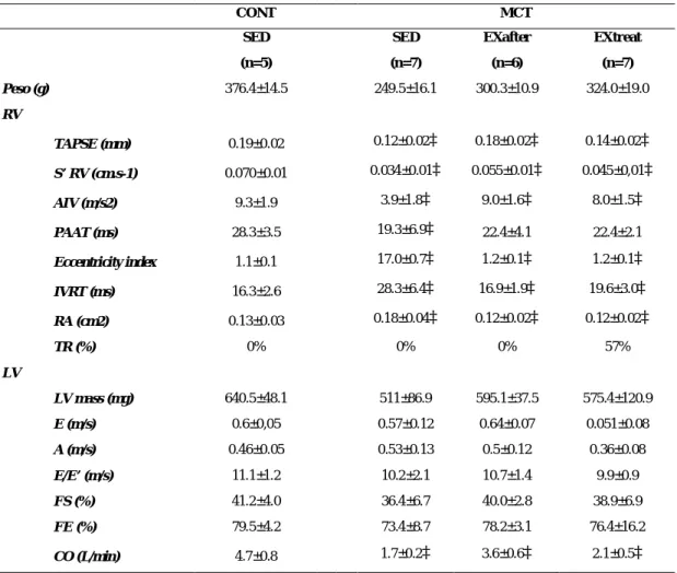
Table 1 summarizes the results from bi-ventricular echocardiography evaluation and table 2 the hemodynamics evaluation.
Right ventricular systolic function evaluated by TAPSE, S’ and AIV (Figure 1a) was significantly impaired in SED-MCT when compared with trained groups (P<0.001 vs. all groups). The eccentricity index is a ratio that relates to ventricular interdependence and it was significantly increased (Figure 1b and image 1a) in SED+MCT (P<0.001 vs all groups).
Figure 1a: Echocardiography evaluation of right ventricular systolic function ‡ P<0.05 vs.SED+MCT
Figure 1b: Results of the eccentricity index ‡ P<0.05 vs.SED+MCT
SED +CO NT SED +M CT EX afte r+M CT EX treat +M CT 0.0 0.5 1.0 1.5 2.0 E c c e n tr ic it y i n d e x ‡ ‡ ‡
Image 1a: Representative example of LV in PSAX
A significant decrease in PAAT was observed in SED+MCT (P<0.05 vs. SED+Control), being noted that only in the SED+MCT group was observed a typical elevated pulmonary artery pressure flow with a shortened acceleration time and late systolic notching (Figure 1c and image 1b).
Figure 1c: Results of the Pulmonary Artery Acceleration Time ‡ P<0.05 vs.SED+MCT
SED +C ON T SE D+M CT EXaf ter+ MC T EXtre at+M CT 0 10 20 30 40 P A A T ( m s ) ‡
Only in the SED+MCT group the presence of TR was recognizable, being present in 57% of the rats (Image1c).
Image 1c: Representative example of Color Doppler tricuspid regurgitation in SED+MCT animals
Through hemodynamic evaluation it was recognized an RVPmax increased in SED+MCT (+99%), EXafter+MCT (+71%) and EXtreat+MCT (+73%) groups (P<0.001 vs. respective control group). Exercise training induced a decrease in Ea but significance was obtained only in EXafter+MCT as compared with SED-MCT group (P<0.05). A smaller increase in ESPVR (P<0.05 vs. SED+MCT) was achieved in EXafter+MCT and EXtreat+MCT .
Regarding LV systolic function LVCO and SV were significantly reduced (Figure 2) in the SED+MCT group (P<0.05 vs. all groups). Although not significant, FS and EF had an improvement in all MCT-trained groups. In hemodynamics data SED+MCT presented a significant decrease in Pmax that was normalized in all MCT-trained groups.
Exercise has been shown to be able to maintain left ventricular mass in patients with PH [126]. A non significant reduction in LV Mass was seen in SED+MCT group when compared with all MCT- trained groups.
Table 1. Echocardiography evaluation parameters
CONT MCT
SED SED EXafter EXtreat
(n=5) (n=7) (n=6) (n=7) Peso (g) 376.4±14.5 249.5±16.1 300.3±10.9 324.0±19.0 RV TAPSE (mm) 0.19±0.02 0.12±0.02‡ 0.18±0.02‡ 0.14±0.02‡ S’ RV (cm.s-1) 0.070±0.01 0.034±0.01‡ 0.055±0.01‡ 0.045±0,01‡ AIV (m/s2) 9.3±1.9 3.9±1.8‡ 9.0±1.6‡ 8.0±1.5‡ PAAT (ms) 28.3±3.5 19.3±6.9‡ 22.4±4.1 22.4±2.1 Eccentricity index 1.1±0.1 17.0±0.7‡ 1.2±0.1‡ 1.2±0.1‡ IVRT (ms) 16.3±2.6 28.3±6.4‡ 16.9±1.9‡ 19.6±3.0‡ RA (cm2) 0.13±0.03 0.18±0.04‡ 0.12±0.02‡ 0.12±0.02‡ TR (%) 0% 0% 0% 57% LV LV mass (mg) 640.5±48.1 511±86.9 595.1±37.5 575.4±120.9 E (m/s) 0.6±0,05 0.57±0.12 0.64±0.07 0.051±0.08 A (m/s) 0.46±0.05 0.53±0.13 0.5±0.12 0.36±0.08 E/E’ (m/s) 11.1±1.2 10.2±2.1 10.7±1.4 9.9±0.9 FS (%) 41.2±4.0 36.4±6.7 40.0±2.8 38.9±6.9 FE (%) 79.5±4.2 73.4±8.7 78.2±3.1 76.4±16.2 CO (L/min) 4.7±0.8 1.7±0.2‡ 3.6±0.6‡ 2.1±0.5‡
.
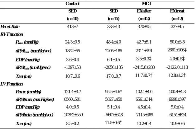
Table 2. Hemodynamic evaluation parameters
Control MCT
SED SED EXafter EXtreat
(n=10) (n=15) (n=12) (n=12) Heart Rate 413±7 333±13 370±15 327±15 RV Function Pmax (mmHg) 24.3±0.5 48.4±4.0 42.7±5.1 50.0±3.8 dP/dtmax (mmHg/sec) 1852±55 2205±185 2311±191 2661±106‡ EDP (mmHg) 3.6±0.4 6.1±0.5 3.5±0.3‡ 4.0±0.5‡ dP/dtmin (mmHg/sec) -1397±53 -2056±185 -2415.8±288 -2122.0±113 Tau (ms) 10.7±0.6 17.0±0.7 11.7±0.7‡ 12.8±1.3‡ LV Function Pmax (mmHg) 121.4±3.7 95.5±4.6* 102.1±4.0 100.4±4.3 dP/dtmax (mmHg/sec) 8560±501 5827±450 6561±314 6998±597 EDP (mmHg) 4.0±0.5 5.1±0.4 4.5±0.4 5.0±0.4 dP/dtmin (mmHg/sec) -10352±559 -5607±648 -7115±489 -6151±824 Tau (ms) 8.5±0.2 11.5±0.6* 10.2±0.4 10.9±0.6
Pmax: maximum pressure; dP/dtmax: peak rate of pressure rise; dP/dtmin: peak rate of pressure fall; EDP: end-diastolic pressure; Tau: time constant of ventricular pressure decay. Data is presented as mean±SEM. *P<0.05 vs. SED+Control, ‡ P<0.05 vs. SED+MCT.
4.2 Effects of exercise on bi-ventricular diastolic function
Diastolic function of the right ventricle was markedly impaired in SED+MCT group, namely there was an increase in end-diastolic pressure and a longer RV time constant tau (P<0.01 vs. all other groups). Exercise training normalized both end-diastolic pressure and tau in all MCT-trained groups.
In the ECHO data we identified (Figure 3a) a significant and prolonged IVRT in SED+MCT (P<0.05 vs. all groups) with a significant enlargement of RA area in SED+MCT (P<0.05 vs. all groups).
Figure 3a: Results of IVRT and RA area ‡ P<0.05 vs.SED+MCT
The role of exercise in the LV diastolic function was less clear. SED+MCT group had an increase in tau (P<0.01 vs. SED+Control), that was averted by exercise in all MCT-trained groups.
In the ECHO data we identified a tendency to 1 the E/A ratio in the SED+MCT group with no significant difference in the E/E’ ratio between all groups.
SE D+C ON T SE D+M CT EX afte r+M CT EX treat +M CT 0 10 20 30 40 IV R T ( m s ) ‡ ‡ ‡ SED +CO NT SED +M CT EXaf ter+ MC T EXtre at+M CT 0.00 0.05 0.10 0.15 0.20 0.25 R A ( c m 2 ) ‡ ‡ ‡
4.3 Exercise training improved exercise tolerance in MCT-treated rats
Exercise training promoted a significant improvement in exercise tolerance in both MCT trained groups, with a longer time till exhaustion (P<0.05 vs.SED+MCT). SED+MCT had a marked lower capacity when compared with SED+CONT (p<0.05).
SED +CO NT SED +M CT EX afte r+M CT EX trea t+M CT 0 55 110
‡
‡
‡
‡
T im e t o e x h a u s ti o n ( m in )5. DISCUSSION
The study of pulmonary hemodynamics is of great importance in many diseases directly or indirectly involving the cardiopulmonary apparatus. Over the past few years, evolution in ultrasonographic imaging has allowed the development of new indexes to noninvasively estimate many parameters previously measurable only with right-heart catheterization. It is now an important and imperative modality in the noninvasive assessment of PH and has been used to screen for the disease, determine right and left heart structure and function, and assess response to therapy in persons with PH. In this study it proved to be an important tool in assessing the repercussion of the effect of chronic exercise in bi-ventricular function.
All ECHO RV systolic evaluated parameters as TAPSE, AIV and S’ RV showed an improvement in MCT-trained groups, with decreased interventricular septum shift toward the left and no TR identified. Pulmonary artery acceleration time values in our study were similar to the values observed in earlier studies of rats with MCT-induced PAH[127]. .Short PAAT values are likely due to reduced capacitance and the increased impedance of the pulmonary vascular bed causing the deceleration to begin at early systole[128]. [32]. These data support the evidence of a reduced pathological remodeling of the RV when submitted to chronic pressure overload. It is also to emphasize the improvement in RV diastolic dysfunction with normalized both end-diastolic pressure and tau, with reduction in IVRT time and RA dimensions in all MCT-trained groups. The influence of RV failure in LV function is well known, leading to a decrease in LV preload, and low cardiac output states. This happens not only by the concept of ventricular interdependence but also because long-standing PH presents with autocrine/paracrine system activation in the absence of a direct hemodynamic stress that takes to a disturbed contractile phenotype[10]. In this study exercise training averted LV function with a significantly improve in LVCO and SV, regardless the absence of any major improvement in left diastolic function. On this point it is important to emphasize that ECHO may be preferable as an easier, more reproducible and non-invasive measure of cardiac output since it is well recognized that the presence of moderate to severe TR, as observed in advanced PAH, may lead to inaccurate invasive thermodilution derived cardiac outputs whereas TR has no effect on the ECHO derived measurements.
measure. This failure may be explained by the fact that rats have a much thinner right ventricular wall and a higher heart rate than humans. This restrictions may have conditioned a more accurate assessment of E' which may partly explain the results of left diastolic function. Another restriction was the quantification of TR velocity and has described in previous studies (30) it is usually unsuccessful in rats with systolic PAP below 65 mmHg. Our data supports this observation as we were able to assess the presence of TR but unable to quantify TR velocity. This was also true for higher velocities due to the limitation of the linear probe. Finally, it also should be emphasized that observed values of echocardiographic parameters in this study can be assumed valid only in rats.
Regardless echocardiography showed that it may have a promising role in the evaluation of cardiac response to exercise in patients with PAH.
The present study demonstrates that exercise training at different time-points exerts a positive impact in the RV response to chronic pressure overload, protecting from cardiac dysfunction and improving survival in an experimental model of PAH.
Exercise training ameliorated RV function in all different time points of MCT-induced PAH. That overlies of great importance since it is widely accepted that it is the main prognostic factor in PAH. It seems to provide a cardioprotective phenotype that allows the RV to work under overloading conditions with better tolerance.
This suggests that exercise can act as an upstream modulator of several pathological pathways activated in the RV during PAH. The benefits of exercise training may be associated with the prevention of calcium handling disturbances, alpha to beta-MHC shift, decreased neurohumoral activation, collagen deposition and inflammation and preserved oxidative phosphorylation through the reduction of mitochondrial oxidative damage. In fact, as already known in left HF the modulation of these pathways leads to improvements in cardiac function, promotes a cardiac adaptive phenotype and improves survival. Results from our laboratory suggest that similar changes can also be present in RV.
From the clinical perspective the EXtreat+MCT is likely to be the group that more closely matches the clinical condition of a patient with right overload pressure, that is, with PAH already established. These results suggest that even in these patients, exercise may be safe, ameliorating RV function, functional capacity and eventually improving survival.
6.
CONCLUSIONS
The findings from the present study indicate that exercise performed during or after the establishment of RV chronic pressure overload secondary to MCT-induced PAH averts RV systolic and diastolic dysfunction and improves survival. Also by preventing RV pathological remodeling, LV function is improved resulting in higher cardiac outputs. Altogether, these data highlight that exercise training can be a new modulator of RV function and can represent an important adjunctive therapeutic option in the management of PAH patients. The recognition of benefits of exercise training on the overloaded RV justifies the urgent need of further investigation on clinical settings.
7.
REFERENCES
1. Members, W.C., et al., ACCF/AHA 2009 Expert Consensus Document on Pulmonary Hypertension. Circulation, 2009. 119(16): p. 2250-2294.
2. D'Alonzo, G.E., et al., Survival in Patients with Primary Pulmonary HypertensionResults from a National Prospective Registry. Annals of Internal Medicine, 1991. 115(5): p. 343-349.
3. Voelkel, N.F., et al., Right Ventricular Function and Failure. Circulation, 2006. 114(17): p. 1883-1891.
4. Bogaard, H.J., et al., Chronic Pulmonary Artery Pressure Elevation Is Insufficient to Explain Right Heart Failure. Circulation, 2009. 120(20): p. 1951-1960.
5. Correia-Pinto, J., et al., Time course and mechanisms of left ventricular systolic and diastolic dysfunction in monocrotaline-induced pulmonary hypertension. Basic Research in Cardiology, 2009. 104(5): p. 535-545.
6. Piao, L., G. Marsboom, and S. Archer, Mitochondrial metabolic adaptation in right ventricular hypertrophy and failure. Journal of Molecular Medicine, 2010. 88(10): p. 1011-1020.
7. Kögler, H., et al., Mechanical Load-Dependent Regulation of Gene Expression in Monocrotaline-Induced Right Ventricular Hypertrophy in the Rat. Circulation Research, 2003. 93(3): p. 230-237.
8. Falcão-Pires, I., et al., Apelin decreases myocardial injury and improves right ventricular function in monocrotaline-induced pulmonary hypertension. American Journal of Physiology - Heart and Circulatory Physiology, 2009. 296(6): p. H2007-H2014.
9. Henriques-Coelho, T., et al., Endogenous production of ghrelin and beneficial effects of its exogenous administration in monocrotaline-induced pulmonary hypertension. American Journal of Physiology - Heart and Circulatory Physiology, 2004. 287(6): p. H2885-H2890.
10. Lourenço, A.P., et al., Myocardial dysfunction and neurohumoral activation without remodeling in left ventricle of monocrotaline-induced pulmonary hypertensive rats. American Journal of Physiology - Heart and Circulatory Physiology, 2006. 291(4): p. H1587-H1594.
11. Campian, M.E., et al., Early inflammatory response during the development of right ventricular heart failure in a rat model. European Journal of Heart Failure, 2010. 12(7): p. 653-658.
12. Redout, E.M., et al., Antioxidant treatment attenuates pulmonary arterial hypertension-induced heart failure. American Journal of Physiology - Heart and Circulatory Physiology, 2010. 298(3): p. H1038-H1047.
13. Bogaard, H.J., et al., The Right Ventricle Under Pressure. Chest, 2009. 135(3): p. 794-804.
14. Drake, J.I., et al., Molecular Signature of a Right Heart Failure Program in Chronic Severe Pulmonary Hypertension. American Journal of Respiratory Cell and Molecular Biology, 2011. 45(6): p. 1239-1247.
15. Boissiere, J., et al., Moderate exercise training does not worsen left ventricle remodeling and function in untreated severe hypertensive rats. Journal of Applied Physiology, 2008. 104(2): p. 321-327.