Contents lists available atScienceDirect
Acta Tropica
journal homepage:www.elsevier.com/locate/actatropica
Clinical, laboratorial and immunological aspects of severe malaria in
children from Guinea-Bissau
Janine Domingos
a,1, Anaxore Casimiro
a,1, Daniela Portugal-Calisto
a,b,1, Luís Varandas
a,c,
Fátima Nogueira
a, Marcelo Sousa Silva
a,d,e,f,⁎aGlobal Health and Tropical Medicine, Instituto de Higiene e Medicina Tropical, Universidade Nova de Lisboa, Portugal bInstitute of Medical Microbiology, University of Zurich, Zurich, Switzerland
cInfectious Disease Unit, Hospital Dona Estefânia. Lisbon, Portugal
dImunoparasitology Laboratory, Department of Clinical and Toxicological Analysis, Health Sciences Centre, Federal University of Rio Grande do Norte, Natal, Brazil ePrograma de Pós-graduação em Bioquímica, Universidade Federal do Rio Grande do Norte, Natal, Brazil
fPrograma de Pós-graduação em Ciências Farmacêuticas, Universidade Federal do Rio Grande do Norte, Natal, Brazil
A R T I C L E I N F O Keywords: Plasmodium falciparum Severe malaria Malaria antigens P. falciparum antibodies Humoral immunity A B S T R A C T
Malaria is a parasitic disease of which Plasmodium falciparum causes the most severe form of the disease. The immune response against Plasmodium spp. is complex and remains unclear. The present report aimed to better understand the humoral immune response in severe malaria and analyse new immunodominant antigen can-didates as possible serological marker in severe malaria in children.
This study included children aged 0–16 years from Guinea-Bissau with clinical signs of severe malaria. Serological and immunochemical characterisation of different anti-P. falciparum antibodies were made by ELISA and immunoblotting using a crude protein extract of P. falciparum.
Sera from 12 children with severe malaria were analysed. Nine samples were positive for total anti-P. falci-parum antibodies, seven for IgM and eight for total IgG anti-P. falcifalci-parum. There was a predominance of IgG1 response, suggesting a cytophilic action in severe malaria and a major role of IgG1 over other immunoglobulins. The antigenic profile of P. falciparum showed a consistent immunoblotting pattern of approximately 180 kDa, 100 kDa and around 50–40 kDa.
The serological reactivity found in protein bands makes them as immunodominant antigens and promising candidates for serological markers in the context of severe malaria.
1. Introduction
Plasmodium falciparum causes the most serious form of the disease. Infections with this parasite can be fatal in the absence of prompt re-cognition of the disease, appropriate treatment and patient manage-ment (Anon., 2012). The World Health Organization (WHO) have es-tablished a set of clinical and laboratory criteria that defines severe malaria (Anon., 2014).
Immunity seems to require humoral and cellular immune responses, as the passive transfer of purified immunoglobulin (Ig) G from hyper-immune sera to infected subjects has demonstrated that IgG mediates anti-parasitic activity and overall protection against malaria (Cohen et al., 1961). Also, a study of individuals from Ivory Coast and Burkina Faso infected with P. falciparum demonstrated that a predominance of
IgG1 and IgG3 cytophilic antibodies was associated with protection against the disease, whereas a predominance of IgG2 and IgG4 non-cytophilic antibodies were related to an increased susceptibility to malaria infection (Bouharoun-Tayoun and Druilhe, 1992). However, the question about which cytophilic antibody is producedfirst or which one is more relevant remains unclear (Pinto et al., 2001).
On the other hand, IgM has been known to neutralize antigens very effectively, acting as part of an early immune response. Several studies suggest that IgM has a protective role against malaria infection (Boudin et al., 1993). For instance, IgM antibodies and mononuclear cells act synergistically to inhibit the growth of P. falciparum in vitro more ef-fectively than IgG (Brown et al., 1986). However, further evidence in-dicates that the binding of IgM antibodies to the surface of infected erythrocytes via the Fc receptor has a positive correlation with the
https://doi.org/10.1016/j.actatropica.2018.04.020
Received 17 July 2017; Received in revised form 17 April 2018; Accepted 17 April 2018
⁎Corresponding author at: Global Health and Tropical Medicine, Instituto de Higiene e Medicina Tropical, Universidade Nova de Lisboa, Rua da Junqueira 100, 1349-008, Lisbon,
Portugal.
1These authors Contributed equally to this manuscript.
E-mail address:mssilva@ihmt.unl.pt(M.S. Silva).
Available online 21 April 2018
0001-706X/ © 2018 Elsevier B.V. All rights reserved.
formation of rosettes (Rowe et al., 2002; Ghumra et al., 2008), sug-gesting a possible role of IgM in the severity of falciparum malaria.
The role of IgE antibodies in protection and/or pathogenesis is not well established. Studies conducted in some islands of Burkina Faso and in the frontiers of Thailand-Myanmar and Thailand – Cambodia in-dicated that IgE may be related with the severity of malaria, as they showed high levels of IgE in subjects with severe malaria (Calissano et al., 2003). On the other hand, another study from a holoendemic area of Tanzania, suggested that higher levels of malaria-specific IgE in children were associated with a statistically significantly reduced risk of acute malaria (Bereczky et al., 2004).
The wide gap in understanding the biology of P. falciparum infection makes difficult to identify the most effective vaccine candidate from among the many of potential antigens of P. falciparum. The malaria vaccines that present themselves as current candidates are directed towards stages of the parasite's life cycle in humans and mosquitoes, but until now, relatively few proteins have been studied towards devel-oping a potential vaccine (Crompton et al., 2010). Nevertheless, a yet large number of antigens have presented a major challenge towards the identification of the protective responses and their targets, and it may be that efficient immunity is mediated by responses to multiple anti-gens.
Despite the global importance of P. falciparum, much work is yet to be done regarding antigens characterisation. Therefore, the present study aims to identify and characterise immunochemically the P. fal-ciparum antigens involved in the induction and maintenance of humoral immunity, in children with clinical signs of severe malaria caused by P. falciparum.
2. Material and methods
2.1. Description of the studied population
This study included 12 children from 0 to 16 years old, with signs of severe malaria or malaria requiring hospitalization due to oral intol-erance. All the children included were clinically observed in the pae-diatric consultations of the Hospital São José de Bor, in the village of Bor, Bissau (Guinea-Bissau). The severe malaria diagnosis was assessed through clinical evaluation and children fulfilling the following inclu-sion criteria were enrolled: i) clinical diagnosis of severe malaria, ac-cording to the clinical criteria established by WHO (2); ii) positive microscopic examination. Children presenting the following medical conditions were excluded: i) diarrhoea and severe dehydration; ii) se-vere malnutrition; iii) pneumonia; iv) meningitis; v) encephalitis; vi) tuberculosis; vi) diagnosed HIV infection.
An informed consent was applied to every participant and signed by the parent or legal guardian. The participation of patients in the study was voluntary. The study protocol was approved by the ethical com-mittee (protocol number 3–2012-TM) of Instituto de Higiene e Medicina Tropical, Lisbon, Portugal.
2.2. Samples collection
Blood samples were collected by venepuncture into heparin tubes. Approximately 100μl of total blood was absorbed in Whatman™ filter paper (GE Healthcare, UK) and let dry at room temperature (RT). The remaining sample was stored at−20 °C until needed.
2.3. Laboratorial confirmation of Plasmodium falciparum infection In order to confirm the positive infection of malaria, rapid diag-nostic test– RDT (Rapid Malaria pf/pan Antigen Test, Boson Biotech, China), microscopic examinations and Polymerase Chain Reaction (PCR) were performed. The RDT applied uses P. falciparum histidine-rich protein 2 (HRP-2) and lactate dehydrogenase (LDH) and was per-formed according to the supplier’s instructions.
Microscopic examination consisted on the visualization of thick blood smears,fixed with 10% methanol (v/v) for 1 min, stained with 1% Giemsa’s solution (Merck, Germany) in Phosphate Buffered Saline (PBS) (v/v) for 10 min, washed with water and allowed to dry at RT.
Genomic DNA of P. falciparum was extracted using QIAamp DNA Mini Kit (Quiagen, USA), according to manufacturer’s instructions. DNA was afterwards amplified by nested-PCR, using primers designed bySnounou et al. (1993), recognising the small subunit (18S) of ribo-somal RNA gene, and synthesised by Thermo Scientific (Germany). For PCR reactions the following reagents’ concentration was used: a) 10 x NH4 buffer, 50 mM MgCl, 100 mM mixed dNTP, 100 pmol/μl of sense and antisense primers; 5U/μl Taq polymerase, 3 μl DNA and water up to 50μl. Amplification conditions consisted of a) initial denaturation at 94 °C for 2 min; b) denaturation at 94 °C for 0.30 min; c) annealing at 55 °C for 1.30 min; d) extension at 72 °C for 2 min; repeat step b) to d) for 40 times; e)final extension at 72 °C for 10 min. A negative control (no template added) and a positive control (sample with infected blood) were used. The amplified products were observed in 2% agarosis gel (w/v).
2.4. Plasmodium falciparum parasites culture and total protein extraction Cultures of Dd2 P. falciparum clones were developed in complete RPMI 1640 culture medium with 5% haematocrit, 37 °C and an atmo-sphere with 5% of CO2as described in the work ofCosta et al. (2013).
To extract P. falciparum proteins, parasite cultures were transferred to 15 ml tubes, centrifuged at 2500g for 2 min at RT and the supernatant was discarded. Cells were suspended in lysis buffer (5 mM sodium phosphate, pH 8 + protease inhibitor [Complete Ultra Tablets, Roche, Switzerland]),five times the pellet volume. The tubes were placed on ice for 15 min and then centrifuged at 18000g, at 4 °C for 20 min. The supernatant was discarded. The pellet was washed three times with ice cold PBS. Every wash cycle comprised the addition of PBS and a cen-trifugation at 18000g for 10 min at 4 °C. The supernatant was discarded at every wash cycle. The parasite lysis was accomplished with four times the pellet volume of lysis buffer (0.1% Triton X-100 in PBS + protease inhibitor [Complete ULTRA Tablets, Roche, Switzer-land]). The tubes were placed on ice for 30 min with gently shaking, and homogenised by vortex every 10 min. After, the tubes were placed at−70 °C for 5 min and then at 37 °C for 5 min. This process was re-peated two more times. After this cycle, the samples were centrifuged at 18000g for 10 min at 4 °C. The supernatant was recovered and stored at −20 °C. The quantification of total protein of P. falciparum was per-formed with Bicinchoninic Acid Protein kit (Sigma, USA), according to the manufacturer’s protocol.
2.5. Serological measurement of anti-Plasmodium falciparum antibodies Total antibodies (IgG + A + M), IgM, total IgG and its subclasses and IgE were detected by enzyme-linked immunosorbent assay (ELISA) using crude protein extract of P. falciparum to coat 96-well microplates (Thermo Fisher Scientific, USA). The assay was developed according to Costa’s protocol (Costa et al., 2013), with few changes: wells were coated with 200 ng/well of crude protein extract of P. falciparum in bicarbonate buffer (0.1 M, pH 8.5). The primary antibody was diluted 1/100, except when measuring total IgG, which was diluted 1/200. The secondary antibody consisted of several different antibodies (used in separated assays), with different dilutions, different substrates and stop solutions applied.
All samples were tested in duplicate and positive and negative controls were used (adults with acute malaria and children without stays in endemic areas of malaria, respectively). For the serological measurement of IgE and IgG subclasses, neither positive nor negative controls were used, for lack of truly representative control samples.
The cut-off was established as the mean of the negative controls plus a stand-deviation (SD), obtained for the different antibody analysed.
The results were expressed in OD/cut-off to be compared between as-says. Samples with OD/cut-off ratio above 1.1 were considered as po-sitive and under 0.9 were considered to be negative for malaria anti-bodies. Values in between 0.9 and 1.1 were repeated and, when the result remained, were considered undetermined.
2.6. Characterisation of Plasmodium falciparum proteins by immunoblotting
Western Blot was performed as described inCalisto (2015), using 23 ng/well of crude protein extract of P. falciparum. The primary anti-body was diluted 1/100 (v/v) in antianti-body buffer (1% blocking buffer in wash buffer). The determination of the protein’s molecular weight was estimated by comparison with a 10–190 kDa commercial molecular weight marker (HyperPAGE Prestained Protein Marker, Bioline, UK). As negative controls, sera from subjects who have never been in malaria-endemic areas were used. As positive controls, sera from patients in-fected with P. falciparum were used.
2.7. Ethics approval and consent to participate
This study protocol was approved by the ethical committee of Instituto de Higiene e Medicina Tropical, Universidade Nova de Lisboa. Lisbon, Portugal. Protocol number 3–2012-TM.
3. Results
In this study 12 children were included with a mean age of 6.75 years (SD = 1.194); min = 2, max = 14. The majority of the children was female, representing 58% of patients (n = 7). The clinical and la-boratory results of the examination at admission are summarised in Tables S1 and S2.
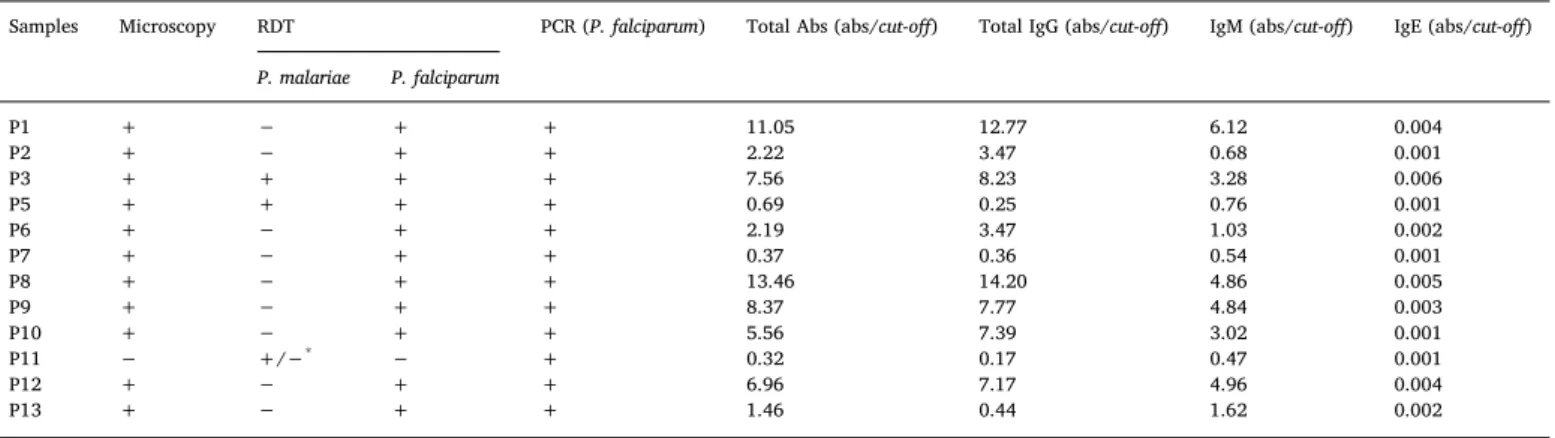
To confirm the infection by P. falciparum, RDT, microscopy and PCR were performed. The results are presented inTable 1. Eleven samples were positive for P. falciparum in RDT and microscopy, but all the 12 samples were positive when tested by PCR. Children denominated of P3 and P5 showed a possible co-infection with P. falciparum and P. ma-lariae according to the results obtained by RDT. The sample P11 was weakly positive for P. malariae by RDT and it was the only sample that was negative in microscopy. PCR was performed for the amplification of P. falciparum DNA and all samples were positive.
The levels of total antibodies (IgG + IgA + IgM), IgM, IgG and its subclasses and IgE were determined in the twelve infected children with severe malaria. The results are presented inFig. 1A. From the twelve samples, nine (75%) were positive for total P. falciparum anti-bodies, seven (58.3%) were positive for IgM, with one sample con-sidered as undetermined. IgG anti-P. falciparum showed eight (66.7%)
positive samples (Table 2,Fig. 1A). The levels of total IgG were almost always higher than IgM, except for sample P13. The levels of IgG subclasses were assessed in the 12 Plasmodium-infected children, in order to characterise the nature of the humoral response in the popu-lation studied. A predominance of the cytophilic IgG1 response was observed, with no serologic reactivity of any other subclasses (Error! Reference source not found.1B). None of the samples tested was positive for IgE antibody (Table 2).
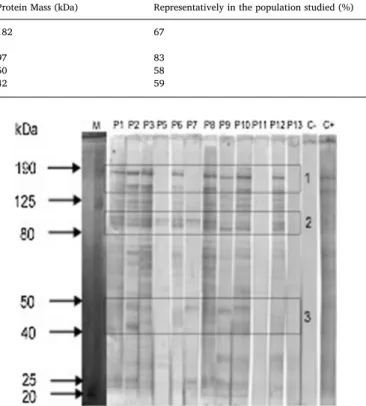
The P. falciparum antigens responsible for the serological reactions by ELISA assays were detected by immunoblotting. The antigenic pro-file of the population studied is presented inFig. 2. The analysis of the antigens profile of the samples from children with severe malaria showed a consistent reactive pattern around the molecular weights of Table 1
Laboratory confirmation of malaria infection and results of serologic assays.
Samples Microscopy RDT PCR (P. falciparum) Total Abs (abs/cut-off) Total IgG (abs/cut-off) IgM (abs/cut-off) IgE (abs/cut-off) P. malariae P. falciparum P1 + − + + 11.05 12.77 6.12 0.004 P2 + − + + 2.22 3.47 0.68 0.001 P3 + + + + 7.56 8.23 3.28 0.006 P5 + + + + 0.69 0.25 0.76 0.001 P6 + − + + 2.19 3.47 1.03 0.002 P7 + − + + 0.37 0.36 0.54 0.001 P8 + − + + 13.46 14.20 4.86 0.005 P9 + − + + 8.37 7.77 4.84 0.003 P10 + − + + 5.56 7.39 3.02 0.001 P11 − +/−* − + 0.32 0.17 0.47 0.001 P12 + − + + 6.96 7.17 4.96 0.004 P13 + − + + 1.46 0.44 1.62 0.002 *Weakly positive.
Fig. 1. Serological levels of anti-Plasmodium falciparum antibodies, namely total antibodies (IgG, IgA, IgM), total IgG, total IgM, IgG isotypes (IgG1, IgG2, IgG3, IgG4), and IgE performed by indirect ELISA.
190 kDa and 100 kDa (Error! Reference source not found.2, region 1 and 2). Notwithstanding, another (less relevant) pattern seems to emerge in the molecular weight of approximately 50 kDa and 40 kDa (Fig. 2, region 3).
Comparing the protein molecular weight marker with the most re-active proteins obtained, it is possible to estimate potential P. falci-parum antigens that were responsible for the IgG anti-P. falcifalci-parum antibodies detected through ELISA. That analysis is presented in Table 2.
4. Discussion
The present study aimed to characterise the mechanism of an early humoral immune response of severe malaria, by serologic and antigenic reactivity during thefirst/former Plasmodium infections. The levels of IgG measured indicated its importance for parasite control.
A group of 12 children from Bissau, Guinea-Bissau, with severe malaria were included. Laboratorial diagnosis confirmed the infection with P. falciparum. Sample P11 may seem contradictory but several explanations may exist: (i) sensitivity/specificity of the methods; (ii) low parasite load; (iii) poor quality of the sample. Also, the poly-morphism in the hrp-2 gene of P. falciparum may affect the sensitivity of the HRP-2 based RDT (Baker et al., 2010;Baker et al., 2005; Kumar et al., 2012). For LDH, there is no polymorphism described, but the detection of this antigen may fail due to the low sensitivity of the test in cases of low parasitemia (Jang et al., 2013). Due to the higher sensi-tivity and specificity of PCR regarding the other two methods, sample P11 was still considered for further analyses (Mahende et al., 2016).
The results obtained by ELISA showed that the majority of the children included have started developing the humoral response al-ready. The three negative samples may correspond to recent infection, with antibodies at undetectable levels in in vitro assays or with no time
to orchestrate the immune response.
Comparing the levels of those IgG and IgM antibodies, IgG antibody was presented in higher levels than IgM, suggesting that IgG is im-portant for parasite control, as indicated byRichards et al. (2010). Only sample P13 had a higher level of IgM than IgG, indicating that child did not develop a specific humoral response until blood collection.
In this present work, the mean age of the children with anti-malaria antibodies was 9.4 years, and without anti-malaria antibodies was 4.5 years, allowing to speculate that older children can mount stronger humoral responses, perhaps due to more exposures to the parasite in the past. Analogous results were described in the study ofRichards et al. (2010).
The nature of the humoral response in severe P. falciparum malaria seems to be characterised by a predominance of the cytophilic IgG1. Pinto et al. also reported a dominance of cytophilic antibody rather than non-cytophilic subclasses, when evaluating the response of IgG subclasses anti-P. vivax in 34 children from 0 to 15 years old. In par-ticular, patients showed a predominant IgG1 response, where 28 of these children had never had any previous history of malaria and 6 had one or two episodes of malaria (Pinto et al., 2001).
Since cytophilic antibodies are known to promote phagocytosis and antibody-dependent cell-mediated cytotoxicity (ADCC) processes (Bouharoun-Tayoun and Druilhe, 1992;Ferrante et al., 1990), the re-sults of this present report suggest that these antibodies are involved in opsonisation of infected red cells, as indicated elsewhere (Groux and Gysin, 1990;Tebo et al., 2002). Furthermore, there is also evidence that these subclasses antibodies act synergistically with monocytes through antibody-dependent cellular inhibition (ADCI) processes, strongly in-hibiting parasite growth (Theisen et al., 1998;Singh et al., 2005;Shi et al., 1999; Tebo et al., 2001). There was no reactivity with the others IgG subclasses studied, suggesting that they are not implicated in pro-tective responses in severe malaria.
Regarding IgE anti-P. falciparum antibodies, all the twelve samples were negative and unfortunately, no suggestions can be made con-cerning the controversy between the protective role (Bereczky et al., 2004;Duarte et al., 2007) or the contribution to the severity of malaria (Calissano et al., 2003;Perlmann et al., 1994). As the use of biotine-streptavidine complex increases the sensitivity of the technique, more analyses could be performed to discard the possibility of IgE levels are being underestimated. The confirmation of the serological assays with a larger sample size and with populations in different stages of the dis-ease would be useful to better understand the role of IgE in malaria.
The results obtained in ELISA assays are concordant with the re-activity presented in immunoblotting. Almost all samples showed ser-ologic reactivity, with the exception of P5, P7 and P11 that, similarly to ELISA results, showed a very modest reaction in Western Blot or no reactivity at all. These observations suggest that IgG is required for protection against the parasite in severe cases of the disease. Similar results were found in studies of immunoglobulin transfer of immune donors to patients with acute P. falciparum malaria (Cohen et al., 1961). The analysis of the antigens profile of the samples from children with severe malaria showed a consistent reactive pattern of approxi-mately 100 kDa and 180 kDa. These results are similar to those obtained in a study of 321 uninfected adults that showed IgG antibodies many years after their last stay in endemic areas (Calisto, 2015). Notwith-standing, another pattern seems to emerge between the molecular Table 2
Potential P. falciparum antigens responsible for the positivity for total IgG antibodies. Only the most reactive protein bands in immunoblotting were considered.
Protein Mass (kDa) Representatively in the population studied (%) Potential antigen candidates References
182 67 MSP-1 precursor 175 K antigen Freeman and Holder (1983) Camus and Hadley (1985)
97 83 96tR antigen Jouin et al. (1987)
50 58 51 kDa glycosylated antigen Epping et al. (1988)
42 59 MSP-1 (MSP1)42 Blackmann et al. (1991)
Fig. 2. Protein pattern of the reactivity detected by immunoblotting using 23μg/well of Plasmodium falciparum crude extract. M: molecular weight marker HyperPage (Bioline, UK). C-: negative control; C+: positive control.
weights of 50–40 kDa, having similarities with a study of Costa and colleagues with 227 adults with imported uncomplicated malaria (Costa et al., 2013). It is interesting to notice that the antigenic re-activity founded in the present report shares two different patterns, consistent with two different populations.
The antigenic reactivity in P5 and P7 samples by immunoblotting while negative in ELISA is probably the reflect of the higher sensitivity and specificity of the immunoblotting over ELISA (Palaeya et al., 2013; Cheong et al., 2013). However, we know that the sample population in this study is reduced and therefore this information should be explored in future studies. These results, though only suggestions towards the identification of proteins that reacted with IgG antibodies specific for P. falciparum, can prove to be relevant in the development of new protein targets as vaccine candidates.
Further in silico analysis of those antigens should be performed in order to fully characterised them and use them in new serological markers and/or vaccine designs. The knowledge of serological markers, specifically in severe malaria, is important to understand the evolution of the disease and avoid severity. The characterisation of the immune response and the antigen candidates achieved with this work can be useful to develop diagnostic tests in order to identify the severe stage of malaria as well as new and more promising vaccine candidates.
On the other hand, we also understand the importance of in-flammation, lymphocytes migration and cytokines involved in the pa-thogenesis of severe malaria, however we intend to evaluate these components in future studies.
Authors’ contributions
Conceived and designed the experiments: AC, LV, MSS. Performed the experiments: JD, AC, DPC, FN.
Analysed the data: JD, AC, DPC, LV, MSS. Wrote the paper: JD, AC, DPC, MSS.
All authors read and approved thefinal version of the manuscript. Funding
This work was supported by Global Health and Tropical Medicine– Lisbon [GHTM-UID/multi/04413/2013]. MSS thanks to Programa Ciências Sem Fronteiras– Capes Brazil [Grant 019/2013].
Acknowledgments
The authors want to thank to Tiago Vaz and Fernanda Murtinheira for all the laboratory support.
References
Anon, 2012. World Health Organization;1; Management of Severe Malaria– A Practical Handbook, 3rd ed. . Available at:http://www.who.int. (Accessed 28 September 2016).
Anon, 2014. World Health Organization Tropical Medicine and International Health, vol. 19. John Wiley & Sons, pp. 7–131(Suppl.1), Available at:http://onlinelibrary.wiley. com/doi/10.1111/tmi.12313_2/epdf(Accessed 21.12.2016).
Baker, J., McCarthy, J., Gatton, M., Kyle, D.E., Belizario, V., Luchavez, J., et al., 2005. Genetic diversity of Plasmodium falciparum histidine-rich protein 2 (PfHRP2) and its effect on the performance of PfHRP2-based rapid diagnostic tests. J. Infect. Dis. 192, 870–877.
Baker, J., Ho, M.-F., Pelecanos, A., Gatton, M., Chen, N., Abdullah, S., et al., 2010. Global sequence variation in the histidine-rich proteins 2 and 3 of Plasmodium falciparum: implications for the performance of malaria rapid diagnostic tests. Malar. J. 9, 129.
http://dx.doi.org/10.1186/1475-2875-9-129.
Bereczky, S., Montgomery, S.M., Troye-Blomberg, M., Rooth, I., Shaw, M.A., Färnert, A., 2004. Elevated anti-malarial IgE in asymptomatic individuals is associated with re-duced risk for subsequent clinical malaria. Int. J. Parasitol. 34, 935–942.
Blackmann, M.J., Whittle, H., Holder, A.A., 1991. Processing of the Plasmodium falci-parum major merozoite surface protein-1: Identification of a 33-kilodalton secondary processing product which is shed prior to erythrocyte invasion. Mol. Biochem. Parasitol. 49, 35–44.
Boudin, C., Chumpitazi, B., Dziegiel, M., Peyron, F., Picot, S., Hogh, B., et al., 1993. Possible role of specific immunoglobulin M antibodies to Plasmodium falciparum
antigens in immunoprotection of humans living in a hyperendemic area, Burkina Faso. J. Clin. Microbiol. 31 (3), 636–641.
Bouharoun-Tayoun, H., Druilhe, P., 1992. Plasmodium falciparum malaria: evidence for an isotype imbalance which may be responsible for delayed acquisition of protective immunity. Infect. Immun. 60 (4), 1473–1481.
Brown, J., Greenwood, B.M., Terry, R.J., 1986. Cellular mechanisms involved in recovery from acute malaria in Gambian children. Parasite Immunol. 8, 551–564.
Calissano, C., Modiano, D., Sirima, B.S., Konate, A., Sawadogo, A., Perlmann, H., et al., 2003. IgE antibodies to Plasmodium falciparum and severity of malaria in children of one ethnic group living in Burkina Faso. Am. J. Trop. Med. Hyg. 69 (1), 31–35.
Calisto, D.C.P., 2015. Master Thesis. Instituto de Higiene e Medicina Tropical, Universidade Nova de Lisboa, Lisbon.
Camus, D., Hadley, T.J., 1985. A Plasmodium falciparum antigen that binds to host er-ythrocytes and merozoites. Science 230 (4725), 553–556.
Cheong, F.W., Fong, M.Y., Lau, Y.L., Mahmud, R., 2013. Immunogenicity of bacterial-expressed recombinant Plasmodium knowlesi merozoite surface protein-142 (MSP-142). Malar. J. 12, 454.http://dx.doi.org/10.1186/1475-2875-12-454.
Cohen, S., Mcgregor, I.A., Gamma-Globulin, Carrington S., 1961. Acquired immunity to human malaria. Nature 192, 733–737.
Costa, R.M., Nogueira, F., de Sousa, K.P., Vitorino, R., Silva, M.S., 2013.
Immunoproteomic analysis of Plasmodium falciparum antigens using sera from pa-tients with clinical history of imported malaria. Malar. J. 12, 100.http://dx.doi.org/ 10.1186/1475-2875-12-100.
Crompton, P.D., Pierce, S.K., Miller, L.H., 2010. Advances and challenges in malaria vaccine development. J. Clin. Invest. 120 (12), 4168–4178.
Duarte, J., Deshpande, P., Guiyedi, V., Mécheri, S., Fesel, C., Cazenave, P.-A., et al., 2007. Total and functional parasite specific IgE responses in Plasmodium falciparum-in-fected patients exhibiting different clinical status. Malar. J. 6, 1.http://dx.doi.org/ 10.1186/1475-2875-6-1.
Epping, R.J., Goldstone, S.D., Ingram, L.T., Upcroft, J.A., Ramasamy, R., Cooper, J.A., et al., 1988. An epitope recognised by inhibitory monoclonal antibodies that react with a 51 kilodalton merozoite surface antigen in Plasmodium falciparum. Mol. Biochem. Parasitol. 28, 1–10.
Ferrante, A., Beard, L.J., Roberton, D.M., 1990. IgG subclass deficiency. Pedriatr. Allergy Immunol. 2 (2), 49–62.
Freeman, R.R., Holder, A.A., 1983. Surface antigens of malaria merozoites–a high mo-lecular weight precursor is processed to an 83,000 mol wt form expressed on the surface of plasmodiumfalciparum merozoites. J. Exp. Med. 158, 1647–1653.
Ghumra, A., Semblat, J.-P., McIntosh, R.S., Raza, A., Rasmussen, I.B., Braathen, R., et al., 2008. Identification of residues in the Cu4 domain of polymeric IgM essential for interaction with Plasmodium falciparum erythrocyte membrane protein 1 (PfEMP1). J. Immunol. 181 (3), 1988–2000.
Groux, H., Gysin, J., 1990. Opsonization as an effector mechanism in human protection against asexual blood stages of Plasmodium falciparum: functional role of IgG sub-classes. Res. Immunol. 141, 529–542.
Jang, J., Cho, C., Han, E., An, S.S.A., Lim, C., 2013. pLDH level of clinically isolated Plasmodium vivax and detection limit of pLDH based malaria rapid diagnostic test. Malar. J. 12, 181.http://dx.doi.org/10.1186/1475-2875-12-181.
Jouin, H., Dubois, P., Gysin, J., Fandeur, T., Mercereau-Puijalon, O., da Silva, L.P., 1987. Characterization of a 96-kilodalton thermostable polypeptide antigen of Plasmodium falciparum related to protective immunity in the squirrel monkey. Infect. Immun. 55 (6), 1387–1392.
Kumar, N., Singh, J.P., Pande, V., Mishra, N., Srivastava, B., Kapoor, R., et al., 2012. Genetic variation in histidine rich proteins among Indian Plasmodium falciparum population: possible cause of variable sensitivity of malaria rapid diagnostic tests. Malar. J. 11 (1), 298.
Mahende, C., Ngasala, B., Lusingu, J., Yong, T.-S., Lushino, P., Lemnge, M., et al., 2016. Performance of rapid diagnostic test, blood-film microscopy and PCR for the diag-nosis of malaria infection among febrile children from Korogwe District, Tanzania. Malar. J. 15, 391.
Palaeya, V., Lau, Y.L., Mahmud, R., Chen, Y., Cloning, Fong M.Y., 2013. expression, and immunocharacterization of surface protein containing an altered thrombospondin repeat domain (SPATR) from Plasmodium knowlesi. Malar. J. 12, 182.http://dx.doi. org/10.1186/1475-2875-12-182.
Perlmann, H., Helmby, H., Hagstedt, M., Carlson, J., Larsson, P.H., Troye-Blomberg, M., et al., 1994. IgE elevation and IgE anti-malarial antibodies in Plasmodium falciparum malaria: association of high IgE levels with cerebral malaria. Clin. Exp. Immunol. 97, 284–292.
Pinto, A.Y. das N., da S. Ventura, A.M.R., de Souza, J.M., 2001. Resposta de anticorpos IgG anti-plasmodium vivax em crianças expostas à malária, antes e após tratamento específico. J. Pediatr. (Rio. J) 77 (4), 299–306.
Richards, J.S., Stanisic, D.I., Fowkes, F.J.I., Tavul, L., Dabod, E., Thompson, J.K., et al., 2010. Association between naturally acquired antibodies to erythrocyte binding an-tigens of plasmodium falciparum and protection from malaria and high-density parasitemia. Clin. Infect. Dis. 51 (8), e50–60.
Rowe, J.A., Shafi, J., Kai, O.K., Marsh, K., Raza, A., 2002. Nonimmune IgM, but not IgG binds to the surface of plasmodium falciparum-infected erythrocytes and correlates with rosetting and severe malaria. Am. J. Trop. Med. Hyg. 66 (6), 692–699.
Shi, Y.P., Udhayakumar, V., Oloo, A.J., Nahlen, B.L., Lal, A.A., 1999. Differential effect and interaction of monocytes, hyperimmune sera, and immunoglobulin G on the growth of asexual stage Plasmodium falciparum parasites. Am. J. Trop. Med. Hyg. 60 (1), 135–141.
Singh, S., Soe, S., Roussilhon, C., Druilhe, P., Corradin, G., 2005. Plasmodium falciparum merozoite surface protein 6 displays multiple targets for naturally occurring anti-bodies that mediate monocyte-dependent parasite killing. Infect. Immun. 73 (2), 1235–1238.
Snounou, G., Viriyakosola, S., Zhu, X.P., Jarra, W., Pinheiro, L., do Rosario, V.E., et al., 1993. High sensitivity of detection of human malaria parasites by the use of nested polymerase chain reaction. Mol. Biochem. Parasitol. 61, 315–320.
Tebo, A.E., Kremsner, P.G., Luty, A.J.F., 2001. Plasmodium falciparum: a major role for IgG3 in antibody-dependent monocyte-mediated cellular inhibition of parasite growth in vitro. Exp. Parasitol. 98, 20–28.
Tebo, A.E., Kremsner, P.G., Luty, A.J.F., 2002. Fcgamma receptor-mediated phagocytosis
of Plasmodium falciparum-infected erythrocytes in vitro. Clin. Exp. Immunol. 130, 300–306.
Theisen, M., Soe, S., Oeuvray, C., Thomas, A.W., Vuust, J., Danielsen, S., et al., 1998. The glutamate-rich protein (Glurp) of plasmodium falciparum is a target for antibody-dependent monocyte-mediated inhibitionof parasite growth in vitro. Infect. Immun. 66 (1), 11–17.