ABSTRACT
The correction or improvement of common vision problems (such as, myopia, hypermetropia and astigmatism) through refractive laser surgery, as a permanent alternative to eyeglasses or contact lenses, has been growing in demand over recent years. The most common refractive laser surgery is the laser in-situ keratomileusis (LASIK) and its recent technological variants with improvements. Nevertheless, the generality (if not all), these ophthalmic laser procedures are based in the same physical principle: the reshaping of the cornea and thus altering its optical properties in order to improve vision. Here, the authors attempt to describe in general terms, a very promising, refractive laser procedure much less invasive that doesn’t alter the shape or thickness of the cornea and probably overcomes some limitations of usual LASIK procedure. Keywords: LASIK, PRK, FLEX, SMILE, LIRIC, Refractive surgery, Cornea.
RESUMO
A correção ou melhoria de problemas de visão comuns (como, miopia, hipermetropia e astigmatismo) realizados através da cirurgia refractiva por laser, como alternativa permanente para óculos ou lentes de contato, tem tido um aumento razoável nos últimos anos. A cirurgia refractiva a laser mais comum é a queratomileusis in-situ assistida por laser (designada por LASIK) e suas variantes tecnológicas melhoradas mais recentes. No entanto, a generalidade (se não todos), estes procedimentos oftalmológicos com laser são baseados no mesmo princípio físico: a remodelação geométrica da córnea, alterando assim as suas propriedades ópticas por forma a melhorar a visão. Neste artigo, os autores tentam descrever, em termos gerais, um procedimento refrativo por laser muito promissor, muito menos invasivo, e que não altera a forma geométrica ou a espessura da córnea e, que potencialmente, supera algumas das limitações do procedimento LASIK tradicional.
INTRODUCTION
Refractive laser surgery has becoming one of the fastest growing clinical procedures in ophthalmology over the last two decades, due mainly to the advance of laser and optical imaging technologies allowing a greater precision to the surgeon. Photorefractive keratectomy (PRK), laser in-situ keratomileusis (LASIK), laser-assisted in-situ keratomileusis (LASEK), small incision lenticule extraction (SMILE)1, refractive lenticule extraction (ReLEx)2 through SMILE, intrastromal femtosecond lasers (INTRACOR)3 and all the variants of refractive laser surgery are essential similar in the goal, which is the visual correction of refractive errors through ablation of corneal tissue (usually corneal stroma) by changing the shape (that is, the curvature and thickness) of the cornea anterior surface, thus altering its refractive properties and optical power (that is, the diopter)4. Although the two main refractive components of the eye are the cornea and the crystalline lens, and thus, all the refractive surgery is directed towards changing the integrated refractive power of these eye components.
Nowadays the most popular and widespread type of refractive surgery is the LASIK, that uses an excimer laser emitting at 193 nm central wavelength to ablate corneal stroma using a single-photon absorption mechanism. The surgical procedure, involves first a circular cut of the corneal epithelium to be flapped in order to expose the stroma (which can be done mechanically using a blade of a microkeratome, or using a femtosecond laser)5, and then is optically ablated by the excimer laser radiation. In the case of the photorefractive keratectomy (PRK), the corneal epithelium is totally removed before the stromal ablation procedure. Thus, the LASIK procedure avoids the anterior stromal haze and pain that are frequently associated with PRK procedure (although the haze after PRK can be avoided using intraoperative application of mitomycin C), because the corneal epithelium remains almost entirely intact, lowering the risk of infection and scarring4. The appearance of near infrared (NIR) femtosecond laser technology provided to the ophthalmologist a powerful surgical tool, that is less invasive and allows highly localized corneal surgeries, both in the corneal flap cutting and in the intrastromal vision correction6,7. Due to the ultra-short duration of the femtosecond laser pulses (the order of 10-15 of a second), a significant reduction of the threshold for laser-induced breakdown (LIB) is obtained, minimizing
this way the collateral damage and the formation of large cavitation gas bubbles consisting of carbon dioxide and water6. One technical drawback of using LASIK flap created by a NIR femtosecond laser, is that the surgeon still needs an excimer laser to reshape de corneal stroma, altering this way its refractive power. Thus, the femtosecond flap cutting is a very expensive tool, becoming the major barrier to its adaptation on refractive surgery and increasing significantly the overall cost of the clinical procedure. Nevertheless, there is a new technological solution that does not use a microkeratome or an excimer laser, and makes use a single femtosecond laser to create the corneal flap and the refractive lenticule in one step: the femtosecond lenticule extraction (FLEX) laser refractive surgery8.
The latest worldwide LASIK scientific literature review published considered to that date9, showed very high satisfaction rates, from 96% to 98.7% of all patients were satisfied or very satisfied after their LASIK surgery. With similar clinical results to LASIK in myopic refractive surgery, promising results in hyperopia, less biomechanical corneal impairment10, and with the recent refinements on SMILE procedure is for sure a technique to stay and to increase in clinical practice in the near future11.
As we have described before, all these refractive laser procedure variations rely on the change of the radius of curvature of the anterior surface of the cornea in order to alter the refractive power, however the optical refraction imaging through a spherical surface (such as the cornea) depends also from the index of refraction its surface where the light beam propagates12. Thus, instead of changing the surface curvature of the cornea, if one is able to change its refractive index (RI), this will be a viable alternative way of modifying the refractive power of the element. The present paper is an attempt to spread the concept to a wider ophthalmology community and to explain a procedure that has been recently demonstrated for the first time on the corneas of live cats, by a research group from University of Rochester under the direction of Prof. Wayne Knox, and is now being commercially developed by their joint startup company, Clerio Vision Inc. (Rochester, NY,USA)13. In the following sections, we try to explain the general concept in terms of geometric optics and its surgical procedure, as well as some possible explanations for the physical mechanism behind this induced RI change.
CONCEPT AND RESULTS
Changing the Refractive Power of a
Spherical Surface
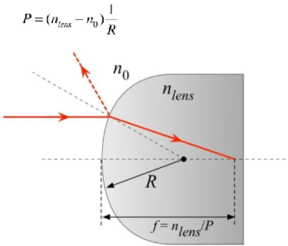
Recalling the geometrical optics from textbooks12, and considering the refraction of a light beam at a curved surface and shown schematically on Fig.1, one can state that the refractive power (P, sometimes called also vergence) of the interface can be mathematically expressed by the following relation:
Figure 1 - Refraction at a curved interface.
Where, R is the spherical radius of curvature of the surface, f the focal length of this interface, n0 and nlens are
the refractive indices of external medium and internal (lens) medium, respectively. The units of the refractive power are diopters (1 D = 1/m). Thus, to change the refractive power of the optical element, one can see by the relation above that is only necessary to change the radius of curvature (that is, R in the Fig.1) or the index of refraction of the element (that is, nlens in the Fig.1). The
conventional refractive laser surgery (such as LASIK) alters the radius of curvature of the cornea’s anterior surface, but if one can change the refractive index of the cornea, instead?
In fact, this idea was experimentally demonstrated for the first time in 2008 by Li Ding. et.al.14, in the corneas of post-mortem adult cats using high-repetition-rate low-energy femtosecond lasers pulses emitted at 800 nm wavelength. Following the work done, the same research group demonstrated for the first time the efficacy of
intra-tissue refractive index shaping (IRIS) directly into cat’s corneas in-vivo15. In this latest development, they used low-energy femtosecond laser pulses emitted at 400 nm wavelength, which has been shown previously that intrinsic two-photon absorption of the cornea allows 400 nm wavelength radiation to be performed with greater efficacy than when using 800 nm femtosecond laser pulses16. The authors of this new refractive laser surgery technique, called their procedure LIRIC, acronym for ‘laser induced refractive index-change’ (previously named IRIS), which we will try to describe in general terms for the ophthalmology professionals on the next section.
LASER INDUCED REFRACTIVE
INDEX-CHANGE (LIRIC)
The use of laser radiation to spatially alter the index of refraction of a material has been used for more than three decades, although not in biological tissue “per se”, but in many transparent optical materials (silica glasses, various polymers, and many others.) and using different laser sources17. The laser induced refractive index change has been demonstrated inside the core of optical fibers (fiber core radius: ~8 μm) using a tunable excimer laser pumped dye laser operated at a wavelength in the 486~500 nm, with a frequency-doubling crystal to provide a UV radiation wavelength at 244 nm18. Fractional refractive index perturbations of ~3x10⁻⁵, have been written in a 4.4 mm length of the fiber core with a 5-minute exposure time. The first demonstration in Portugal, using a similar UV photo-imprinted technique, was done by one of the authors in 199419, with a KrF excimer laser emitting at 248 nm wavelength (laser pulses of 20 ns and repetition rate of 50 Hz). In 1996, femtosecond laser micromachining has attracted increasing interest due to its unique 3D structuring capabilities in transparent materials20,21.
A femtosecond laser micromachining process can have different experimental conditions (e.g., laser pulse energy and width, repetition rate, scanning speed, focal spot size, etc.) in a variety of target materials, but the achieved end-results can be classified into two main categories depending on whether they can induce optical breakdown in the irradiated materials or not. The femtosecond lasers used in micromachining can be operated over a wide range
of parameters, from low-repetition-rates (10 Hz to 1 MHz) with pulse energies of micro-joules (μJ) to milli-joules (mJ), to high-repetition-rates (>1 MHz) with pulse energies of the order of nano-joules (nJ). For low-repetition-rate femtosecond lasers, the radiant exposure is usually well above the material breakdown threshold, and each laser pulse is responsible for a modification of the material in the focal volume region of the laser beam. This high energy focused pulses can originate asymmetric changes in the material. On the other hand, for high-repetition-rate femtosecond lasers, generally the pulse energy is much lower but the thermal diffusion time is longer, and the absorbed laser energy can accumulate within the focal volume. As a result, the size of the modification region will increase and a more symmetric structure is created.
In the case of corneal refractive surgery, although femtosecond lasers in the near infrared can pass through transparent corneal tissue without significant one-photon absorption, only when the pulses are focused inside the cornea is the intensity of the laser beam sufficient to cause a highly localized modification to the tissue with minimal effects on the surroundings. This unique precision is the primary reason for the application of corneal flap cutting LASIK surgery. Because these low-repetition-rate femtosecond lasers can induce optical breakdown of corneal tissue, generally associated with high-density micro-plasma generation, micro-bubble formation and sock-wave emission, these destructive effects extend beyond the focal region within the target corneal tissue. In contrast, with the use of high-repetition-rate femtosecond lasers it’s possible to alter the shape or optics properties without causing tissue destruction. In fact, with the use of high-repetition-rate (≥80 MHz) femtosecond laser with ultra-short pulse widths (≤100 fs) and nano-joule pulse energies, which allows to operate below the optical breakdown threshold22, it’s possible to alter the optical properties of the cornea (that is, its index of refraction) without changing the shape or thickness of the cornea14 or destroying the tissue.
The laser induced refractive index-change (LIRIC) procedure when compared with the LASIK procedure, uses 100 to 1000 times lower laser pulse energy (<1 nJ on the focal point at the tissue) at a high-repetition-rate (~80 MHz) and ultra-short laser pulses (≤ 100 fs), and
alters with highly spatial control the index of refraction of the cornea (recall the above equation, changes the parameter nlens in the Fig.1) and not its shape. This highly
spatial control of the laser beam is obtained with high-numerical aperture (NA =1.0) water immersion microscope objective, mounted on a 3-axis high precision translation stage15. Adding to this, the LIRIC procedure does not need any flap-cutting nor ablation, thus no tissue damage and thus without inducing wound-healing reaction in the cornea. Although the exact mechanism of refractive index (RI) change in cornea is not yet fully understood, some of its features seem consistent with low-density electron plasma model for femtosecond laser-material interactions23, but more research work is need to obtain more conclusions. From the first experimental results, the LIRIC technique seems to only increase the RI, which results in an increase of the power of cornea, and thus be only able to correct moderate amounts of hyperopia.
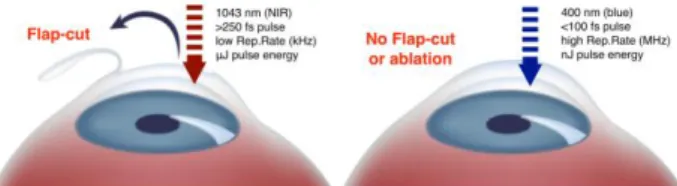
Figure 2, shows schematically the main differences between the two laser procedures: FLEX (which is a more advanced LASIK variation that uses a single femtosecond laser to create the corneal flap and the refractive lenticule in one step) and the LIRIC.
Figure 2 – FLEX (left) and LIRIC (right) laser procedures.
On figure 3, it is shown schematically what happen to the cornea when FLEX and LIRIC are used. In the FLEX procedure, the shape and thickness of the cornea is changed, and in the case of LIRIC the change of the optical properties of the cornea is achieved by altering locally its refractive index (RI).
The LIRIC system implemented and recently demonstrated by the research group of University of Rochester, which they designated by Blue-IRIS (Blue Intra-tissue Refractive Index Shaping), uses a frequency-double mode-locked femtosecond Ti:Sapphire laser (model Vitesse from Coherent Corp, USA) emitting at 400 nm wavelength, 100 fs laser pulses at a repetition rate of 80 MHz, with an average output power of 250 mW. The laser beam is then attenuated and focused with a high numerical aperture water immersion objective (20x, NA=1.0), into the corneal mid-stroma region of live cats (3-layer depths from 280 μm to 300 μm below the surface of the epithelium) providing pulsed energies on the focal region around 0.8 nJ15. Using a micrometer 3-axis translation-stage delivery system, they were able to inscribe into the cornea of the live cats an approximately area of 2.5 x 2.5 mm2, -1 D (diopter) cylinder lenses (slightly eccentric relative to the pupillary center of each eye), and which remained stable for up to 12 months. In these laser-inscribed cat eyes, no statistically significant changes to the corneal curvature or thickness were observed within 1 month after the surgical intervention. Figure 4 shows these experimental results in live cat cornea using the LIRIC procedure. The micro-bubbles that appear across the inscribed patterns (Fig.4A) immediately after the LIRIC procedure, have dissipated within several minutes, making the patterns almost invisible.
Figure 4 – Photographs of Blue-IRIS patterns (A) inscribed in the cornea of live cats in-vivo, and (B) Shack-Hartmann wavefront sensor spot arrays patterns. [reproduced with permission from the publisher15].
Subsequent work by the same authors24 demonstrated the capability of the LIRIC technique to write freeform optics (that is, optical devices with an arbitrary optical profile) in hydrogel-based contact lenses. This laser writing customization capability works well also in FDA-approved ophthalmic polymers25, which are in the materials of intraocular lenses (IOL) that ophthalmic surgeons use in
cataract surgery for the replacement of the clouded crystalline lens of the eye (see figure 5). A joint European-American start-up initiative (PerfectLens, LLC – www.perfectlens.com) lead scientifically by Prof. Josef Bille from University of Heidelberg, is also developing technology to modify an implanted cataract lens using a femtosecond laser, which can modify spherical aberration, asphericity, toricity, and multi-focality in a ten second in office procedure26,27.
Figure 5 – LIRIC procedure in hydrogel IOL.
In the case of a possible “erase-reaction” from the body’s natural healing process, results have shown that the laser induced RI patterns maintain their state for at least two years in the case of corneas, and five years on the hydrogel IOLs13, nevertheless a long-term study is still needed, as well as, LIRIC procedure in humans corneas. Although, the physical mechanism of locally change the refractive index of cornea is still not well understood, it’s explanation could be in fact result of several contribution mechanisms, such as multiphoton ionization, cascade ionization23, or some kind of photo disruption of collagen fibers originating irregular region (lakes) where there is no collagen present and thus large fluctuation in fiber density28. Very recently (May, 2017), the research group of University of Rochester have demonstrated for the first time the technique in postmortem human corneas29, demonstrating successfully the capability of the laser to write microstructures (in this case, phase gratings) directly in the human cornea. Figure 6 shows a differential interference contrast (DIC) microscope images of the LIRIC writing of identical phase grating lines (covering a 3 mm by 8 mm area and line spacing of 5 μm) where the RI change is visible relative to the background of the
cornea, both on cat and on human for performance comparison.
Figure 6 – DIC images of LIRIC phase grating lines written in (a) cat and (b) human corneas [reproduced with permission from the publisher29]
Although the human cornea average RI change obtained in this first demonstration were relatively low for clinical refractive correction purposes, due to the laser writing scan speed (5 mm/s) and only one corneal layer, it’s possible increase the RI change value by creating multiple layers, high density Fresnel lenses and increasing the scan speed. For sure, we will see more improved results in the coming months, and the first human blind eye trials will occur later this year (private communication
from Prof. W. Knox with the authors of this article).
CONCLUSIONS
Refractive laser surgery is growing every year around the world, in particular the ‘new’ LASIK modalities, such as FLEX (Femtosecond Lenticule Extraction) or SMILE (Small Incision Lenticule Extraction), are gaining popularity with expectations that by the end of 2017 about 1 million SMILE procedures will have been performed30. Although these ‘new’ techniques are less-invasive (small-incision surgery) and promise a better end-results, they are still fundamentally based on the 20-years old LASIK procedure of reshaping the size of the cornea, which has its clinical drawbacks. Here we have described a fundamentally different approach for possible correction of refractive errors where the laser radiation from a femtosecond laser emitting at 400 nm wavelength, with high-repetition-rate (~MHz) and very low pulse energy (~nJ), alters locally with high spatial precision the refractive index of the cornea, maintaining its the shape and thickness, and also avoiding the traditional cutting-&-flap of the LASIK procedure. The experimental results so
far indicate the capability of the technique, which will be entering the human trials tests later this year and will prove its validity for refractive surgery eye correction.
BIBLIOGRAPHY
1. Reinstein DZ, Archer TJ, Gobbe M. Small incision lenticule extraction (SMILE) history, fundamentals of a new refractive surgery technique and clinical outcomes. Eye Vis. 2014 Dec 16;1(1):3.
2. Ağca A, Demirok A, Yıldırım Y, Demircan A, Yaşa D, Yeşilkaya C, et al. Refractive lenticule extraction (ReLEx) through a small incision (SMILE) for correction of myopia and myopic astigmatism: current perspectives. Clin Ophthalmol. 2016 Oct;Volume 10:1905–12. 3. Krueger RR, Talamo JH, Lindstrom RL. Textbook of
Refractive Laser Assisted Cataract Surgery (ReLACS). Krueger RR, Talamo JH, Lindstrom RL, editors. New York, NY: Springer New York; 2013.
4. Kugler LJ, Wang MX. Lasers in refractive surgery: history, present, and future. Appl Opt. 2010;49(25):F1–9. 5. Azar DT, Koch DD. LASIK: Fundamentals, Surgical Techniques, and Complications. Dekker, Marcel; 2003. 6. Juhasz T, Loesel FH, Kurtz RM, Horvath C, Bille JF,
Mourou G. Corneal refractive surgery with femtosecond lasers. IEEE J Sel Top Quantum Electron. 1999;5(4):902– 10.
7. Kim P, Sutton GL, Rootman DS. Applications of the femtosecond laser in corneal refractive surgery. Curr Opin Ophthalmol. 2011;22(4):238–44.
8. Blum M, Kunert K, Schröder M, Sekundo W. Femtosecond lenticule extraction for the correction of myopia: Preliminary 6-month results. Graefe’s Arch Clin Exp Ophthalmol. 2010;248(7):1019–27.
9. Sandoval HP, Donnenfeld ED, Kohnen T, Lindstrom RL, Potvin R, Tremblay DM, et al. Modern laser in situ keratomileusis outcomes. J Cataract Refract Surg. Elsevier; 2017 May 29;42(8):1224–34.
10. Garcia-Porta N, Fernandes P, Queiros A, Salgado-Borges J, Parafita-Mato M, González-Méijome JM. Corneal Biomechanical Properties in Different Ocular Conditions and New Measurement Techniques. ISRN Ophthalmol. 2014;2014:1–19.
11. Borges JM, Charles HC, Lee CM, Smith RT, Cunha-Vaz JG, Goldberg MF, et al. A clinicopathologic study of dye
laser photocoagulation on primate retina. Retina. 1987;7(1):46–57.
12. Herman IP. Physics of the Human Body. Berlin, Heidelberg: Springer Berlin Heidelberg; 2007. (Biological and Medical Physics, Biomedical Engineering).
13. Zheleznyak L. LIRIC: Next-generation refractive laser surgery. Vol. 9, BioOptics World. 2016. p. 45–9. 14. Ding L, Knox WH, Bu¨hren J, Nagy LJ, Huxlin KR.
Intratissue Refractive Index Shaping (IRIS) of the Cornea and Lens Using a Low-Pulse-Energy Femtosecond Laser Oscillator. Investig Opthalmology Vis Sci. 2008 Dec 1;49(12):5332.
15. Savage DE, Brooks DR, DeMagistris M, Xu L, MacRae S, Ellis JD, et al. First Demonstration of Ocular Refractive Change Using Blue-IRIS in Live Cats. Investig Opthalmology Vis Sci. 2014 Jul 25;55(7):4603.
16. Xu L, Knox WH, DeMagistris M, Wang N, Huxlin KR. Noninvasive Intratissue Refractive Index Shaping (IRIS) of the Cornea with Blue Femtosecond Laser Light. Investig Opthalmology Vis Sci. 2011 Oct 17;52(11):8148. 17. Misawa H, Joudkazis S. 3D Laser Microfabrication.
WILEY-VCH Verlag GmbH & Co; 2006.
18. Meltz G, Morey WW, Glenn WH. Formation of Bragg gratings in optical fibers by a transverse holographic method. Opt Lett. 1989 Aug;14(15):823.
19. Araújo FM, Sousa JM, Lobo Ribeiro AB. Redes de Difracção em Fibra Óptica. In: 9a Conferência Nacional de Física. Covilhã, Portugal: Sociedade Portuguesa de Física; 1994. p. 182–3.
20. Davis KM, Miura K, Sugimoto N, Hirao K. Writing waveguides in glass with a femtosecond laser. Opt Lett. 1996;21(21):1729.
21. Glezer EN, Milosavljevic M, Huang L, Finlay RJ, Her T-H, Callan JP, et al. Three-dimensional optical storage inside transparent materials. Opt Lett. 1996;21(24):2023. 22. Kolozsvári L, Nógrádi A, Hopp B, Bor Z. UV
Absorbance of the Human Cornea in the 240- to 400-nm Range. Invest Ophthalmol Vis Sci. 2002 Jul 1;43(7):2165–8.
23. Noack J, Vogel A. Laser-induced plasma formation in water at nanosecond to femtosecond time scales: calculation of thresholds, absorption coefficients, and energy density. IEEE J Quantum Electron. 1999;35(8):1156–67.
24. Gandara-Montano GA, Ivansky A, Savage DE, Ellis JD, Knox WH. Femtosecond laser writing of freeform
gradient index microlenses in hydrogel-based contact lenses. Opt Mater Express. 2015 Oct 1;5(10):2257. 25. Xu L. Femtosecond Laser Processing of Ophthalmic
Materials and Ocular Tissues: A Novel Approach for Non-invasive Vision Correction. University of Rochester; 2013.
26. Sahler R, Bille JF, Enright S, Chhoeung S, Chan K. Creation of a refractive lens within an existing intraocular lens using a femtosecond laser. J Cataract Refract Surg. 2016 Aug;42(8):1207–15.
27. Bille JF, Engelhardt J, Volpp H-R, Laghouissa A, Motzkus M, Jiang Z, et al. Chemical basis for alteration of an intraocular lens using a femtosecond laser. Biomed Opt Express. 2017;8(3):1390.
28. Benedek GB. Theory of Transparency of the Eye. Appl Opt. 1971 Mar 1;10(3):459.
29. Wozniak KT, Gearhart SM, Savage DE, Ellis JD, Knox WH, Huxlin KR. Comparable change in stromal refractive index of cat and human corneas following blue-IRIS. J Biomed Opt. 2017 May 24;22(5):55007.
30. Stephenson M. SMILE expected to gain popularity as myopia treatment in the U.S. EyeWord [Internet]. 2017 Jun;64–7. Available from: http://www.ascrs.org
CONTACT
António Lobo
Faculdade de Ciências da Saúde Universidade Fernando Pessoa R. Carlos da Maia, 296 4200-150 Porto Portugal
E-mail: [email protected] Acknowledgements:
The authors would like thank to Prof. Wayne Knox (from University of Rochester, USA) for the written permission, as requested by the publishers, the use of two figures included on the manuscript.
Funding
This research did not receive any specific grant from funding agencies in the public, commercial, or not-for-profit sectors
Declaration of Interest statement:

![Figure 6 – DIC images of LIRIC phase grating lines written in (a) cat and (b) human corneas [reproduced with permission from the publisher 29 ]](https://thumb-eu.123doks.com/thumbv2/123dok_br/17296236.790666/6.871.67.418.210.378/figure-images-grating-written-corneas-reproduced-permission-publisher.webp)
