R E S E A R C H
Open Access
Cloning, structural modelling and
characterization of VesT2s, a wasp venom
hyaluronidase (HAase) from
Vespa tropica
Prapenpuksiri Rungsa
1, Paroonkorn Incamnoi
2, Sophida Sukprasert
3, Nunthawun Uawonggul
4,
Sompong Klaynongsruang
1, Jureerut Daduang
5, Rina Patramanon
1, Sittiruk Roytrakul
6and Sakda Daduang
1,7*Abstract
Background:Wasp venom is a complex mixture containing proteins, enzymes and small molecules, including some of the most dangerous allergens. The greater banded wasp (Vespa tropica) is well-known for its lethal venom, whose one of the major components is a hyaluronidase (HAase). It is believed that the high protein proportion and activity of this enzyme is responsible for the venom potency.
Methods:In the present study, cDNA cloning, sequencing and 3D-structure of Vespa tropica venom HAase were described. Anti-native HAase antibody was used for neutralization assay.
Results: Two isoforms, VesT2a and VesT2b, were classified as members of the glycosidase hydrolase 56 family with high similarity (42–97 %) to the allergen venom HAase. VesT2a gene contained 1486 nucleotide residues encoding 357 amino acids whereas the VesT2b isoform consisted of 1411 residues encoding 356 amino acids. The mature VesT2a and VesT2b are similar in mass and pI after prediction. They are 39119.73 Da/pI 8.91 and 39571.5 Da/pI 9.38, respectively. Two catalytic residues in VesT2a, Asp107 and Glu109 were substituted in VesT2b by Asn, thus impeding enzymatic activity. The 3D-structure of the VesT2s isoform consisted of a central core (α/β)7barrel and two disulfide bridges. The five putative glycosylation sites (Asn79, Asn99, Asn127, Asn187 and Asn325) of VesT2a and the three glycosylation sites (Asn1, Asn66 and Asn81) in VesT2b were predicted. An allergenic property significantly depends on the number of putativeN-glycosylation sites. The anti-native HAase serum specifically recognized to venom HAase was able to neutralize toxicity ofV. tropicavenom. The ratio of venom antiserum was 1:12.
Conclusions:The wasp venom allergy is known to cause life-threatening and fatal IgE-mediated anaphylactic reactions in allergic individuals. Structural analysis was a helpful tool for prediction of allergenic properties including their cross reactivity among the vespid HAase.
Keyword:Wasp venom,Vespa tropica, Hyaluronidase (HAase)
Background
Vespidae venom consists of complex mixtures of en-zymes, proteins, peptides and small molecules respon-sible for many of the non-allergic and mild allergic reactions–such as local pain, inflammation and itching–
as well as moderate and serious allergic reactions–such
as anaphylaxis, and delayed hypersensitivity – including systemic toxic reactions, coagulopathy, acute renal failure and hepatotoxicity [1, 2]. Wasp venom contains many biological active compounds [3, 4]. The major allergens are phospholipase A1, hyaluronidase (HAase) and antigen 5 [5–8].
Venom HAase is an enzyme that hydrolyses hyalur-onic acid (HA), one of the primary components of the extracellular matrix of vertebrates, which facilitates venom toxin diffusion into the tissue and blood circula-tion of the prey [9, 10]. HAase mainly acts as a“ spread-ing factor” to enhance venom action. It has been
* Correspondence:[email protected]
1Protein and Proteomics Research Center for Commercial and Industrial
Purposes (ProCCI), Department of Biochemistry, Faculty of Science, Khon Kaen University, Khon Kaen 40002, Thailand
7Division of Pharmacognosy and Toxicology, Faculty of Pharmaceutical
Sciences, Khon Kaen University, Khon Kaen, Thailand
Full list of author information is available at the end of the article
identified in the venom of animals including snakes, bees, scorpions, fish, spiders, ants, wasps, caterpillars etc. [11–16]. Clinical studies have demonstrated that HAase is an “allergic factor” due to its ability to initiate pathogenic reactions in the majority of venom allergic patients [17–19]. It is also able to induce several ana-phylactic IgE-mediated reactions in humans and has been suggested to be involved in the difficulties in the clinical diagnosis of venom allergic individuals [20–22]. The wasp venom HAase belongs to the hyaluronate gly-canohydrolase family (EC 3.2.1.35), which degrades hya-luronic acid (HA) [23, 24]. Wasp venom HAase is responsible for the cross-reactivity of wasp and bee venom sera in patients as well [2, 25].
The greater banded wasp (Vespa tropica) is mostly distributed in the forest throughout Indochina penin-sula including Thailand. It has a body length of up to 5 cm and its nest is usually found underground [26].V. tropica is among the most venomous known insects. The lethal dose of its pure venom in experimental ani-mals (LD50 of approximately 2.8 mg/kg in mice) is
more potent than that ofV. affinisvenom [26, 27]. The potency of V. tropica venom has been reported to nearly stop the end plate potentials of Drosophila lar-vae in nerve-muscle preparation in response to treat-ment with this venom [28]. HAase was reported to be a major protein inV. tropicavenom, where it is found by 2.5-fold the proportion observed in V. affinis venom [26]. The understanding of HAase in terms of bio-chemical and structural characterization of these wasps is important for the development of new tools for treating multiple stings and for diagnosis and therapy of allergic reactions caused by this venom. Therefore,
the present study aimed to characterize HAase isoforms in the venom ofV. tropicaby analyzing its sequence and 3D modelling.
Methods
Animals
The wasps were collected from Siang Sao Village, Sri Songkram district, Nakorn Panom Province, northeastern Thailand [26]. The worker wasps were immediately shocked on ice. The venom reservoirs were removed from the sting apparatus by removing them from the bodies with forceps and squeezing. The droplets of venom and specimens ofV. tropicawere collected in a 1.5-mL micro-centrifuge tube and then keep at−80 °C until use.
RT-PCR and rapid amplification of cDNA ends (5′ and 3′ RACE)
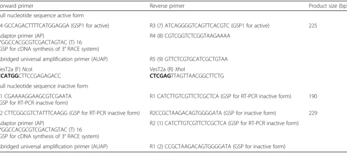
Total RNA was extracted from the venom gland ofV. tro-pica with TRIzol® reagent (Invitrogen, Life technologies, USA). RT-PCR was performed using the RevertAid First strand cDNA synthesis kit (Thermo Scientific, USA) as described in the instruction manual. PCR primers for the amplification of VesT2 were designed based on the se-quence similarity of the conserved region of HAase from vespid venom and conserved nucleotide sequences corre-sponding to peptide sequences obtained from LC-MS/MS analysis (Table 1) [26]. The PCR was performed using green master mix reagent kits with Taq DNA polymerase (Promega, Singapore). The reaction contained 2 μg of cDNA, 1 UTaq DNA polymerase, 2.0 mM dNTP, 2.0 mM MgCl2and 2μM of primerin to a final volume of 25μL
under the following conditions: initial denaturation for 5 min at 94 °C, followed by 35 cycles at 94 °C (30 s); 55 °C
Table 1Primer design of gene-specific primers and PCR product size
Forward primer Reverse primer Product size (bp)
Full nucleotide sequence active form
F4 GCCAGACTTTTCATGGAGGA (GSP1 for active) R3 (7) ATCAGGGGTCAGTTCACGTC (GSP1 for active) 225
Adaptor primer (AP)
5′GGCCACGCGTCGACTAGTAC (T) 16 (GSP for cDNA synthesis of 3′RACE system)
R4 (8) CGTCGGTCTCGGTAAGAAAA
Abridged universal amplification primer (AUAP) R5 (9) GTTCTCGTGCATCGCTGTAA
VesT2a (F)NcoI
CCATGGCTTCCGAGAGACC
VesT2a (R)XhoI
CTCGAGTTAGTTAACGGCTTCTG
Full nucleotide sequence inactive form
F1 CGAAAAGGAAGCGTCGAATA (GSP for RT-PCR inactive form)
R1 CATCTTGTCGTTCTCGCTCA (GSP for RT-PCR inactive form) 190
F2 CTTCGGCGTCTATTTCAAGG (GSP for RT-PCR inactive form) R2CCGCTAAGACAGTGGGGATA (GSP for inactive form) 229
Adaptor primer (AP)
5′GGCCACGCGTCGACTAGTAC (T) 16 (GSP for cDNA synthesis of 3′RACE system)
R2 (1) CATCTTGTCGTTCTCGCTCA (GSP for RT-PCR inactive form)
Abridged universal amplification primer (AUAP) R1 (2) CCGCTAAGACAGTGGGGATA (GSP for inactive form)
(30 s); 72 °C (1 min) and a final extension at 72 °C for 5 min. The rapid amplification of cDNA ends (RACE) was performed with the RACE system (Invitrogen, Life Technologies, USA). The RACE PCR products were cloned into the pGEM®-T easy vector (Promega, USA) for sequencing [29].
Sequence analysis and structure modelling
The basic characterizations of the gene and protein se-quences were analyzed using NCBI (http://www.ncbi.nlm. nih.gov/Database/index.html) and the basic local alignment search tool BLAST (http://www.ncbi.nlm.nih.gov/BLAST/). The phylogenic tree was created using CLUSTAL-X soft-ware analysis using the Neighbour-Joining method [30]. The three-dimensional models were created using the SWISS-MODEL program, the automated protein hom-ology modelling template at ExPASY (Switzerland) and a template search with the Alignment Mode program from the protein database (http://swissmodel.expasy.org/) [31, 32]. The model was elucidated as a PDB file, and the structure was previewed and analyzed using Swiss-Pdb Viewer Deep View v4 software (http://www.expasy.org/). The molecular mass and isoelectric points were computed using the Compute pI/MW tool of ExPASy Bioinformatics (http://web.expasy.org/compute_pi/). The N-glycosylation sites were predicted using the CBS prediction severs (http://www.cbs.dtu.dk/services/NetNGlyc/) and com-pared with other wasp and bee venom HAases.
Zymographic HAase activity assay
TheV. tropicavenom HAase activity was detected using 10 % SDS-PAGE containing hyaluronic acid as a sub-strate. Proteins were separated at 15 mA. The gel was incubated in 3 % Triton X-100 for 1 h with agitation in order to remove SDS and then transferred to the HAase
assay buffer (0.15 M NaCl in 0.1 M formate buffer), rinsed twice with assay buffer, and then incubated on a rotating shaker for 16 h at 37 °C. The gels were rinsed twice with distilled water and stained in 0.5 % Alcian blue solution for 1 h. The destain was performed with 7 % acetic acid that was changed every 1 h until clear bands appeared on a pale blue background [33].
Turbidity HAase activity assay
The turbidity HAase method followed the one by Pukrit-tayakamee et al. [34] with slight modifications. We mixed 0.5 mg/mL HA and buffer containing 0.15 M NaCl to a final volume of 100 μL and incubated for 30 min at 37 °C. The reaction was stopped using 200μL of 2 % CTAB containing 2.5 % NaOH. The absorbance was measured at 405 nm. The turbidity reducing activity was expressed as the percentage of remaining HA by taking the absorbance of the tube at 100 % in which no enzyme was added. The optimal pH of the venom HAase was determined by changing the buffers of the enzymatic turbidimetric venom HAase activity assay as follows: 0.2 M formate buffer, pH 2–4; 0.2 M acetate buffer, pH 5–6; 0.2 M Tris–HCl buffer, pH 7–10.
Mouse anti-hyaluronidase serum
The HAase band from zymographic gel were cut and frozen at−70 °C overnight, the gel was freeze-dried and
ground. Anesthetized mice were subcutaneously immu-nized with gel swollen in PBS buffer (135 mM NaCl, 1.5 mM KH2PO4, 2.5 mM KCl, and 8 mM Na2HPO4)
emulsified with Freud’s complete adjuvant. Mice were four times boosted with the antigen emulsified with in-complete Freund’s adjuvant. After retro-orbital plexus bleeding, blood was kept at 4 °C for 12 h and centrifuged at 10000 ×gfor antiserum collection.
Western immunoblotting
Proteins were separated by SDS-PAGE and blotted onto a nitrocellulose membrane (Bio-Rad, USA). After being eletrotransferred, the membrane was incubated with 5 % nonfat dry milk for 1 h, anti-HAase antibody for 1 h and goat anti-mouse IgG linked alkaline phosphatase (1:500) for 1 h. The blotted bands were detected by a substrate kit (GE Healthcare, Sweden). The membrane was inten-sive washed before the next incubation.
Neutralization assay
Crickets (Gryllus sp.) were abdominally injected with venom pre-incubated with anti-HAase serum 10 min
before considered paralyzed. The paralyzed crickets were defined as those that could return from the overturned position.
Results
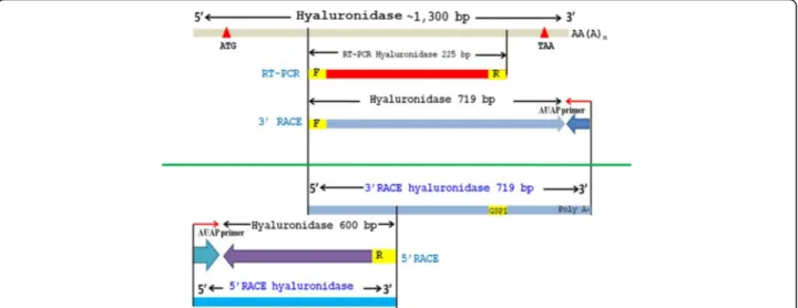
Sequence and structural modelling analysis of VesT2s The completed cDNA sequence was designed according to the peptide sequences obtained by LC-MS/MS and the sequence similarities of the conserved region of the other wasp venom hyaluronidases [26]. The primers were designed from nucleotide sequences based on the conserved region corresponding to the peptide sequence. The nucleotide fragment was obtained via RT-PCR. The
3′and 5′end were determined using RACE. They were completely overlapped (Fig. 1). Two HAase isoforms, VesT2a and VesT2b, were obtained.
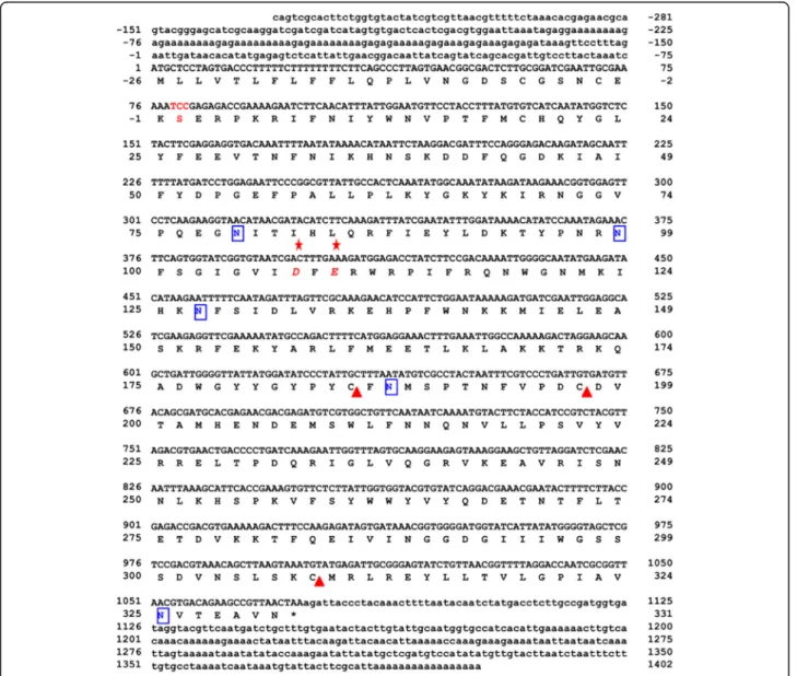
The full length VesT2a cDNA was 1,683 bp in length and contained 281 bp of the 5′-untranslated region (UTR) and 328 bp of the 3′-UTR; 1,074 bp of an open reading frame (ORF) encoded a protein of 357 amino acids (Fig. 2). The primary sequence of the deduced VesT2a contained 357 amino acid residues including a predicted signal peptide (26 amino acid residues) that was rich in the amino acids Asn, Lys, Ile and Leu, with a predicted mature pI and molecular mass of 8.91 and 39,119.73 Da, respectively. The five potentially immuno-genic N-glycosylated sites (Asn-Xaa-Thr/Ser, where Xaa is any amino acid residue except proline) on residues Asn79, Asn99, Asn127, Asn187 and Asn325 were
predicted. The two disulfide bridges (Cys19-Cys308 and Cys185-Cys197) were responsible for the stabilization of protein structure (Fig. 2).
Additionally, a putative HAase isoform was recently suggested as another component appearing in the 2D-PAGE profile from the corresponding cDNA of VesT2b [26]. It had an experimental mass of approximately 46 to 47 kDa. After amplification using several strategies (Fig. 1), the VesT2b precursor contained a 195-bp 5′ -UTR, a 145-bp 3′-UTR and an 1146-bp ORF. The ORF consisted of a 57-bp predicted signal sequence, which corresponded to 19 amino acid residues, and a 1089-bp mature sequence encoding 337 amino acids. The pri-mary sequence of the deduced VesT2b mature protein contained 337 amino acid residues (996 bp) and was rich in the amino acids Lys, Asn and Ile with a theoretical pI
9.38 and a predicted molecular mass of 39571.5 Da. The three potentially immunogenic N-glycosylated sites (Asn1, Asn66 and Asn81) and the two disulfide bridges (Cys21-Cys310 and Cys187-Cys199) were predicted (Fig. 3). The VesT2s mature amino acid sequence in these studies had 61.52 % homology; the two catalytic residues in VesT2a, Asp107 and Glu109, were substituted byAsn in VesT2b (Fig. 3).
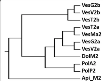
The multi-sequence alignment of venom HAases (Fig. 4) showed the highest BLAST homology score (>90 % identity) of VesT2a to many HAases, VesV2a of Vespula vulgaris, VesG2a of Vespa germanica, VesMa2 of Vespa magnifica, and Dol m 2 of Dolichovespula maculata, suggesting high evolutionary conservation among these species. The catalytic residues (Asp107 and Glu109) were conserved in active venom VesT2a [20, 22, 35, 36]. The phylogenetic tree analysis revealed the highest similarity of VesT2a to VesMa2, which was higher than that of the VesT2b depicted in the phylogenetic tree of the insect HAase (Fig. 5). VesT2s contained cysteine residues that were conserved among venom HAases and also formed two disulfide bonds (Fig. 6a and b). For VesT2a, these were Cys19-Cys308 and Cys185-Cys197, whereas they were Cys21-Cys310 and Cys187-Cys199 in the structure of VesT2b.
VesV2 (PDB ID: 2ATM) was used as a template for computational homology modelling. VesT2a and VesT2b showed 92.28 % and 62.69 % sequence identity to VesV2,
respectively, with an E value of 6.19e−153. Based on the
model, VesT2a and VesT2b displayed a central core (α/ β)7consisting of seven helices and seven beta-sheets
be-longing to family 56 of glycoside hydrolases [37] (Fig. 7).
HAase activity of wasp venom VesT2a
V. tropica venom VesT2a was tested for specific HAase activity using zymographic method at 37 °C, pH 3.7, under reducing conditions. The result showed a trans-parent band (Fig. 8a). The turbidity method was used to determine the optimal pH of venom HAase, with hyalur-onic acid as substrate. The V. tropica HAase, VesT2a, had an optimal pH of about 3 (Fig. 8b). It clearly dis-played a higher HAase activity between pH 2 and 5.
Neutralization assay
The Western immunoblotting revealed the specificity of antibodies to their antigens when the titer was 1:100 (Fig. 9). The anti-HAase serum was able to reduce venom toxicity (Table 2). Non-paralyzed crickets were observed at the ratio 1:12 (venom: antiserum).
Discussion
In this study, we described the identification, biochemis-try, bioactivity and structural characteristics of the HAase from the venom of greater banded wasp V. tro-pica. This study describes the existence of two isoforms of VesT2s, VesT2a and VesT2b. The primary sequence of VesT2a and VesT2b were clearly isoenzymes with 61.52 % similarity but with different molecular masses and pIs of the mature sequence (357 amino acids/ 39119.73 Da/pI 8.91 and 337 amino acids/39571.53 Da/
(See figure on previous page.)
Fig. 4Sequence alignment of the deduced amino acid sequence ofVespa tropicaHAase with other allergen venom HAases. VesT2s was aligned with the known HAases; VesV2a and VesV2b (Vespula vulgaris; active and inactive forms), VesG2a and VesG2b (Vespula germanica; active and inactive forms),Vespa magnifica(VesMa2), DolM2 (Dolichovespula maculata), PolA2 (Polistes annularis), PolP2 (Polybia paulista) and Api_M2 (Apis mellifera). The shaded yellow alignment corresponds to conserved residues in HAase. The N-terminus was shown in the underlined amino acid sequence obtained by Edman sequencing. The catalytic residues (D and E letters) are indicated with the red stars (✩). The conserved cysteine positions among the HAase are indicated with blue triangles (△)
Fig. 5The phylogenic tree of HAases from insect venoms
pI 9.38, respectively). Mass differences were mainly esti-mated from amino acid variations, including the degree of glycosylation of VesT2s. However, they were classified into the same family of glycoside hydrolase family 56 by sequence similarity. This phenomenon also occurs with HAases in many species, such asVesV2a and VesV2b, the HAase isoenzymes in Vespula vugaris venom. VesV2a and b share 58 % amino acid identity to each other [5, 20].
Rungsa et al. [26] indicated that the mass of HAase in V. tropica venom was approximately 43 kDa after
analysis by denaturing two-dimensional electrophoresis, which was confirmed by peptide mass fingerprinting. However, the mature sequence of HAase in this study, VesT2s, was smaller in size, with approximately 39 kDa. The molecular mass of about 43 kDa of native VesT2s was not surprising, since wasp venom HAase is a glyco-protein whose differences in estimated values of theoret-ical pI and molecular masses are frequent [9, 38, 39].
The phylogenetic tree demonstrated that VesT2a is found in the same cluster of active HAase from insect venoms. VesT2b is also found in a cluster of inactive
Fig. 7The predicted three-dimensional structural modelling of VesT2s. TheVespa tropicaHAase [VesT2a (a) and VesT2b (b)] modelling used VesV2 as a template (Vespula vulgaris, PDB accession number 2ATM_A). VesT2s was generated with SWISS-MODEL automated software and was visualized by the Swiss-Pdb Viewer Deep View v4.0 program. The two catalytic sites of VesT2a (Glu109 and Asp 107) were changed to Asn in VesT2b
HAase from insect venoms [2, 20, 35, 38, 40]. The en-zyme function of VesT2s is different because of two catalytic residues in VesT2a, Asp107 and Glu109. Both are substituted by Asn in VesT2b that has no HAase en-zymatic activity towards various substrates [20, 35, 41]. The less acidic Asn cannot act as a proton donor as the acidic amino acids, Asp and Glu [36, 37].
Glycosylation sites are the most common post-translational modification of many insect venom proteins as they contribute to biological activity, immunogenicity, and solubility, stability and protease resistance. VesT2s represents one of the strongest conserved hymenoptera venom allergens in wasps, yellow jackets and honeybees [42, 43]. VesT2a is highly similar to VesMa2 (Vespa mag-nifica HAase) while VesT2b is close to VesV2b (Vespula vugarisHAase b).V. vugarisandV. magnificaalso belong
to the Vespidae family [20, 35, 40]. Therefore, we presume that the VesT2s isoform might have a similar structure and allergic properties.
Insect venom allergies are known to cause life-threatening and sometimes fatal IgE-mediated anaphyl-actic reactions in allergic individuals. Approximately 30 to 50 % of patients with insect venom allergies have IgE antibodies that react with both honeybee and yellow jacket venom [44]. Previous studies have demonstrated that human IgE antibodies share cross reactive B-cell epitopes with various venom HAases to VesV2 [2, 25]. Honeybee and yellow jacket venom HAases with a mo-lecular mass of approximately 42–45 kDa are considered to be major allergen proteins and are responsible for cross-reactivity with allergen patient sera [44]. The venom HAase in insects are classical allergens responsible for cross-reactivity. Nevertheless, the cross-reactivity of venom HAase was identified by cross reactive carbohy-drate determinants (CCD) [42, 45]. Previous studies showed that VesV2s and VesMa2 were isoallergens that significantly differed in the number of putative N-glycosylation sites (Table 3) [9, 22, 37, 40]. Accord-ing to the sequencAccord-ing analysis of VesT2s, it contains five N-glycosylation sites in VesT2a (Asn79, Asn99, Asn127, Asn187 and Asn325) and three N -glycosyla-tion sites in VesT2b (Asn1, Asn66 and Asn81). Based on this data, we speculated about a high degree of CCD. These data are potentially relevant, especially regarding to the cross-reaction [40, 46].
Via the turbidity method,V. tropicavenom HAase was clearly active at a pH ranging from 2 to 5 (more than 80 % of relative activity) with an optimal pH of approxi-mately 3 to 4. At pH 6 to 10, the activity reduced and no detectable activity was observed within the range of basic pH (8–10). Therefore, VesT2a was predicted as a strong acid HAase. However, the optimal pH (3 to 4) in this study was quite different from those of other wasp venoms, such as V. vulgaris (pH 5–6), V. germanica (pH 5–6) andD. maculata(pH 5–6) [47]. Generally, the
Fig. 9Western immunoblotting analysis of venom HAase with an anti-HAase serum. Lane 1: molecular weight marker. Lanes 2–4: HAase was incubated with different dilutions of anti-HAase serum. Venom HAase is indicated by the arrow
Table 2The neutralization assay ofV. tropicavenom against anti-HAase serum in crickets (Gryllussp.)
V. tropicavenom: Anti-HAase serum (μL/μL)
Neutralized crickets/total crickets after injections withV. tropicavenom and anti-HAase serum
1:4 2/4
1:8 1/4
1:12 0/4
Table 3N-glycosylation in wasp venom HAase. Asn-Xaa-Ser/Thr residues represent the possibleN-glycosylation sites predicted by NetNGlyc 1.0 Server (N-glycosylation inV. vulgarisandV. magnifica
HAase was obtained in the experiment in the native form)
V. tropica(this study) V. vulgaris[22] V. magnifica[9]
VesT2a (active HAase)
VesT2b (inactive HAase)
VesV2a (active HAase)
VesV2b (inactive HAase)
VesMa2 (active HAase)
Asn79 Asn1 Asn79 Asn66 Asn105
Asn99 Asn66 Asn99 Asn81 Asn125
Asn127 Asn81 Asn127 Asn153
Asn187 Asn351
activity of HAases to degrade hyaluronic acid (HA) have an optimal pH ranging from 3 to 4, which is in accord-ance with VesT2a in this study (Table 4) [48].
A previous study showed the high potency of V. tro-picavenom (PD50~ 3μg/g body weight of cricket) [26].
Venom HAase, a “spreading factor”, is well-known for its toxin-enhancing activity. Therefore, the anti-HAase serum was produced. The anti-HAase serum shows neu-tralizing efficiency against crude venom by ratio the ratio of 1:12 (venom:antiserum). Inhibition of HAase activity not only prevents local tissue damage, but also retards the venom toxin diffusion into the tissues and blood circulation, resulting in the delay of fatal outcomes in several cases [13]. HAase activity may play a vital role in allergenicity and toxicity of venoms.
Conclusions
Hymenoptera venom showed cross-reactivity with bee and wasp venoms [2]. The allergic responses to wasp venom are known to cause life-threatening and fatal IgE-mediated anaphylactic reactions in sensitive individ-uals. The cross reactivity among the hyaluronidase from yellow jacket and bee venom are presumably induced by CCDs, but less often shared by peptide epitopes [19]. Knowledge on the structural determinants responsible for the allergic potency is expected to have important clinical implications.
Funding
This work was mainly supported by the Higher Education Research Promotion and National Research University (NRU) Project of Thailand, Office of the Higher Education Commission (CHE), through the Food and Functional Food Research Cluster of Khon Kaen University (KKU). It was also partially supported by the “The Thailand Research Fund–Master Research Granted (TRF–MAG)”year 2008 (MRG-WII515S069),“TRF–CHE jointly funded Research Grant for Mid-Career University Faculty”, fiscal years 2007–2009; and KKU Research Fund, fiscal years 2007–2010.
Authors’contributions
PR conducted most of the experiments, coordinated the data analysis and drafted the manuscript. PI and SS contributed to bioinformatics analyses. NU conducted Western blotting experiments. SK contributed to the study design and writing of the manuscript. JD performed the molecular analyses and contributed to the writing of the manuscript. RP contributed to writing and editing of the manuscript. SR performed the proteomic study. SD designed the research and the experiments, coordinated the study, wrote and edited the manuscript. All authors read and approved the final manuscript.
Competing interests
The authors declare that there are no competing interests.
Ethics approval and consent to participate
The present study was approved by the Animal Ethics Committee of Khon Kaen University based on the Ethics for Animal Experimentation of the National Research Council of Thailand (reference. 0514.1.12.2/1).
Author details
1Protein and Proteomics Research Center for Commercial and Industrial
Purposes (ProCCI), Department of Biochemistry, Faculty of Science, Khon Kaen University, Khon Kaen 40002, Thailand.2Department of Chemistry,
Faculty of Engineering, Rajamangala University of Technology Isan, Khon Kaen Campus, Khon Kaen, Thailand.3Chulabhorn International College of
Medicine, Thammasat University (Rangsit Campus), Pathumthani, Thailand.
4Division of Chemistry, Faculty of Science, Nakhon Phanom University,
Nakhon Phanom, Thailand.5Department of Clinical Chemistry, Faculty of
Associated Medical Sciences, Khon Kaen University, Khon Kaen, Thailand.
6Genome Institute, National Center for Genetic Engineering and
Biotechnology, National Science and Technology Development Agency (NSTDA), Pathumthani, Thailand.7Division of Pharmacognosy and Toxicology,
Faculty of Pharmaceutical Sciences, Khon Kaen University, Khon Kaen, Thailand.
Received: 20 April 2016 Accepted: 29 September 2016
References
1. King TP, Kochoumian L, Joslyn A. Wasp venom proteins: phospholipase A1 and B. Arch Biochem Biophys. 1984;230(1):1–12.
2. King TP, Lu G, Gonzalez M, Qian N, Soldatova L. Yellow jacket venom allergens, hyaluronidase and phospholipase: sequence similarity and antigenic cross-reactivity with their hornet and wasp homologs and possible implications for clinical allergy. J Allergy Clin Immunol. 1996;98(3):588–600.
3. Jalaei J, Fazeli M, Rajaian H, Shekarforoush SS. In vitro antibacterial effect of wasp (Vespa orientalis) venom. J Venom Anim Toxins Incl Trop Dis. 2014; 20(22):1–6.
4. Santos LD, Pieroni M, Menegasso ARS, Pinto JRAS, Palma MS. A new scenario of bioprospecting of Hymenoptera venoms through proteomic approach. J Venom Anim Toxins Incl Trop Dis. 2011;17:364–77.
5. King TP, Alagon AC, Kuan J, Sobotka AK, Lichtenstein LM. Immunochemical studies of yellowjacket venom proteins. Mol Immunol. 1983;20(3):297–308. 6. Abe T, Sugita M, Fujikura T, Hiyoshi J, Akasu M. Giant hornet (Vespa mandarinia)
venomous phospholipases. The purification, characterization and inhibitory properties by biscoclaurine alkaloids. Toxicon. 2000;38(12):1803–16. 7. Kreil G. Hyaluronidases - a group of neglected enzymes. Protein Sci. 1995;
4(9):1666–9.
8. Sukprasert S, Rungsa P, Uawonggul N, Incamnoi P, Thammasirirak S, Daduang J, et al. Purification and structural characterisation of phospholipase A1 (Vespapase, Ves a 1) from Thai banded tiger wasp (Vespa affinis) venom. Toxicon. 2013;61:151–64.
9. Justo Jacomini DL, Campos Pereira FD, Santos Pinto JR A d, dos Santos LD, da Silva Neto AJ, Giratto DT, et al. Hyaluronidase from the venom of the social waspPolybia paulista(Hymenoptera, Vespidae): cloning, structural modeling, purification, and immunological analysis. Toxicon. 2013;64:70–80. 10. Bordon KCF, Perino MG, Giglio JR, Arantes EC. Isolation, enzymatic
characterization and antiedematogenic activity of the first reported rattlesnake hyaluronidase fromCrotalus durissus terrificusvenom. Biochimie. 2012;94(12):2740–8.
11. Feng L, Gao R, Gopalakrishnakone P. Isolation and characterization of a hyaluronidase from the venom of Chinese red scorpionButhus martensi. Comp Biochem Physiol C Toxicol Pharmacol. 2008;148(3):250–7. 12. Girish KS, Shashidharamurthy R, Nagaraju S, Gowda TV, Kemparaju K.
Isolation and characterization of hyaluronidase a“spreading factor”from Indian cobra (Naja naja) venom. Biochimie. 2004;86(3):193–202. 13. Wahby AF, Mahdy e-SM, El-Mezayen HA, Salama WH, Abdel-Aty AM, Fahmy
AS. Egyptian horned viperCerastes cerastesvenom hyaluronidase: Purification, partial characterization and evidence for its action as a spreading factor. Toxicon. 2012;60(8):1380–9.
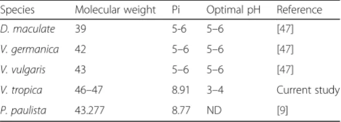
Table 4The biochemical and physiological characterization of vespid venom
Species Molecular weight Pi Optimal pH Reference
D. maculate 39 5-6 5–6 [47]
V. germanica 42 5–6 5–6 [47]
V. vulgaris 43 5–6 5–6 [47]
V. tropica 46–47 8.91 3–4 Current study
14. Magalhães MR, da Silva Jr NJ, Ulhoa CJ. A hyaluronidase fromPotamotrygon motoro(freshwater stingrays) venom: isolation and characterization. Toxicon. 2008;51(6):1060–7.
15. Nagaraju S, Devaraja S, Kemparaju K. Purification and properties of hyaluronidase fromHippasa partita(funnel web spider) venom gland extract. Toxicon. 2007;50(3):383–93.
16. Hoffman DR, Dove DE, Jacobson RS. Allergens in Hymenoptera venom: XX. Isolation of four allergens from imported fire ant (Solenopsis invicta) venom. J Allergy Clin Immunol. 1988;82(5 Pt 1):818–27.
17. Xia X, Liu R, Li Y, Xue S, Liu Q, Jiang X, et al. Cloning and molecular characterization of scorpionButhus martensivenom hyaluronidases: a novel full-length and diversiform noncoding isoforms. Gene. 2014;547(2):338–45. 18. Kaneiwa T, Mizumoto S, Sugahara K, Yamada S. Identification of human
hyaluronidase-4 as a novel chondroitin sulfate hydrolase that preferentially cleaves the galactosaminidic linkage in the trisulfated tetrasaccharide sequence. Glycobiology. 2010;20(3):300–9.
19. Jin C, Focke M, Léonard R, Jarisch R, Altmann F, Hemmer W. Reassessing the role of hyaluronidase in yellow jacket venom allergy. J Allergy Clin Immunol. 2010;125(1):184–90. e1.
20. Kolarich D, Leonard R, Hemmer W, Altmann F. The N-glycans of yellow jacket venom hyaluronidases and the protein sequence of its major isoform inVespula vulgaris. FEBS J. 2005;272(20):5182–90.
21. Justo Jacomini DL, Gomes Moreira SM, Campos Pereira FD, de Zollner RL, Brochetto Braga MR. Reactivity of IgE to the allergen hyaluronidase from
Polybia paulista(Hymenoptera, Vespidae) venom. Toxicon. 2014;82:104–11. 22. Seppälä U, Selby D, Monsalve R, King TP, Ebner C, Roepstorff P, et al. Structural
and immunological characterization of the N-glycans from the major yellow jacket allergen Ves v 2: The N-glycan structures are needed for the human antibody recognition. Mol Immunol. 2009;46(10):2014–21.
23. Arming S, Strobl B, Wechselberger C, Kreil G.In vitromutagenesis of PH-20 hyaluronidase from human sperm. Eur J Biochem. 1997;247(3):810–4. 24. Jin C, Hantusch B, Hemmer W, Stadlmann J, Altmann F. Affinity of IgE and
IgG against cross-reactive carbohydrate determinants on plant and insect glycoproteins. J Allergy Clin Immunol. 2008;121(1):185–90. e2.
25. King TP, Joslyn A, Kochoumian L. Antigenic cross-reactivity of venom proteins from hornets, wasps, and yellow jackets. J Allergy Clin Immunol. 1985;75(5):621–8.
26. Rungsa P, Incamnoi P, Sukprasert P, Uawonggul N, Klaynongsruang S, Daduang J, et al. Comparative proteomic analysis of two wasps venom,
Vespa tropicaandVespa affinis. Toxicon. 2016;119:159–67.
27. Schmidt JO, Yamane S, Matsuura M, Starr CK. Hornet venoms: lethalities and lethal capacities. Toxicon. 1986;24(9):950–4.
28. Gawade SP. The effect of venom from the Indian tropical waspVespa tropica
on nerve - muscle preparations fromDrosophila larvae. Toxicon. 1983; 21(6):882–6.
29. Incamnoi P, Patramanon R, Thammasirirak S, Chaveerach A, Uawonggul N, Sukprasert S, et al. Heteromtoxin (HmTx), a novel heterodimeric phospholipase A2 fromHeterometrus laoticusscorpion venom. Toxicon. 2013;61:62–71.
30. Saitou N, Nei M. The neighbor-joining method: a new method for reconstructing phylogenetic trees. Mol Biol Evol. 1987;4(4):406–25. 31. Bordoli L, Kiefer F, Arnold K, Benkert P, Battey J, Schwede T. Protein
structure homology modeling using SWISS-MODEL workspace. Nat Protoc. 2009;4:1–13.
32. Arnold K, Bordoli L, Kopp J, Schwede T. The SWISS-MODEL workspace: a web-based environment for protein structure homology modelling. Bioinformatics. 2006;22(2):195–201.
33. Mio K, Stern R. Reverse hyaluronan substrate gel zymography procedure for the detection of hyaluronidase inhibitors. Glycoconj J. 2000;17(11):761–6. 34. Pukrittayakamee S, Warrell DA, Desakorn V, McMichael AJ, White NJ, Bunnag
D. The hyaluronidase activities of some Southeast Asian snake venoms. Toxicon. 1988;26(7):629–37.
35. Kolarich D, Loos A, Leonard R, Mach L, Marzban G, Hemmer W, et al. A proteomic study of the major allergens from yellow jacket venoms. Proteomics. 2007;7(10):1615–23.
36. Marković-Housley Z, Miglierini G, Soldatova L, Rizkallah PJ, Müller U, Schirmer T. Crystal Structure of hyaluronidase, a major allergen of bee venom. Structure. 2000;8(10):1025–35.
37. Skov LK, Seppala U, Coen JJ, Crickmore N, King TP, Monsalve R, et al. Structure of recombinant Ves v 2 at 2.0 Angstrom resolution: structural analysis of an
allergenic hyaluronidase from wasp venom. Acta Crystallogr D Biol Crystallogr. 2006;62(Pt 6):595–604.
38. Lu G, Kochoumian L, King TP. Sequence identity and antigenic cross-reactivity of white face hornet venom allergen, also a hyaluronidase, with other proteins. J Biol Chem. 1995;270(9):4457–65.
39. Pinto JR, Santos LD, Arcuri HA, Dias NB, Palma MS. Proteomic characterization of the hyaluronidase (E.C. 3.2.1.35) from the venom of the social waspPolybia paulista. Protein Pept Lett. 2012;19(6):625–35.
40. An S, Chen L, Wei JF, Yang X, Ma D, Xu X, et al. Purification and characterization of two new allergens from the venom ofVespa magnifica. PLoS One. 2012;7(2):e31920.
41. El-Safory NS, Fazary AE, Lee C-K. Hyaluronidases, a group of glycosidases: current and future perspectives. Carbohydr Polym. 2010;81(2):165–81. 42. King TP, Spangfort MD. Structure and biology of stinging insect venom
allergens. Int Arch Allergy Immunol. 2000;123(2):99–106.
43. King TP, Wittkowski KM. Hyaluronidase and hyaluronan in insect venom allergy. Int Arch Allergy Immunol. 2011;156(2):205–11.
44. Seismann H, Blank S, Braren I, Greunke K, Cifuentes L, Grunwald T, et al. Dissecting cross-reactivity in hymenoptera venom allergy by circumvention ofα-1,3-core fucosylation. Mol Immunol. 2010;47(4):799–808.
45. Hemmer W, Focke M, Kolarich D, Dalik I, Gotz M, Jarisch R. Identification by immunoblot of venom glycoproteins displaying immunoglobulin E-binding N-glycans as cross-reactive allergens in honeybee and yellow jacket venom. Clin Exp Allergy. 2004;34(3):460–9.
46. Hemmer W, Focke M, Kolarich D, Wilson IBH, Altmann F, Wöhrl S, et al. Antibody binding to venom carbohydrates is a frequent cause for double positivity to honeybee and yellow jacket venom in patients with stinging-insect allergy. J Allergy Clin Immunol. 2001;108(6):1045–52.
47. Girish KS, Kemparaju K. The magic glue hyaluronan and its eraser hyaluronidase: a biological overview. Life Sci. 2007;80(21):1921–43.
48. Hokputsa S, Jumel K, Alexander C, Harding SE. Hydrodynamic characterisation of chemically degraded hyaluronic acid. Carbohydr Polym. 2003;52(2):111–7.
• We accept pre-submission inquiries
• Our selector tool helps you to find the most relevant journal • We provide round the clock customer support
• Convenient online submission • Thorough peer review
• Inclusion in PubMed and all major indexing services • Maximum visibility for your research
Submit your manuscript at www.biomedcentral.com/submit

![Fig. 7 The predicted three-dimensional structural modelling of VesT2s. The Vespa tropica HAase [VesT2a (a) and VesT2b (b)] modelling used VesV2 as a template (Vespula vulgaris, PDB accession number 2ATM_A)](https://thumb-eu.123doks.com/thumbv2/123dok_br/15915082.674206/8.892.89.810.131.433/predicted-dimensional-structural-modelling-modelling-template-vespula-accession.webp)