R E S E A R C H
Open Access
Identification, expression and
characterization of the recombinant Sol g
4.1 protein from the venom of the tropical
fire ant
Solenopsis geminata
Hathairat Srisong
1, Sophida Sukprasert
2, Sompong Klaynongsruang
1, Jureerut Daduang
3and Sakda Daduang
1,4*Abstract
Background:Fire ant venom is a complex mixture consisting of basic piperidine alkaloids, various biologically active peptides and protein components, including a variety of major allergenic proteins. Tropical fire antSolenopsis geminatais an important stinging ant species that causes anaphylaxis and serious medical problems. Although the biological activities of allergenic venom proteins that are unique to ant venom, particularlySolenopsis2 and 4, are still unknown, these proteins are believed to play important roles in mediating the effects of the piperidine derivatives in the venom.
Methods:In the present study, the cDNA cloning, sequencing and three-dimensional structure of Sol g 4.1 venom
protein are described. The recombinant Sol g 4.1 protein (rSol g 4.1) was produced inE. coli, and its possible function as a hydrophobic binding protein was characterized by paralyzing crickets using the 50% piperidine dose (PD50). Moreover, an antiserum was produced in mice to determine the allergenic properties of Sol g 4.1, and the antiserum was capable of binding to Sol g 4.1, as determined by Western blotting.
Results:The molecular weight of Sol g 4.1 protein is 16 kDa, as determined by SDS-PAGE. The complete cDNA is
414 bp in length and contains a leader sequence of 19 amino acids. The protein consists of six cysteines that presumably form three disulfide bonds, based on a predicted three-dimensional model, creating the interior hydrophobic pocket and stabilizing the structure. The rSol g 4.1 protein was expressed in inclusion bodies, as determined by SDS-PAGE. Dialysis techniques were used to refold the recombinant protein into the native form. Its secondary structure, which primarily consists ofα-helices, was confirmed by circular dichroism analysis, and the
three-dimensional model was also verified. The results of allergenic analysis performed on mice showed that the obtained protein was predicted to be allergenically active. Moreover, we report on the possible role of the Sol g 4.1 venom protein, which significantly reduced the PD50from 0.027 to 0.013% in paralyzed crickets via synergistic effects after interactions with piperidine alkaloids.
Conclusions:The primary structure of Sol g 4.1 showed high similarity to that of venom proteins in theSolenopsis
2 and 4 family. Those proteins are life-threatening and produce IgE-mediated anaphylactic reactions in allergic individuals. The possible function of this protein is the binding of the interior hydrophobic pockets with piperidine alkaloids, as determined by the analysis of the structural model and PD50test.
Keywords:Fire ant, Sol g 4.1 protein, Allergen, Venom protein, Stinging ant
* Correspondence:[email protected] 1
Protein and Proteomics Research Center for Commercial and Industrial Purposes (ProCCI), Department of Biochemistry, Faculty of Science, Khon Kaen University, Khon Kaen 40002, Thailand
4Division of Pharmacognosy and Toxicology, Faculty of Pharmaceutical
Sciences, Khon Kaen University, Khon Kaen 40002, Thailand Full list of author information is available at the end of the article
Background
Fire ants of the genusSolenopsis, which originally came
from South and Central America, are distributed in tropical regions across the globe [1–4]. According to international reports, ant hypersensitivity is currently one of major causes of severe systemic reactions or ana-phylaxis [5, 6]. The majority of fire ant venom consists of 90–95% basic piperidine alkaloids, which are pro-duced in the venom glands, stored in the poison sac and dispensed through the stinging apparatus [7,8]. The al-kaloids are mainly hydrophobic piperidines composed of different combinations of the same 2,6-dialkylpiperidines [9]. These alkaloids function primarily in defense, colony hygiene, and food procurement and have physiological functions such as histamine-releasing, antibacterial, anti-fungal, insecticidal, phytotoxic and hemolytic properties [10–13]. The alkaloid causes the formation of the char-acteristic pustule, burning sensation and sterile necrotic lesions at the site of envenomation [14].
The small aqueous phase of venom contains four major proteins that are responsible for the allergenic ac-tivity [15]. A single fire ant sting contains only 10– 100 ng of protein and can cause specific IgE antibody production [1]. Four allergenic proteins have been iso-lated from Solenopsis invicta (S. invicta) venom and
characterized [15, 16]. Sol i 1 is a phospholipase A1and belongs to the lipoprotein lipase family; it is similar to a version found in wasp venom [17]. Sol i 3 is a member of an antigen 5 protein family with an unknown bio-logical function [18]. Sol i 2 and Sol i 4 are unique to ant venoms and do not seem to be homologous to any bee or vespid venom proteins [16]; their biological func-tions are still unknown.
Sol i 2 makes a covalent bond to form a homodimer. Each molecule consists of seven Cys residues: six cysteines form three intramolecular disulfide bonds that stabilize the structure, and the seventh cysteine (Cys22) links two monomers by a disulfide bond [15,19,20]. Proteins simi-lar to Sol i 2 are found in the venom of other Solenopsis
species, includingSolenopsis geminata(Sol g 2),Solenopsis richteri(Sol r 2),Solenopsis saevissima(Sol s 2), and Sole-nopsis xyloni(Sol × 2) [3,21]. Sol i 4 is related to Sol i 2,
sharing 37% sequence identity, and is 118 amino acids long. It lacks the dimerizing cysteine and carbohydrate and is present in venom as a monomer [22]. Sol i 4 com-prises 8–10% of the venom protein and is the most basic protein component [1,22]. Proteins similar to Sol i 4 have been identified in the venom ofS. geminataspecies (Sol g
4). Sol g 4 has two isoforms that are 97% identical, and other isoforms are 90% identical to Sol i 4. Venom toxicity is expected to be caused by solenopsins and methyl-, alkyl- or alkenyl-substituted piperidines [23]. The venom has cytotoxic, insecticidal, antibiotic and antimicrobial properties as well [11,24].
The morphology and venom composition ofS. invicta
are similar to those of Solenopsis species in tropical
areas, including S. geminata [1, 6]. The venom of the
tropical fire ant S. geminata produces anaphylaxis and
serious medical problems in Taiwan, Indonesia and many Asian islands and in Thailand [5]. S. geminata is
widely distributed throughout all areas in Thailand, and these ants are commonly found in houses and fields [25,
26]. Major components are piperidine alkaloids [1, 27]. Although other components, including unidentified sol-uble insect proteins, comprise a small proportion of venom, they play important roles in venom action. Therefore, in this study, we identified and sequenced Sol g 4.1, a major protein component ofS. geminatavenom,
using a comparative study. We produced the recombin-ant Sol g 4.1 protein in E. coli and characterized it to
better understand its properties, including allergenic properties, and possible functions.
Methods
Fire ant venom collection and gland extraction
Solenopsis geminata is normally found throughout
Thailand. Adult S. geminata workers were collected
from suburban areas of Khon Kaen City, Khon Kaen Province, in the dry season from January to April 2013. Venom from the tips of the stingers was collected with capillary tubes under a magnifying glass and stored at− 20 °C in PBS until use. All bottom insect parts were chopped for a single large-scale extraction, with a hom-ogenate: PBS ratio of 1:200 w/v. The extract was
centri-fuged at 10,000 rpm for 10 min, and the supernatant was separated and stored at−80 °C until use. The pro-tein contents were quantitatively determined by the Bradford method [28] using bovine serum albumin as the standard.
Isolation of mRNA and first strand cDNA synthesis
Approximately 1 g of wholeS. geminatabodies was frozen
at−80 °C until use. RT-PCR was performed to synthesize the first-strand cDNAs with oligo (dT)18 primer and RevertAid First strand cDNA synthesis kit (Thermo Scien-tific, USA), as described in the instruction manual.
Protein identification by liquid chromatography coupled with mass spectrometry (LC-MS/MS)
In-gel digestion and mass spectrometry techniques were performed using the methods described by Sukprasert et al. [26]. Briefly, the endogenous Sol g 4.1 protein and the purified recombinant protein were separated by native-PAGE and SDS-PAGE (sodium dodecyl sulfate polyacrylamide gel electrophoresis), respectively. Both natural and recombinant Sol g 4. 1 proteins were ex-cised, washed and digested with 20 ng/spot of modified trypsin (Promega, USA) in 50% acetonitrile/10 mM am-monium bicarbonate at 37 °C for 3 h. Peptides were ex-tracted by washing the gel pieces three times with 200μL of 50% acetonitrile/0.1% formic acid. The
super-natant was dried at 37 °C for 3 h, dissolved in 0.1% (v/v)
formic acid and stored at 30 °C until the mass spectrom-etry analysis.
The sample was then subjected to the Ultimate 3000 LC System (Dionex) coupled with an ESI-Ion trap MS (HCTultra PTM Discovery System, Bruker Daltonik). A database was generally searched to identify peptides identification using a local MASCOT server and the fol-lowing search parameters: NCBI proteins and SwissProt for protein databases, a specified trypsin enzymatic cleavage with one possible missed cleavage, ±0.6 Da mass tolerances for MS and MS/MS, a peptide tolerance of ±0.5 Da, 1+, 2+, and 3+ ions, methionine oxidation as the variable modification, carbamidomethyl (C) as the fixed modification, and monoisotopic mass.
Polymerase chain reaction amplification
A degenerate sense oligonucleotide primer was designed according to the sequence similarity of the conserved re-gion of Solenopsis 4 venom proteins and the nucleotide sequences corresponding to peptide sequences obtained from previous studies [26]. The RACE procedures were performed using the RACE System (Invitrogen, Life Tech-nologies, USA). The 3΄-RACE and 5΄-RACE reactions
were performed with a gene-specific primer and common primers listed in Table1. PCR was performed for 30 cycles: 30 s at 94 °C, 1 min at 58 °C, and 1 min at 72 °C. The final extension step was performed for 7 min. The DNA frag-ment was verified with a sense primer (Fsol4_Nco) and an antisense primer (Rsol4_Xho). All sequences were verified by sequencing dependently derived clones.
The PCR product of Sol g 4.1 lacking a leader se-quence was ligated into the pGEM-T easy vector (Pro-mega Inc., USA) and transformed into competent DH5α E. coli t cells (Invitrogen, USA). After transformation,
positive colonies were screened by colony PCR using the conditions described above. The transformants were confirmed by extracting recombinant plasmids, digesting them with restriction enzymes and performing agarose gel electrophoresis. Moreover, the coding sequences of the recombinant plasmids were confirmed by First BASE Laboratory (Seri Kembangan, Selangor, Malaysia), which used the T7 promoter forward and T7 terminator re-verse primers.
Sequence analysis and structural modeling
The basic characterization of gene and protein sequences was performed using the NCBI database (http:// www.ncbi.nlm.nih.gov/) and the basic local alignment search tool BLAST (https://blast.ncbi.nlm.nih.gov/). The molecular weight and isoelectric points were computed using the Compute pI/MW tool provided by ExPASy Bioinformatics (https://www.expasy.org/). The three-dimensional structure was modeled using the Swiss-Model System and the auto-mated protein homology modeling server at ExPASy (Switzerland) [29]. The X-ray crystal structure of the venom allergen 2 (Sol i 2) monomer from S. invicta (PDB code:
2ygu) venom was used as a template for computational homology modeling. The three-dimensional models were vi-sualized and compared using the UCSF Chimera program (http://www.cgl.ucsf.edu/chimera/). The stereochemical quality validation of model was performed by PROCHECK tool including Ramachandran plot.
Expression of the rSol g 4.1 protein
The Sol g 4.1 gene was subcloned from the pGEM-T easy vector into the pET-32a expression vector (Invitro-gen, UK). Briefly, the vectors were double digested with NcoI and XhoI restriction enzymes, and the Sol g 4.1 gene was ligated into the same restriction sites of the pET-32a expression vector. Recombinant plasmids were transformed into E. coli BL21 (DE3) pLysS competent
cells (Promega, Malaysia). A single colony from a freshly
Table 1List of primers used in PCR and RACE-PCR
Degenerate
F2 5΄-AAWGTATRAAWACAVHAYC-3΄
R3 5΄-CKTYTBYCAWTKATTRGTSCC-3΄
RACE
3RACE 5΄-CGCAGCTGATATTAAGG-3΄
5RACE 5΄-GTCAATTCGAGCACACCC-3΄
PCR
FSol4_Nco 5΄-CCATGGCTGCTGATATTAAGGA-3΄
RSol4_Xho 5΄-CTCGAGTCATTTTTTTTTGCCATAC-3΄
Common
streaked plate was picked, inoculated in LB (Sigma-Al-drich, USA) starter media containing 50μg/mL
ampicil-lin and incubated at 37 °C overnight with shaking until the culture was turbid but not saturated.
The 5-mL starter cultures were transferred into 500 mL of LB expression medium containing 100μg/mL
ampicillin and incubated at 37 °C until the cell density reached to OD600 of ~ 0.5. Afterwards, the temperature was reduced to 30 °C and the culture was induced with 0.4 mM IPTG. The induced cultures were grown for 8 h. Cell pellets were collected and washed with 10 mL of lysis buffer (20 mM sodium phosphate, pH 7.4, 100 mM NaCl, 1 mM DTT, and 0.1 mM PMSF) and dis-rupted by sonication on ice. After centrifugation at 15,000×g for 20 min at 4 °C, the supernatants were sepa-rated on 13% SDS-PAGE gels.
Refolding and purification of the rSol g 4.1 protein
The rSol g 4.1 protein with the polyhistidine tag was de-tected as an insoluble protein; therefore, the induced cell pellets were sonicated with lysis buffer on ice, dissolved in 20 mL of buffer A (20 mM sodium phosphate pH 7.4, 8 M urea, and 1 mM DTT) and incubated with shaking for 3 h. After centrifugation at 15,000×g for 10 min at 4 ° C, the rSol g 4.1 protein was refolded into the conform-ation with the correct intramolecular associconform-ations by dia-lysis against 50 volumes of buffer B (20 mM sodium phosphate buffer pH 7.4, 10% glycerol, 0.1 mM EDTA, 1 mM DTT, 100 mM NaCl, 0.1 mM PMSF) and in solu-tions with gradually reduced urea concentrasolu-tions until the buffer was urea-free for 3 h at 4 °C in each buffer. Finally, the protein was dialyzed against buffer C (20 mM sodium phosphate buffer pH 7.4, 10% glycerol, 1 mM DTT, 100 mM NaCl, and 0.1 mM PMSF) overnight.
The rSol g 4.1 protein was purified using a His Gravi-Trap column (GE Healthcare, USA) according to the manufacturer’s instructions. Briefly, the column was equilibrated with 10 mL of binding buffer (20 mM so-dium phosphate, 500 mM NaCl and 20 mM imidazole, pH 7.4). After the sample was loaded, the column was washed with 10 column volumes of binding buffer to re-move the contaminating proteins and eluted with 5 mL of elution buffer (20 mM sodium phosphate, 300 mM NaCl and 300 mM imidazole, pH 7.4). Each eluted frac-tion was analyzed by 13% SDS-PAGE and dialyzed against 10 mM sodium phosphate buffer, pH 7.4.
The rSol g 4.1 protein was cleaved with enterokinase (Sigma-Aldrich, USA) to remove the tag from the pro-tein, according to the manufacturer’s instructions. En-zyme aliquots of 0.1, 0.2, 0.5 or 1 U were mixed with reaction buffers and 1 mg of rSol g 4.1 protein, and all reactions were then incubated for 2, 4, 7 or 16 h at room temperature. Each reaction was analyzed by 13%
SDS-PAGE. Finally, the rSol g 4.1 protein lacking the tag was separated using a His GraviTrap column.
SDS-PAGE and western immunoblotting
One-dimensional SDS-PAGE was performed according to a standard method using a 13% (w/v) separating gel and a 4% (w/v) stacking gel. Phosphorylase B (97 kDa), bovine serum albumin (66 kDa), chicken ovalbumin (45 kDa), carbonic anhydrase (30 kDa), trypsin inhibitor (20 kDa) and α-lactalbumin (14.4 kDa) were used as
standards. After samples were applied to the gel, the proteins were resolved at 150 V for 1 h. Gels were stained with Coomassie brilliant blue R-250 (CBB).
For blots of the IgE reactivity test, the gel was placed in a blotting apparatus after electrophoresis, and proteins were electro-transferred to a nitrocellulose membrane for 1 h. The membrane was incubated with blocking solution (5% nonfat dry milk in TBST buffer). It was also incubated with antiserum diluted in blocking solution for 1 h, washed three times with TBST with shaking, and incubated with a 1:50 dilution of alkaline phosphatase-conjugated rat anti-mouse IgE (SouthernBiotech, USA) with rocking. The membrane was washed three times with TBST and TBS and then de-veloped with BCIP/NBT (GE Healthcare, Sweden). The membrane was rinsed with water to stop the color develop-ment and allowed to dry. For blotting to confirm the size of the rSol g 4.1 protein, we used a 1:1,000 dilution of an anti-His tag antibody (Sigma-Aldrich, USA) as the primary antibody, and the protein was detected by incubating the membrane with a 1:8,000 dilution of an alkaline phosphatase-conjugated goat anti-mouse IgG (Sigma-Al-drich, USA).
Polyclonal antibody production
The method reported by Dearman et al. [30] to produce antibodies in mice was applied here to study antiserum production in the sera of BALB/c strain mice. Crude venom proteins were separated by native-PAGE, and the band of Sol g 4.1 protein is proposed to be 16 kDa, as reported by Sukprasert et al. [26]. The band at this size was excised from the gel and frozen at−70 °C. The gel was dried by lyophilization and then ground into a fine powder. The powder was rehydrated in 1–2 mL of PBS buffer (137 mM NaCl, 2 mM KH2PO4, 2.7 mM KCl and 10 mM Na2HPO4, pH 7.4). This protein suspension was mixed with an equal volume of Freund’s complete adju-vant (Sigma-Aldrich, USA) for emulsification. Mice were subcutaneously immunized with approximately 100 μL
of emulsion. After 10 days, they were again boosted with protein and Freund’s incomplete adjuvant and injected 2–3 times every 10 days. Three days after every injec-tion, blood was collected from the retro-orbital plexus using a 100-μL micropipette coated with 1 U/mL
The serum was collected by centrifugation at 10,000×g for 10 min, and the supernatant containing the serum was pooled. The titer and specificity of the anti-serum was determined by ELISA and Western blotting techniques. An acrylamide gel fragment lacking protein was used as a control.
Circular dichroism (CD) measurements
Estimations of the secondary structure were performed using CD with a 1 mg/mL solution in a quartz cell cu-vette with a path length of 0.5 cm, a scanning rate of 100 nm min−1and a range of 190
–260 nm on a Jasco
J-815 CD Spectrometer (JASCO, Japan) at the Faculty of Science, Khon Kaen University. Excitation and emission spectra were recorded using a slit width of 5 nm, and absorption spectra were measured using an Agilent HP 8453 spectrophotometer. The CD spectra were analyzed to compare the secondary structures of the proteins using Spectra Manager II software. The CD spectra of refolded and non-refolded rSol g 4.1 proteins without the tag were compared. The non-refolded rSol g 4.1 pro-tein was solubilized with 8 M urea and the refolded rSol g 4.1 protein was solubilized in 0.1 mM sodium phos-phate buffer, pH 7.4, and CD spectra were recorded.
Paralytic dose 50 (PD50) assay with piperidine derivatives
The PD50test was used to determine possible functions of the refolded rSol g 4.1 protein lacking the tag, which may affect the interaction with piperidine alkaloids in paralyzed crickets (Gryllussp.). A cricket body weight of
0.35 ± 1 g was used. The PD50 was defined as the con-centration of piperidines (Sigma-Aldrich, USA) that par-alyzed 50% of injected crickets; crickets that could not turn over from the dorsal, upright position were consid-ered paralyzed. We designed experiments using three groups: one–injection with piperidine (2-methylpiperi-dine, C6H13N) alone, two–injection with the rSol g 4.1 protein alone, and three–injection with both piperidine and the rSol g 4.1 protein.
First, various concentrations of piperidine were mixed with PBS, pH 7.4, quantified, and then injected into the cricket abdomen. After 10 min, paralyzed crickets were counted and analyzed for PD50P1 [32]. Second, various concentrations of the rSol g 4.1 protein concentrations were injected alone, as described above. Finally, the opti-mal concentrations of rSol g 4.1 proteins that did not paralyze crickets were mixed with various piperidine concentrations. The PD50values for the mixtures in par-alyzed crickets were recorded and determined as PD50P2. All concentration tests used six crickets and were performed in triplicate. For the statistical analysis, the results are presented as means ± SEM (standard er-rors of the mean). According to reports adhering to the central limit theorem [33,34], the sample data exhibited
an approximately normal distribution and were sub-jected to unpaired t-test analysis.
Results
Full-length Sol g 4.1 protein
We utilized RT-PCR, PCR and standard cloning tech-niques to obtain the complete cDNA sequence of theS. geminata venom allergen Sol g 4.1. The middle section
of the cDNA was cloned using degenerate primers (Table1). PCR products were cloned, sequenced and an-alyzed. The sequence was used to choose exact primers for 3΄and 5΄-RACE, as shown in Table1. Amplification
of the 3΄-fragments was performed using both oligo dT
primers and the primer 3RACE. The 5΄-fragments were
obtained with a matched known sequence from 3΄-RACE results using 5RACE and AAP primers. All
se-quences resulted from positive clones that were merged and identified. The full-length nucleotide sequence from 5΄UTR through the poly-A tail (3΄UTR) and the
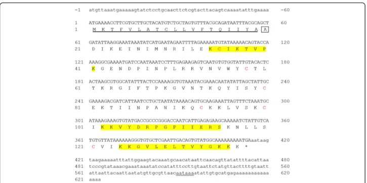
de-duced amino acid sequence are shown in Fig.1.
The complete coding sequence of the cDNA is 414 nucleotides to the stop codon, corresponding to 137 amino acids, including six cysteine residues after deduc-tion, which are related to other published Solenopsis 4 venom proteins (Sol i 4.01, Sol i 4.02, and Sol i 4q) [35– 37]. The signal sequence was analyzed using the Signal P program and identified 57 bp encoding 19 amino acids. The primary sequence of the deduced mature Sol g 4.1 protein contains 118 amino acid residues and starts with alanine (A), as confirmed by automated Edman degrad-ation sequencing (data not shown).
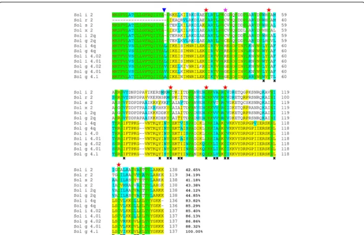
Although the leucines (L) and valines (V) observed in Solenopsis 4 venom proteins are substituted with A resi-dues in the Sol g 4.1 protein, these amino acids are clas-sified into hydrophobic groups and followed by DIKE sequences that were all highly conserved, as shown in Fig.2. The protein was rich in the amino acids K, N and P, with a theoretical isoelectric point of 9.87 and a pre-dicted molecular weight of 13,527.50 Da. GenBank Blastx searches revealed that the Sol g 4.1 protein closely resembles a member of the unique Solenopsis 2 and 4 venom proteins, whose biological functions remain unknown.
Comparison to Solenopsis 2 and 4
The alignment of the amino acid sequences of the Sol g 4.1 protein with the published Solenopsis 2 and 4 venom protein sequences from Solenopsis species is shown in
identity with the Sol g 4.01 and 4.02 allergens (GenBank ID: AAF65312 and GenBank ID: AAF65313), respect-ively; therefore, we designated this venom protein the Sol g 4.1 protein to differentiate between these proteins. The protein showed similarity to Sol i 4.01 and Sol i 4.02 (GenBank ID: AAC97369 and GenBank ID: AAC97370, respectively) (both 85%) [22, 36]. The iden-tity among all sequenced Solenopsis 4 proteins ranged from 83.8 to 88.3%, illustrating that the Solenopsis 4 proteins are rarely diverse and on average exhibit 86.0% identity among all Solenopsis 4 venom proteins. These sequences are highly conserved across species but are still poorly understood. Only 28 of the 118 mature amino acid sequences closely matched Solenopsis 2 and 4 venom proteins, in contrast to other published reports. Interestingly, the signal peptides of both groups are highly conserved and contain the greatest number of hydrophobic amino acid groups.
TheSolenopsis venom proteins were used to construct
a phylogenetic tree and analyzed using MEGA6 software [38] to confirm these results (Additional file 1). The major finding of this analysis is the conservation of six cysteines among all Solenopsis 2 and 4 venom proteins, but the seventh cysteine was only present in group 2; it forms a disulfide bond identical to that in other mole-cules [39]. Interestingly, Sol g 4q (GenBank ID: AAY32927) is more similar to Sol i 4.01 (99.3%) than Sol g 4.01 (88%). Although S. geminata 4 venom proteins
are found in tropical regions, different habitation sites
have important effects, due to food, natural enemies and survival skills, which have led to various evolutionary ad-aptations [40].
Expression and purification of the rSol g 4.1 protein
The molecular weight of the expressed recombinant pro-tein was approximately 34 kDa on SDS-PAGE. The ex-pression levels of the recombinant clones were determined after an incubation with 0.2, 0.4, 0.6, 0.8 or 1.0 mM IPTG for 2, 4, 6, 8, and 10 h or overnight. The growth patterns were significantly different in terms of both IPTG concentration and induction times (data not shown). Therefore, the optimal conditions for culture growth were 0.4 mM IPTG and 8 h, as shown in Fig.3a, lane 2. The rSol g 4.1 protein was expressed in inclusion bodies. Moreover, protein induction was confirmed by blotting lysates from induced and non-induced cultures with an anti-His tag antibody. The expressed protein strongly bound to antibodies, whereas proteins from non-induced culture did not bind (Fig.3b). After purifi-cation, the rSol g 4.1 protein was dialyzed with a 12-kDa molecular weight cutoff membrane. The fusion protein was expressed as a monomer, and the purity was con-firmed as a single band that represented 37% of the total proteins in Fig.4, lane 1. The results for the cleavage of the tag from the rSol g 4.1 protein are not shown. The optimal conditions for the removal of the tag from the protein is one unit of enzyme and incubation for 7 h (Fig. 4, lane 2). The rSol g 4.1 protein was separated Fig. 1Full-length DNA sequence and translation of the region encoding the Sol g 4.1 protein. Yellow-shaded areas were verified by LC-MS/MS of a partial amino acid sequence. The leader sequence is underlined. The 5΄and 3΄UTRs are indicated by small letters, and the poly (A) tail initiation
Fig. 2Alignment of the deduced amino acid sequences of the Sol g 4.1 protein with other Solenopsis 2 and 4 venom proteins fromS. invicta,S. geminata,S. saevissima,S. xyloniandS. richteri: conserved (red letters, green region), identical (yellow region), and groups of similar (turquois region) or non-similar (black letters, no color region) residues are shown. The end of the signal sequence is indicated by a blue triangle ( ). Alignment of the six cysteines (red stars) between all Solenopsis 2 and 4 genes and the alignment of the seventh cysteine in the Sol 2 genes (pink star). Residues lining the interior face of the Sol g 4.1 protein are indicated byx. The sequences were submitted to GenBank with the following accession numbers: Solenopsis 2 proteins: P35775 for Sol i 2, P35776 for Sol r 2, ABC58726 for Sol s 2, ALM98859 for Sol × 2, AAY32928 for Sol i 2q and AAY32926 for Sol g 2q; and Solenopsis 4 proteins: AAC97369 for Sol i 4.01, AAC97370 for Sol i 4.02, AAF65312 for Sol g 4.01, AAF65313 for Sol g 4.02, AAY32927 for Sol g 4q and AAY32929 for Sol i 4q
using a His GraviTrap column and analyzed on 13% SDS-PAGE gels, as shown in Fig.4, lane 3. The purified protein represented approximately 2% of the total fusion protein.
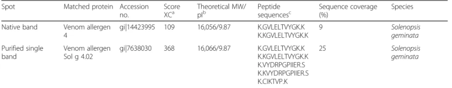
LC-MS/MS was performed to ascertain the definite rSol g 4.1 protein. A single band representing the puri-fied protein was trypsinized and subsequently identipuri-fied using LC-MS/MS. The peptides were identified with the protein MASCOT search engine using the NCBI protein and SwissProt databases. A similarity search using FASTA revealed a very high homology to S. geminata
venom allergen Sol g 4 for both the native and purified proteins, with molecular weights of 16,056 and 16,066 Da, respectively, as shown in Table2, which also corresponds to the experimental weight of 16 kDa from Sol g 4,S. geminatavenom, as described in our previous
reports [26]. The software predicted that the Sol g 4.1 protein was a member of the allergenic protein family. Representative partial amino acid sequences of the Sol g 4.1 protein from the fragment spectra of the unassign-able peptides in the tryptic digest were a 100% match after alignment and are shown in the yellow shaded re-gion in Fig.1.
Secondary structure analysis
The structure of the rSol g 4.1 protein lacking the tag consisted of 41.3% α-helices and 13.8% β-sheets after
refolding, and unidentified structures constituted ap-proximately 21.8% of the protein (Additional file2). The denatured protein only exhibited 16.5% α-helices and
10.2%β-sheets; unidentified structures comprised 48.5%
of the structure. Moreover, the secondary structure of the rSol g 4.1 protein showed 37% similarity to the S. invicta2 monomer (Additional file3), as predicted from
schematic diagrams (PDBsum), which exhibits seven helices from the N-terminus to C-terminus in the overall structure. Thus, the refolded rSol g 4.1 protein likely adopts the native structure.
Three-dimensional modeling of the predicted structure of the sol g 4.1 protein
The Sol i 2 (PDB code: 2ygu) chain A with a 2.60 Å reso-lution was used as a template; its X-ray structure is com-posed of two identical monomers [39]. The template showed the highest identity/similarity (35.90% with an E value of 1.0e−26) to the Sol g 4.1 sequence. They are found
in the sameSolenopsisspecies venom. The Ramachandran
plot displays the psi and phi backbone conformation angles for each amino acid residue in the Sol g 4.1 protein as shown in Additional file4. The plot statistics for the model displayed residues falling in 95% of most favored regions, 4% of additional allowed regions, 0% of generously allowed regions and 1% of disallowed regions. Overall plot showed over 90% of residues within the most favorable region. Therefore, Sol g 4.1 model was an acceptable good quality model and can be used for further analysis.
Moreover, G-factor value from PROCHECK tool used for evaluation probability of all dihedral angles showed 0.14. Based on the model, the Sol g 4.1 protein consists of three disulfide bonds, which were predicted to stabilize structures (Cys16-Cys39, Cys61-Cys74 and Cys81-Cys102), and seven α-helices, which presumably
surround the interior hydrophobic region. The compari-son of the structures of the Sol g 4.1 protein and the template revealed that the Sol g 4.1 protein is present as a monomer, and the overall structure seems similar to part of the venom allergen 2 molecule (Fig. 5a and b). As reported in the study by Borer et al. [39], two alkanes (decane and undecane) and one alkene (β-farnesene),
which are similar to alkane and alkene chains in these compounds, are attached to the sixth position of piperi-dine alkaloids and can bind to the hydrophobic pocket of Sol i 2. Thus, Sol i 2 is also conceivably involved in the transport of alkaloid derivatives from the site of syn-thesis to the venom reservoir or in the formation of a protective complex with the alkaloid in the venom duct.
A comparison of the amino acid residues in the three-dimensional models of the Sol g 4.1 and Sol i 2 structures showed that the interior face of the hydropho-bic region is lined with 17 apolar residues and three polar residues (Fig.2). Moreover, the structure of the Sol g 4.1 protein surface contains an unusually high number of charged residues that are evenly distributed on the surface, as shown in Fig. 5c. Overall, 35% of all residues Fig. 4SDS-PAGE analysis of the purified rSol g 4.1 protein and
on the surface of the Sol g 4.1 protein are charged: Asp, Glu, Lys, and Arg.
Determination of allergenic properties
An antiserum was produced in mice to determine the antigenic properties of the Sol g 4.1 protein. The Sol g 4.1 protein in crude venom was identified as a 16-kDa band on native PAGE gels in Fig. 6a, but the predicted molecular weight of its sequence is approximately 13,340 Da. Western immunoblotting analysis revealed a
clear interaction between the produced antibody and both native and recombinant Sol g 4.1 proteins, which were approximately 16 kDa, while PBS, acrylamide gel and adjuvant controls did not produce bands, as shown in Fig. 6b. This result confirmed that we successfully produced a specific antibody in BALB/c mice (the anti-Sol g 4.1 IgE antibody) that recognized native and recombinant Sol g 4.1 proteins.
The antibody specifically recognized the Sol g 4.1 pro-tein in its native form (Fig. 6b), suggesting that the
Table 2Protein identification of Sol g 4.1 fromS. geminatavenom
Spot Matched protein Accession
no.
Score
XCa Theoretical MW/pIb Peptidesequencesc Sequence coverage(%) Species Native band Venom allergen
4
gi|14423995 109 16,056/9.87 K.GVLELTVYGK.K K.KGVLELTVYGK.K
9 Solenopsis
geminata
Purified single band
Venom allergen Sol g 4.02
gi|7638030 368 16,066/9.87 K.GVLELTVYGK.K
K.KGVLELTVYGK.K K.VYDRPGPIIER.S K.KVYDRPGPIIER.S K.CIKTVP.K
25 Solenopsis
geminata
aScore XC obtained after LC-MS/MS analysis
bTheoretical molecular weight (MW) obtained after the LC-MS/MS analysis. The pI values were calculated using the ExPASy Peptide Mass
program (http://web.expasy.org/compute_pi/)
cDeduced peptide sequence obtained after LC-MS/MS (the number of matching peptides is indicated in parentheses)
antiserum did not show cross-reactivity to other proteins from crude venom. Interestingly, although Sol g 4.1 pro-tein sequences share 42% identity with Sol g 2.1 se-quences (unpublished data), they do not show immunological cross-reactivity, consistent with the re-sults reported by Hoffman [1] for Sol i 2 and Sol i 4, which exhibit 35% sequence homology and no antibody cross-reactivity.
Reduction of PD50activity by the addition of rSol g 4.1
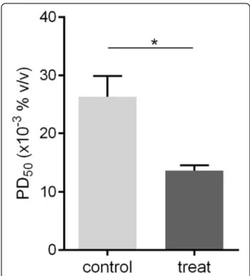
The PD50 was assayed in crickets in vivo to determine whether the refolded rSol g 4.1 protein lacking the tag altered the effects of piperidine alkaloids. The abdomen of the crickets was injected with PBS as a mock control or with a mixture of piperidine and protein and then in-cubated for 30 min. The PD50 of crude venom (positive control) in paralyzed crickets was 89 μg/g body weight, as described in our previous report [26]. First, the PD50 of piperidine in paralyzed crickets was approximately 0.027% (v/v) and designated PD50P1. Second, the injec-tion of the rSol g 4.1 protein in crickets showed that the optimal concentration was 1.0 μg of protein (2.86 μg/g body weight), but the recombinant protein did not in-duce cricket paralysis. Finally, 1.0 μg of rSol g 4.1 pro-tein was mixed with various concentrations of piperidine, and the PD50value was determined to be ap-proximately 0.013% (v/v) and designated PD50P2. There-fore, the rSol g 4.1 protein led to a significant decrease in PD50P1 to PD50P2, from 0.027 to 0.013% (p< 0.05), as shown in Fig.7. The major chemical component of fire ant venom is piperidine alkaloids [41]. Piperidine deriva-tives are the main active components that paralyze prey [42]. Based on the results of these experiments, the Sol g
4.1 protein has important synergistic effect with the pi-peridine derivatives in venom.
Discussion
The complete primary structure of the Sol g 4.1 protein was obtained in this study and showed high homology Fig. 6Allergenic analysis of native and recombinant Sol g 4.1 with anti-Sol g 4.1 IgE antibody.aCrude venom expression pattern, as determined by SDS-PAGE.bDetermination of the allergenic properties of the Sol g 4.1 protein by producing an antiserum in mice and analyzing the product using Western blotting. Recognition of native Sol g 4.1 and rSol g 4.1 proteins by serum IgE in Sol g 4.1 protein-sensitized mice. Serum samples: P1-P3 = individual sera of Sol g 4.1 protein-sensitized mice; N1-N3 = serum from mice injected with PBS, acrylamide gel and adjuvant, respectively, as controls
to Solenopsis 2 and 4 venom proteins, suggesting that they may perform similar functions and exhibit similar localization patterns. Based on three-dimensional model structures, Sol g 4.1 was identical to a portion of the Sol i 2 molecule. According to Borer et al. [39], the overall crystal structure of Sol i 2 is stabilized by three intramo-lecular disulfide bonds and one intermointramo-lecular disulfide bond, which differed from the Sol g 4.1 protein (only contains six cysteines), creating a hydrophobic pocket. Thus, the Sol g 4.1 protein is present as a monomer and that its structure is stabilized by three disulfide bonds. In addition, the Sol g 4.1 protein exhibited 21% identity to the hydrophobic ligand-binding proteins from the pheromone-binding protein/odorant-binding protein (PBP/OBP) family, which is normally composed of pro-teins with molecular weights of 12–16 kDa. The amino acid sequences are extremely diverse but are all distin-guished by a pattern of six cysteines that form three di-sulfide bonds. The three-dimensional structure contains a cluster of six or seven α-helices surrounding the
hydrophobic pocket in which the hydrophobic ligand binds [43,44].
Whole-body extracts contain not only venom compo-nents but also proteolytic enzymes and various other soluble insect proteins. These soluble proteins can react with IgE antibodies that have been induced by proteins from other species, and the proteolytic enzymes can des-troy venom allergens. Moreover, venom contains a sig-nificant concentration of piperidine alkaloids, which are difficult to completely remove from the proteins [41]. Allergenic proteins are also extremely difficult to purify from each other if they have similar pI values [15]. The expression of recombinant proteins will overcome the problem of obtaining large quantities from natural mate-rials. Therefore, the expression and purification of the rSol g 4.1 protein in theE. coli system is a good choice
for heterologous expression of recombinant proteins due to its capacity to produce abundant recombinant protein and easy manipulation.
The Sol g 4.1 protein was cloned into a pET-32a(+) vector containing the thioredoxin (Trx) tag, which can catalyze the formation of disulfides and promote the solubility of the target protein in the cytoplasm ofE. coli
[45]. However, the rSol g 4.1 protein was expressed as an insoluble protein, which may be affected by numer-ous parameters, including temperature [46], and then rSol g 4.1 was refolded by dialysis and we investigated its secondary structure, which was primarily α-helices.
Venom protein expression in E. coli will save research
cost and time, while expression in baculovirus-infected insect cells requires further study. As Solenopsis 4 pro-teins do not have carbohydrate determinants (CCDs) [36,37], this study selected a quick and cheap system to express large concentrations of the Sol g 4.1 venom
protein, which can be applied to allergenic testing of these venom proteins and could reduce the cost of this operation.
Based on the analysis of the allergenic properties, BALB/c mice generated an antibody in response to pro-tein exposure [30] that strongly bound to the native and recombinant Sol g 4.1 proteins, suggesting that, as ex-pected, the Sol g 4.1 protein was immunogenic in mice. This experiment was also supported by the finding that the surface of the Sol g 4.1 protein is composed of 35% charged residues (Asp, Glu, Lys, and Arg), a percentage that is considerably higher than the average value (27%) for normal proteins [47]. Charged amino acids often dis-play significant contributions to the free energy of binding in protein-protein interactions and/or antigen-antibody complexes. The importance of charged surface residues in IgE binding and the allergenicity of the dust mite allergen Blo t 5 and other major allergens has been confirmed in mutagenesis studies [48–50]. Moreover, the sequence of the Sol g 4.1 protein produced in E. coli is highly
con-served and displays greater than 86% identity to Sol i 4.01/ Sol i 4.02 proteins produced using the same protein ex-pression system, as identified as allergic individuals [36]. However, the full characterization of antigen-antibody rec-ognition sites will require the elucidation of the complex structure of Sol g 4.1 protein with its specific antibodies, as allergen epitopes are continuous or discontinuous [51].
Moreover, we studied PD50 values by mixing piperi-dine alkaloids with rSol g 4.1 to verify the hypothetical functions of the Sol g 4.1 protein based on protein se-quences and structural similarity to Sol i 2. The rSol g 4.1 protein may be involved in interactions with hydro-phobic ligands, consistent with the results of the study by Borer et al. [39], who analyzed the role of the hydro-phobic pocket in the allergenic Sol i 2 protein. The high-est binding affinity was observed for hydrophobic ligands such as pheromones, fatty acids, or short-lived hydrophobic primers [52, 53]. Consistent with these findings, Das et al. [54] showed that Sol i 4.02 has an in-terior binding pocket with a size of approximately 0.4 nm3, and the interior pockets of
S. geminatavenom
proteins bind to the alkaloid solenopsin A. Therefore, the Sol g 4.1 protein is also conceivably involved in the interaction with hydrophobic ligands.
Further studies are required to produce large amounts of soluble protein, which will aid in the study of the func-tion of these extremely potent allergens. An analysis of the clear functions of the Sol g 4.1 protein should be per-formed, particularly a study that investigates its interac-tions with alkaloids/ligands and their localization patterns.
Conclusions
geminatavenom. In our study, we describe the
identifi-cation, expression and characterization of rSol g 4.1. Ini-tially, rSol g 4.1 was expressed in inclusion bodies, and the structure of the refolded rSol g 4.1 protein was likely the native form, mainly α-helices, as determined by a
secondary structure analysis. Both native and recombin-ant Sol g 4.1 proteins have a molecular weight of 16 kDa, although the amino acid sequence predicted a molecular weight of 13,340 Da. The predicted three-dimensional model showed three disulfide bonds that stabilized its structure. Solenopsis 2 and 4 venom proteins are unique ant venom proteins, including other Hymenoptera venom proteins [15,19]. Based on a statis-tical analysis of cricket paralysis, Sol g 4.1 resulted in a significant decrease in PD50 values. Thus, similar to Sol g 4.02 [54], Sol g 4.1 seems to function by binding to hydrophobic ligands, such as pheromones and alkaloids. Based on the results of the allergenic test presented here, the anti-Sol g 4.1 IgE antibody responses observed in mice suggest that Sol g 4.1 is an allergenic protein.
Additional files
Additional file 1:Phylogenetic relationships between the amino acid sequences of theSolenopsisspecies groups 2 and 4 proteins.Bombyx moriPBP (GenBank ID: P34174) is an outgroup. The evolutionary tree was analyzed using the neighbor-joining method. The percentage of replicate trees in which the associated taxa clustered together in the 1000 boot-strap character replicates is indicated for groups that appeared in≥50% of bootstrap trees. Horizontal line distances are proportional to calculated phylogenetic differences. (JPG 2218 kb)
Additional file 2:CD spectrum of the Sol g 4.1 protein produced under denatured and refolded conditions. (JPG 34 kb)
Additional file 3:The topology diagram of the Sol g 4.1 protein created using PDBsum software shows the relative locations ofα-helices, which
are presented as red cylinders. The small arrows indicate the directionality of the protein chain from the N-terminus to the C-terminus. Numbers within the secondary structural elements correspond to the residue num-ber in the protein. (JPG 262 kb)
Additional file 4:Ramachandran plot analysis of Sol g 4.1 model. The color codes are: red–most favorable regions, yellow–allowed regions, pale yellow–generously allowed regions; and white–disallowed regions. (JPG 288 kb)
Abbreviations
PBP:Pheromone-binding protein; PD50: 50% of paralytic dose; rSol g 4.1 protein: Recombinant Sol g 4.1 protein;S.:Solenopsisspecies; Sol g 4:Solenopsis geminatavenom allergen number 4; Sol i 2:Solenopsis invicta
venom allergen number 2; Sol i 4:Solenopsis invictavenom allergen number 4
Acknowledgements
We would like to thank the Protein and Proteomics Research Center for Commercial and Industrial Purposes (ProCCI), Department of Biochemistry, Faculty of Science, Khon Kaen University, Thailand, with additional support from the Khon Kaen University Research Fund, fiscal year 2013 to 2015. We also appreciate the advice of Dr. Noppadol Prasertsincharoen, Veterinary Technology Program, Faculty of Veterinary Technology, Kasetsart University, regarding the statistical analyses performed in this study.
Funding
Financial support for this research was provided by The Royal Golden Jubilee Ph.D. Program, The Thailand Research Fund (TRF).
Authors’contributions
HS conducted nearly all of the experiments, coordinated the data analysis, drafted and edited the manuscript for publication. SS partially contributed to the nucleotide sequencing and bioinformatics analyses. SK partially contributed to the experiment designs and writing of the manuscript. JD partially contributed to the nucleotide sequencing and bioinformatics. SD designed the research, coordinated the study, edited the manuscript for publication, and corresponded with the journal editor. All authors read and approved the final manuscript.
Ethics approval and consent to participate
The present study was approved by the Animal Ethics Committee of Khon Kaen University based on the Ethics for Animal Experimentation of the National Research Council of Thailand (reference no. 0514.1.12.2/66).
Consent for publication Not applicable.
Competing interests
The authors declare that they have no competing interests.
Publisher’s Note
Springer Nature remains neutral with regard to jurisdictional claims in published maps and institutional affiliations.
Author details
1Protein and Proteomics Research Center for Commercial and Industrial
Purposes (ProCCI), Department of Biochemistry, Faculty of Science, Khon Kaen University, Khon Kaen 40002, Thailand.2Division of Integrative Medicine, Chulabhorn International College of Medicine, Thammasat University (Rangsit Campus), Pathum Thani 12120, Thailand.3Department of Clinical Chemistry, Faculty of Associated Medical Sciences, Khon Kaen University, Khon Kaen 40002, Thailand.4Division of Pharmacognosy and Toxicology, Faculty of Pharmaceutical Sciences, Khon Kaen University, Khon Kaen 40002, Thailand.
Received: 6 March 2018 Accepted: 3 August 2018
References
1. Hoffman DR. Fire ant venom allergy. Allergy. 1995;50(7):535–44. 2. Tankersley MS. The stinging impact of the imported fire ant. Curr Opin
Allergy Clin Immunol. 2008;8(4):354–9.
3. Hoffman DR. Reactions to less common species of fire ants. J Allergy Clin Immunol. 1997;100(5):679–83.
4. Hoffman DR. Ant venoms. Curr Opin Allergy Clin Immunol. 2010;10(4):342–6. 5. Srisong H, Daduang S, Lopata AL. Current advances in ant venom proteins
causing hypersensitivity reactions in the Asia-Pacific region. Mol Immunol. 2016;69:24–32.
6. Rhoades R. Stinging ants. Curr Opin Allergy Clin Immunol. 2001;1(4):343–8. 7. Arbiser JL, Kau T, Konar M, Narra K, Ramchandran R, Summers SA, et al.
Solenopsin, the alkaloidal component of the fire ant (Solenopsis invicta), is a naturally occurring inhibitor of phosphatidylinositol-3-kinase signaling and angiogenesis. Blood. 2007;109(2):560–5.
8. MacConnell JG, Blum MS, Fales HM. The chemistry of fire ant venom. Tetrahedron. 1971;27(6):1129–39.
9. Chen J, Shang H, Jin X. Interspecific variation of Delta1, 6-piperideines in imported fire ants. Toxicon. 2010;55(6):1181–7.
10. Obin MS, Vander Meer RK. Gaster flagging by fire ants (Solenopsisspp.): functional significance of venom dispersal behavior. J Chem Ecol. 1985; 11(12):1757–68.
11. Jouvenaz DP, Blum MS, MacConnell JG. Antibacterial activity of venom alkaloids from the imported fire ant,Solenopsis invictaBuren. Antimicrob Agents Chemother. 1972;2(4):291–3.
13. Javors MA, Zhou W, Maas JW Jr, Han S, Keenan RW. Effects of fire ant venom alkaloids on platelet and neutrophil function. Life Sci. 1993;53(14): 1105–12.
14. Rakich PM, Latimer KS, Mispagel ME, Steffens WL. Clinical and histologic characterization of cutaneous reactions to stings of the imported fire ant (Solenopsis invicta) in dogs. Vet Pathol. 1993;30(6):555–9.
15. Hoffman DR, Dove DE, Jacobson RS. Allergens in Hymenoptera venom: XX. Isolation of four allergens from imported fire ant (Solenopsis invicta) venom. J Allergy Clin Immunol. 1988;82(5 Pt 1):818–27.
16. dos Santos Pinto JRA, Fox EGP, Saidemberg DM, Santos LD, da Silva Menegasso AR, Costa-Manso E, et al. Proteomic view of the venom from the fire antSolenopsis invictaBuren. J Proteome Res. 2012;11(9):4643–53. 17. Hoffman DR, Sakell RH, Schmidt M. Sol i 1, the phospholipase allergen of
imported fire ant venom. J Allergy Clin Immunol. 2005;115(3):611–6. 18. Schmidt M, McConnell TJ, Hoffman DR. Immunologic characterization of the
recombinant fire ant venom allergen sol i 3. Allergy. 2003;58(4):342–9. 19. Schmidt M, McConnell TJ, Hoffman DR. Production of a recombinant
imported fire ant venom allergen, sol i 2, in native and immunoreactive form. J Allergy Clin Immunol. 1996;98(1):82–8.
20. Schmidt M, Walker RB, Hoffman DR, McConnell TJ. Nucleotide sequence of cDNA encoding the fire ant venom protein Sol i II. FEBS Lett. 1993;319(1–2):138–40. 21. Hoffman DR, Smith AM, Schmidt M, Moffitt JE, Guralnick M. Allergens in Hymenoptera venom. XXII. Comparison of venoms from two species of imported fire ants, Solenopsis invictaandrichteri. J Allergy Clin Immunol. 1990;85(6):988–96.
22. Hoffman DR. Allergens in Hymenoptera venom. XVII. Allergenic components ofSolenopsis invicta(imported fire ant) venom. J Allergy Clin Immunol. 1987;80(3 Pt 1):300–6.
23. Deslippe RJ, Guo YJ. Venom alkaloids of fire ants in relation to worker size and age. Toxicon. 2000;38(2):223–32.
24. Blum MS, Walker JR, Callahan PS, Novak AF. Chemical, insecticidal and antibiotic properties of fire ant venom. Science. 1958;128(3319):306–7. 25. Potiwat R, Sitcharungsi R. Ant allergens and hypersensitivity reactions in
response to ant stings. Asian Pac J Allergy Immunol. 2015;33(4):267–75. 26. Sukprasert S, Uawonggul N, Jamjanya T, Thammasirirak S, Daduang J,
Daduang S. Characterization of the allergen Sol gem 2 from the fire ant venom,Solenopsis geminata. J Venom Anim Toxins incl Trop Dis. 2012;18(3): 325–34. http://www.scielo.br/scielo.php?script=sci_arttext&pid=S1678-91992012000300010
27. Yi GB, McClendon D, Desaiah D, Goddard J, Lister A, Moffitt J, et al. Fire ant venom alkaloid, isosolenopsin A, a potent and selective inhibitor of neuronal nitric oxide synthase. Int J Toxicol. 2003;22(2):81–6. 28. Bradford MM. A rapid and sensitive method for the quantitation of
microgram quantities of protein utilizing the principle of protein-dye binding. Anal Biochem. 1976;72:248–54.
29. Kiefer F, Arnold K, Künzli M, Bordoli L, Schwede T. The SWISS-MODEL repository and associated resources. Nucleic Acids Res. 2009;37:D387–92. 30. Dearman RJ, Stone S, Caddick HT, Basketter DA, Kimber I. Evaluation of
protein allergenic potential in mice: dose-response analyses. Clin Exp Allergy. 2003;33(11):1586–94.
31. Riley V. Adaptation of orbital bleeding technique to rapid serial blood studies. Proc Soc Exp Biol Med. 1960;104:751–4.
32. Uawonggul N, Thammasirirak S, Chaveerach A, Arkaravichien T, Bunyatratchata W, Ruangjirachuporn W, et al. Purification and
characterization of Heteroscorpine-1 (HS-1) toxin fromHeterometrus laoticus
scorpion venom. Toxicon. 2007;49(1):19–29.
33. Kwak SG, Kim JH. Central limit theorem: the cornerstone of modern statistics. Korean J Anesthesiol. 2017;70(2):144–56.
34. Chong SH, Ham S. Protein folding thermodynamics: a new computational approach. J Phys Chem B. 2014;118(19):5017–25.
35. Schmidt M, Hoffman DR. Expression of recombinant fire ant venom allergen Sol i 4. J Allergy Clin Immunol. 2001;107(2):S112–3.
36. Lockwood SA, HaghiPour-Peasley J, Hoffman DR, Deslippe RJ. Identification, expression, and immuno-reactivity of Sol i 2 & Sol i 4 venom proteins of queen red imported fire ants,Solenopsis invictaBuren (Hymenoptera: Formicidae). Toxicon. 2012;60(5):752–9.
37. Han XQ, Lin XM, Chen HJ, Zhang YG, Ye GS, Wu SQ, et al. The prokaryotic expression and bioactivity of the recombinant red fire ant venom allergen Sol i 4. Agr Sci China. 2009;8(2):182–7.
38. Tamura K, Stecher G, Peterson D, Filipski A, Kumar S. MEGA6: molecular evolutionary genetics analysis version 6.0. Mol Biol Evol. 2013;30(12):2725–9.
39. Borer AS, Wassmann P, Schmidt M, Hoffman DR, Zhou JJ, Wright C, et al. Crystal structure of Sol i 2: a major allergen from fire ant venom. J Mol Biol. 2012;415(4):635–48.
40. Schmidt JO. Evolutionary responses of solitary and social Hymenoptera to predation by primates and overwhelmingly powerful vertebrate predators. J Hum Evol. 2014;71:12–9.
41. Chen L, Fadamiro HY. Re-investigation of venom chemistry ofSolenopsisfire ants. II. Identification of novel alkaloids inS. invicta. Toxicon. 2009;53(5):479–86. 42. Chen L, Sharma KR, Fadamiro HY. Fire ant venom alkaloids act as key
attractants for the parasitic phorid fly,Pseudacteon tricuspis(Diptera: Phoridae). Naturwissenschaften. 2009;96(12):1421–9.
43. Sandler BH, Nikonova L, Leal WS, Clardy J. Sexual attraction in the silkworm moth: structure of the pheromone-binding-protein-bombykol complex. Chem Biol. 2000;7(2):143–51.
44. Renthal R. Discovering pheromones of the red imported fire ant (Solenopsis invictaBuren): a review of and proposed new target for pheromone disruption. J Agr Urban Entomol. 2003;20:113–21.
45. Stewart EJ, Aslund F, Beckwith J. Disulfide bond formation in theEscherichia colicytoplasm: anin vivorole reversal for the thioredoxins. EMBO J. 1998; 17(19):5543–50.
46. Shirano Y, Shibata D. Low temperature cultivation ofEscherichia colicarrying a rice lipoxygenase L-2 cDNA produces a soluble and active enzyme at a high level. FEBS Lett. 1990;271(1–2):128–30.
47. Miller S, Janin J, Lesk AM, Chothia C. Interior and surface of monomeric proteins. J Mol Biol. 1987;196(3):641–56.
48. Chan SL, Ong TC, Gao YF, Tiong YS, Wang de Y, Chew FT, et al. Nuclear magnetic resonance structure and IgE epitopes of Blo t 5, a major dust mite allergen. J Immunol. 2008;181(4):2586–96.
49. Metzler WJ, Valentine K, Roebber M, Friedrichs MS, Marsh DG, Mueller L. Determination of the three-dimensional solution structure of ragweed allergen Amb t V by nuclear magnetic resonance spectroscopy. Biochemistry. 1992;31(22):5117–27.
50. Razzera G, Gadermaier G, de Paula V, Almeida MS, Egger M, Jahn-Schmid B, et al. Mapping the interactions between a major pollen allergen and human IgE antibodies. Structure. 2010;18(8):1011–21.
51. Van Regenmortel MH. Immunoinformatics may lead to a reappraisal of the nature of B cell epitopes and of the feasibility of synthetic peptide vaccines. J Mol Recognit. 2006;19(3):183–7.
52. Thode AB, Kruse SW, Nix JC, Jones DN. The role of multiple hydrogen-bonding groups in specific alcohol binding sites in proteins: insights from structural studies of LUSH. J Mol Biol. 2008;376(5):1360–76.
53. Fan J, Francis F, Liu Y, Chen JL, Cheng DF. An overview of odorant-binding protein functions in insect peripheral olfactory reception. Genet Mol Res. 2011;10(4):3056–69.
54. Das T, Alabi I, Colley M, Yan F, Griffith W, Bach S, et al. Major venom proteins of the fire antSolenopsis invicta: insights into possible pheromone-binding function from mass spectrometric analysis. Insect Mol Biol. 2018;