Polyethylene glycol and polyvinylpirrolidone effect on bacterial rRNA
extraction and hybridization from cells exposed to tannins
(1)Pedro Braga Arcuri(2), Michael Larry Thonney(3), Peter Schofield(3) and Alice Nelson Pell(3)
Abstract – In order to detect fluctuations in ruminal microbial populations due to forage tannins using 16S ribosomal RNA (rRNA) probes, recovery of intact rRNA is required. The objective of this work was to evaluate the effect of polyethylene glycol (PEG) and polyvinylpirrolidone (PVP) on extraction of bacterial rRNA, in the presence of tannins from tropical legume forages and other sources, that hybridize with oligonucleotide probes. Ruminococcus albus 8 cells were exposed to 8 g/L tannic acid or 1 g/L condensed tannins extracted from Acacia angustissima, banana (Musa sp.) skin, Desmodium ovalifolium, red grape (Vitis vinifera) skin and Inga edulis, or no tannins. Cells were rinsed with Tris buffer pH 7 containing either 8% PEG or 6% PVP prior to cell lysis. Total RNA samples rinsed with either PEG or PVP migrated through denaturing agarose gels. The 16S rRNA bands successfully hybridized with a R. albus species-specific oligonucleotide probe, regardless of tannin source. The effect of rinsing buffers on the density of 16S rRNA bands, as well as on the hybridization signals was compared. There were significant effects (P<0.01) when the controls were compared to either buffer treatments due to tannin type, buffer used and the interaction of tannin type and buffer. The significant interaction indicates the influence of tannin type on the parameters evaluated.
Index terms: Ruminococcus albus, microbial ecology, animal nutrition.
Efeito de polietilenoglicol e polivinilpirrolidona na extração e hibridização de rRNA bacteriano de células expostas a taninos
Resumo – A recuperação de RNA ribossômico (rRNA) intacto é necessária para a detecção de flutuações na população microbiana ruminal decorrentes dos taninos de forrageiras, utilizando-se sondas para 16S rRNA. O objetivo deste trabalho foi avaliar o efeito de polietilenoglicol (PEG) e polivinilpirrolidona (PVP) na extração de rRNA bacteriano, em presença de taninos de leguminosas forrageiras tropicais e de outras fontes, que possa ser hibridizado com sondas de oligonucleotídeos. Culturas de Ruminococcus albus 8 foram expostas ou não a 8 g/L de ácido tânico ou a 1 g/L de taninos condensados, extraídos de Acacia angustissima, casca de banana (Musa sp.), Desmodium ovalifolium, cascas de uvas vermelhas (Vitis vinifera) e Inga edulis. As culturas foram lavadas com tampão Tris pH 7 contendo 8% PEG ou 6% PVP antes do rompimento das células. Amostras de RNA total lavadas com PEG ou PVP migraram em géis de agarose. Bandas de 16S rRNA hibridizaram com uma sonda de oligonucleotídeos espécie-específica para R. albus, independentemente da fonte de tanino. Comparou-se o efeito dos tampões de lavagem sobre a densidade das bandas de 16S rRNA, assim como sobre a intensidade de hibridização. Ocorreram efeitos significativos para fontes de taninos, tampões e para a interação entre taninos e tampões (P<0.01). A interação significativa indica a influência do tipo de tanino nos parâmetros avaliados.
Termos para indexação: Ruminococcus albus, ecologia microbiana, nutrição animal.
(1)Accepted for publication on June 20, 2003.
Extracted from Ph.D. dissertation presented by the first author to the Dept. of Animal Science, Cornell University, Ithaca, NY, USA.
(2)Embrapa-Centro Nacional de Pesquisa de Gado de Leite, Rua
Eugênio do Nascimento, 610, CEP 36038-330 Juiz de Fo-ra, MG. E-mail: [email protected]
(3)Cornell University, Dept. of Animal Sciences, Morrison Hall,
Ithaca, NY, 14853-4801 USA. E-mail: [email protected], [email protected], [email protected]
Introduction
used by different bacteria to tolerate tannins appear
to differ, but these mechanisms are poorly
under-stood (Jones et al., 1994; Nelson et al., 1997).
The study of ruminal microbial ecology of
ani-mals fed on tannin-rich diets is of fundamental
im-portance for ruminants grazing tropical legumes.
Tropical legumes contain more protein than grasses,
but tannins and other secondary compounds restrict
their use as forages. Tannin-containing forages may
alter the efficiency of use and main site of digestion
of proteins (Barry, 1989). The study of microbial
ecology has been greatly enhanced by the
develop-ment of molecular techniques. However, the
pres-ence of tannins or other polyphenols creates
prob-lems for nucleic acid extraction (Alm et al., 2000;
Krause et al., 2001). Although there are several
meth-ods for isolating nucleic acids in the presence of
polyphenols from soils containing humic acids
(Moré et al., 1994; Ogram, 1998), most of the
meth-ods are related to extraction from plant material (Pich
& Schubert, 1993; MacKenzie et al., 1997). Krause
et al. (2001) looked at extraction methods using
poly-ethylene glycol (PEG) and condensed tannin from
commercial Quebracho (barks from
Quercus
sp.) at
up to 8% of DM. Without PEG, the authors could
only amplify nucleic acid sequences using the
poly-merase chain reaction (PCR) at 0.5% and 1% tannin,
but with PEG they could amplify at all
concentra-tions. They also did some tests of their extraction
protocol on
Calliandra
sp.,
Acacia angustissima
,
A. boliviana
,
A. villosa
,
Leucaena pallida
, L.
diversifolia
and L.
leucocephala
tannins, with
simi-lar results. However, most of these methods were
developed for microbial detection, not enumeration,
so that little emphasis was placed on the
quantita-tive recovery of the nucleic acids. A common
ap-proach to extraction of nucleic acids from
environ-ments containing polyphenols is to add compounds
with high affinity for the polyphenols to reduce
for-mation of nucleic acid-polyphenol complexes
(Loomis, 1974; Krause et al., 2001).
Polyvinylpyrroli-done (PVP) (Molyneux, 1983) and PEG (Molyneux,
1984) are among the chemicals identified as
poten-tially useful as tannin precipitants due to their high
hydrogen bonding affinity, therefore their high
af-finity for polyphenols (Loomis, 1974). In addition,
their low toxicity, water solubility and low cost make
them preferred compounds for complexing
polyphe-nols.
Nucleic acids can be extracted from fresh samples
using a variety of protocols (Ausubel et al., 1996), most
of which are based on the phenol-chloroform-alcohol
(Stahl et al., 1988) or the guanidinium thiocyanate
(Chomczynski & Sacchi, 1987) methods.
The objective of this work was to evaluate the
effect of PEG and PVP on extraction of bacterial
rRNA, in the presence of tannins from tropical
le-gume forages and other sources, that could
success-fully hybridize with oligonucleotide probes.
Material and Methods
Culture and medium
Cultures of Ruminococcus albus 8 initially obtained from the University of Illinois, Urbana-Champaign cul-ture collection, were grown in anaerobically prepared basal medium (Nelson et al., 1997), without the addition of resazurin. Basal medium contained ammonium as the only N source. The energy source used was 0.4% cellobiose (w/v). This medium was chosen to prevent protein pre-cipitation due to tannins. Butyl rubber-stoppered serum bottles (150 mL) or 150x18 mm Balch tubes were used to grow 100 mL or 10 mL cultures at 39oC. After cultures had grown to the desired optical density, they were ex-posed to tannins. Microscopic observations to assess ly-sis and culture purity were performed from fresh mounts in a Nikon Labphoto optical microscope with a contrast phase objective.
Optical densities were measured at 600 nm in a Spectronic 601 spectrophotometer from 1 mL aliquots in quartz cuvettes (1cm length path) or through the 150x18 mm anaerobic culture tubes. Pure culture stocks were maintained in Rum 10 medium (Latham et al., 1978) where the energy source was approximately 0.2 g of acid-swollen cellulose.
Tannin solutions
Isolation of condensed tannins from plant samples
Samples of young leaves were collected from A. angustissima Mill. (Leguminosae) and Inga edulis Mart. (Leguminosae) from plants grown in a greenhouse at Cornell University, Ithaca, NY. Other samples were mature leaves of Desmodium ovalifolium Desv. (Leguminosae) harvested and freeze dried at the Escuela Agricola Panamericana (El Zamorano, Honduras), the skins of ripe red seedless grapes (Vitis vinifera L., Vitaceae) and the skins of unripe bananas (Musa paradisiaca L., Musaceae). Except for the D. ovalifolium sample, all other samples were frozen in liquid nitrogen and ground in a pre-chilled ceramic mortar and pestle. The fruit skins were peeled off and immedi-ately placed in liquid nitrogen. All samples were lyophilized and stored in the dark at 4oC.
Tannins were extracted from plant samples using the procedure described by Hagerman (1988). Variable amounts of plant material (10 to 25 g) were suspended in 10 mL of 80% (v/v) aqueous ethanol per 1.0 g of sample dry matter, stirred for 30 min and stored overnight at 4oC. The suspension was filtered through Whatman filter pa-per no. 40 using slight vacuum. Condensed tannins were separated from low molecular weight tannins, other phe-nolics and pigments using 100 g Sephadex LH-20 dextran resin in 80% aqueous ethanol. After elution with 50% acetone, two extractions with ethyl acetate and lyophiliza-tion, the purified condensed tannin powders were stored at 4oC in the dark.
Spectrophotometric profile of tannin solutions
Standardized (1 mg/mL) aqueous solutions of the con-densed tannins extracted from D. ovalifolium, grape skin and I. edulis as well as tannic acid were diluted (1:50) in water or 0.5M Tris pH 7.0 and their absorbance profile between 240 and 320 nm was recorded from a Beckman DU 500 spectrophotometer using a 100 µL quartz cuvette. The absorbance profile of the 0.5M Tris buffer was also measured.
Tannin-binding buffers
Polyvinylpyrrolidone-40,000 (PVP-40) and polyeth-ylene glycol (PEG) were used to prepare 6% PVP (Loomis, 1974) and 8% (wt/vol) PEG (Makkar et al., 1995) solu-tions in 0.5M Tris buffer (pH 7.0).
Experimental procedure
All treatments were tested in triplicate, except for A. angustissima, where two and one replicates were treated with PEG and PVP buffers, respectively.
Tannin exposure
The absorbance (A600) of overnight cultures was mea-sured. Aliquots in 10 mL volume were transferred into 50-mL polypropylene centrifuge flasks. Tannins were added to yield final concentrations to each aliquot of 8.0 g/L tannic acid or 1.0 g/L purified condensed tannin culture medium. The samples were incubated with tannins for 15 min at room temperature. Both positive and negative control were used. The positive control (T+B-) contained tannin and samples rinsed with Tris buffer without PEG or PVP. The negative control (T-B-) contained neither tannin nor PEG or PVP. The negative control was centrifuged once and kept on ice until all other samples were ready for total RNA extraction.
Rinsing with buffers
Total RNA extraction
The recommendations for use of the RNAqueous
kit (Ambion Inc., Cupertino, CA) were followed. Cells in the presence of beads and lysing/binding solution were beaten for 7 min at maximum speed in a mini-bead beater 8 (Biospec, Bartlesville, OK) at 4oC (Raskin et al., 1997). Maximum lysate volumes were obtained by inserting the pipette tip deep into the bead layer and pipetting two or three times after the bulk volume had been transferred. The lysate was transferred into a disposable 15x75 mm borosilicate glass culture tube. The elution procedure was repeated three times and the three aliquots were saved altogether.
Quantification and quality assessment of RNA
Total RNA concentrations were determined spectro-photometrically at 260 nm, assuming that 1 unit of absor-bance at 260 nm = 40 µg RNA/mL (Ausubel et al., 1995). RNA solutions were stored at -70oC. The integrity of rRNA bands was assessed using electrophoresis with aga-rose denaturing gels (1% agaaga-rose (w/v) in formaldehyde, 10 x MOPS buffer), prepared according to Ausubel et al. (1996). Running conditions were 1 x MOPS buffer (pH 7) at 4.8 to 5.0 volts/cm for approximately 2 hours. Gels were photographed over UV light (254 nm) and digitally recorded using the AlphaImager imaging system with the IS-1000 software version 2.00 (Alpha Innotech Corp., San Leandro, CA).
The density of 16S rRNA bands was measured using the AlphaImager system. The integrated pixel value was obtained using the 1D-multi toolbox from the IS-1000 soft-ware.
Probe hybridization
Oligonucleotide probe S-S-R.al-0196-a-A-18 (5’-GTC ATG CGG CTT CGT TAT- 3’) (Odenyo et al., 1994) was labeled with digoxigenin-11-ddUTP using terminal transferase from the Genius #6 oligonucleotide tailing kit following the recommendations of the manufacturer. Total RNA samples (either 10 µg, 1 µg or 0.1 µg total RNA) were blotted onto a Magna Charge nylon mem-brane using a minifold II slot blot vacuum manifold. Mem-branes were UV-crosslinked in a Spectrolinker XL-1000 UV crosslinker, prehybridized for 2 hours and hybridized overnight at 42oC with 0.5 pmol probe/mL hybridization buffer (5 x sodium-saline-citrate solution, 50% deionized formamide, 0.1% Na-lauroylsarcosine, 0.02% SDS, 2% blocking reagent and 0.1 mg Poly (A)/mL). Stringent washes were performed twice with 2 X SSC solution at room temperature and twice with 0.5 X SSC solution
at 42oC. For detection of hybridized 16S rRNA the Genius #7 luminescent detection kit containing the luminescent substrate disodium 3-(4-methoxyspiro{1,2-dioxetane-3,2’-(5’-chloro) tricyclo [3.3.1.13,7]decan}-4-yl) phenyl phosphate (CSPD®) was used and followed the manufacturer’s recommendations. Results were photographed onto X-ray film after incubation of membranes with substrate solution at 37oC and 45 minutes of film exposure. Hybridized bands were digitally recorded in the AlphaImager imaging system and their densities were mea-sured.
Statistical analysis
Analyses of variance of the integrated pixel values obtained from densitometry of 16S rRNA bands and hy-bridization signals were performed. Factors tested were tannin type, rinsing buffer used and, for the hybridization signals, levels of RNA blotted onto the membranes. The two-way interactions among these factors were also tested. Data were analyzed with ANOVA, the general linear model and SNK test routines of the Minitab software release 11.21 (Minitab Inc. State College, PA).
Results and Discussion
The pH of the suspensions (i.e., culture medium
mixed with tannins or rinsing buffers) ranged from
6.7 to 6.9 for all conditions tested. After
centrifuga-tion, there were differences in the type of cell pellet
formed. Overall, pellets from PEG rinse were smaller
but more difficult to resuspend than the pellets rinsed
with PVP, when compared within the same tannin
type. The linear structure of PEG (Molyneux, 1984)
may explain the difference between buffers in the
firmness of the pellets formed after rinsing and
cen-trifugation.
Bead beating was performed as recommended by
Raskin et al. (1997), except when 2 g of 0.1 mm
zirco-nium beads were used. Microscopic observations
performed with the lysate showed that most cells
were lysed, although in every field from any slide,
1 or 2 intact cells/field where visible. The RNA
ex-traction method using guanidinium thiocyanate and
silica has been used to extract RNA efficiently from
several systems, including nucleic acids from fecal
virus (Hale et al., 1996).
10.9 to 27.8 µg RNA/mL culture. Because the
aque-ous condensed tannin solutions absorb light at
270 nm (Waterman & Mole, 1994), the A
260:A
280ra-tio cannot be used to assess RNA purity from
cul-tures with tannins (Figure 1). Total RNA quality
ex-pressed as A
260:A
280ratios ranged from 2.1 to 2.2 for
T-B-. Tannins also interfered with A
320readings,
of-ten used to indicate the presence of light-scattering
matter (Ausubel et al., 1996).
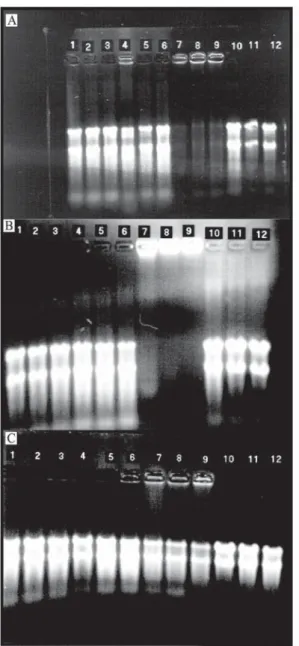
The RNA in the T+B- lanes of gels with
D. ovalifolium
(Figure 2a) and grape skin tannin
(Figure 2b) did not migrate, unlike RNA from the
T+B-treatment for the other tannin sources (Figure 2c).
The RNA could not migrate due to complexation with
condensed tannins. RNA could not migrate in the
presence of
D. ovalifolium
possibly because this
tannin has high biological activity and is a large
poly-mer (Nelson et al., 1997). The complexation is
possi-bly due to the residual negative charge of nucleic
acids (Ausubel et al., 1996) and the ability of tannins
to bind using hydrogen bonds from hydroxyls of the
phenolic residue (Loomis, 1974; Dabo et al., 1993).
Most of the tannin-protein complexes are probably
denatured by guanidinium thiocyanate, since
pro-teins are rapidly denatured in concentrated (4M)
guanidinium solutions (Chomczynski & Sacchi, 1987).
The tannin-RNA complexes probably were retained
in the glass fiber filters and eluted later on.
0 0.1 0.2 0.3 0.4 0.5 0.6 0.7 0.8 0.9
240 250 260 270 280 290 300
Wavelength (nm)
Ab
so
rb
an
ce
u
ni
ts
Figure 1. Absorbance profiles of aqueous solutions (0.01 g/L in 0.5M Tris pH 7) of extracted condensed tannins from Inga edulis ( ), Desmodium ovalifo-lium ( ), grape skin ( ) and tannic acid ( ).
Analysis of variance indicated that band density
means were similar for both buffers tested
(PVP = 3593.8 pixels; PEG = 3223.5 pixels,
P = 0,05). However, band density means differed due
to tannin type and rinsing buffer (Table1). In other
words, the effect of rinsing buffer on band density was
affected by tannin type. For grape skin, the results
were similar because its mean for the PVP treatment
was close (P = 0.0574) to the significance level. The
two buffers had similar effects and yielded less dense
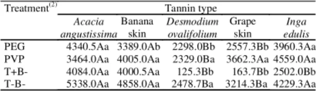
Table 1. Integrated pixel means from densitometric measurements of 16S rRNA bands relative to the interaction between buffers and condensed tannin types(1).
Tannin type Treatment(2)
Acacia angustissima
Banana skin
Desmodium ovalifolium
Grape skin
Inga edulis PEG 4340.5Aa 3389.0Ab 2298.0Bb 2557.3Bb 3960.3Aa PVP 3464.0Aa 4005.0Aa 2329.0Ba 3662.3Aa 4559.0Aa T+B- 4084.0Aa 4000.5Aa 125.3Bb 163.7Bb 2502.0Bb T-B- 5338.0Aa 4858.0Aa 2478.7Ba 3214.3Ba 4229.3Aa
(1)Means followed by the same capital letter within each row and by the
same small letter within each column do not differ in the SNK test for
P>0.05. (2)PEG: polyethylene glycol; PVP: polyvinylpirrolidone;
T+B-: positive control containing tannin and Tris buffer without PEG or PVP; T-B-: negative control containing neither tannin nor PEG or PVP.
Figure 3. Densitometric analysis from denaturing agarose gels after digitally recorded, showing (from left to right
within each Tannin type) the integrated pixel values for 16S rRNA bands from total RNA extracted from cells exposed to tannins and to rinsing with PEG buffer ( ), PVP buffer ( ), 0.5M Tris pH 7, T+B- controls ( ) and not rinsed nor exposed to tannins ( , T-B- controls). The treatments with condensed tannins are represented on the X axis by Acacia angustissima (A), banana skin (B), Desmodium ovalifolium (D), grape skin (G) and Inga edulis (I).
bands than the negative control (T+B+
ver-sus T-B-) (Figure 3).
To evaluate if RNA extracted from cells exposed to
the different conditions tested could be used in
mo-lecular analysis, aliquots of RNA from each of the
treat-ments with the
R. albus
8 species-specific RAL 196
probe were hybridized. The species-specific probe
hybridized to all samples of RNA from cells exposed to
tannins and treated with either PEG or PVP similarly
(T+B+). The results showed less intense hybridization
signals compared to the negative control (T-B-), as
discussed below.
Desmodium ovalifolium
and
I. edulis
leaf tannins were the most and the least effective tannins
tested (Figure 4). Grape skin results were similar to
those
of
D. ovalifolium
leaves, whereas banana skin and
A. angustissima
leaves treatments hybridized similar
to
I. eduli
leaves
.
The RNA of cells exposed to either
D. ovalifolium
leaf or grape skin tannin, but not
ex-posed to PVP or PEG (T+B- controls), did not
hybrid-ize with the probe (Figure 4). Three quantities, 0.1, 1
and 10 µg of total RNA per slot, were blotted. The
significance of tannin effect over integrated pixel
numbers had to be interpreted as confounded with
0 1 2 3 4 5 6
A B D G I
Tannin type
Pi
xe
l num
be
r x 1,
000
12 12
total RNA extraction, membrane and hybridization
conditions due to the experimental design adopted
(samples exposed to each tannin were blotted and
hybridized once). Considering that the membranes
used were cut from adjacent regions of the same lot,
that the solutions used for hybridization and
wash-ing steps were the same as well, and that
hybridiza-tion and washings were done simultaneously, it is
likely that the primary source of variation was the
presence of tannin during RNA extraction. The
ef-fects of tannin type, buffer and level of total RNA
applied were highly significant (P<0.01). Tannin by
buffer interaction was significant (P<0.05). The
sig-nificant tannin by buffer interaction that was
evi-dent from the analysis of the hybridized membrane
data indicates the appropriateness of using buffers
with PVP or PEG to extract RNA in the presence of
tannins. There was a higher hybridization signal for
the
I. edulis
leaf
and grape skin tannins rinsed with
PVP than for the T-B- control (Figure 3). This may be
caused by a complex formation among tannins, probe
and the protein-like structure of residual PVP
mol-ecules eventually attached to the nylon membranes
causing nonspecific binding of the probe (Raskin
et al., 1996). The interaction between tannin type and
RNA factors (P = 0.038) may indicate that the
con-densed tannins could be causing differences in
inte-grated pixel values observed across different levels
of total RNA applied to the membranes. This effect
was probably caused by saturation of the nylon
mem-brane as discussed by Raskin et al. (1996).
The interaction between buffer and RNA level was
not significant (P = 0.44) indicating that the treatment
with buffers did not alter the effect of level of total
RNA blotted.
Conclusions
1. Tannins from different sources must be removed
during RNA extraction for hybridization purposes.
2. Rinsing cells with 0,5M Tris buffers containing
either 6% PVP or 8% PEG before RNA extraction
sig-nificantly reduces tannin effects on gel migration and
species-specific oligonucleotide hybridization with
total RNA samples.
3. Quantitative protocols are possible using total
RNA from cells exposed to different sources of
tan-nin, due to the significant effect of increasing amounts
of RNA blotted to hybridization signals.
Figure 4.Chemiluminescent photographs from
References
ALM, E. W.; ZHENG, D.; RASKIN, L. The presence of humic substances and DNA in RNA extracts affects hy-bridization results. Applied and Environmental Micro-biology, Washington, v. 66, n. 10, p. 4547-4554, 2000.
AUSUBEL, F. M.; BRENT, R.; KINGSTON, R. E.; MOORE, D. D.; SEIDMAN, J. G.; SMITH, J. A.; STRUHL, K. Current protocols in molecular biology.
New York: J. Wiley, 1996. 606 p.
AUSUBEL, F. M.; BRENT, R.; KINGSTON, R. E.; MOORE, D. D.; SEIDMAN, J. G.; SMITH, J. A.; STRUHL, K. (Ed.). Short protocols in molecular biol-ogy. 3rd ed. New York: J. Wiley, 1995. 729 p.
BARRY, T. N. Condensed tannins: their role in ruminant protein and carbohydrate digestion and possible effects upon the rumen ecosystem. In: NOLAN, J. V.; LENG, R. A.; DEMEYER, D. I. (Ed.). The roles of protozoa and fungi in ruminant digestion. Armidale: Penambul
Books, 1989. p. 153-169.
CHOMCZYNSKI, P.; SACCHI, N. Single-step method of RNA isolation by acid guanidinium thiocyanate-phe-nol-chloroform extraction. Analytical Biochemistry, San Diego, v. 162, p. 156-159, 1987.
DABO, S. M.; MITCHELL, E. D.; MELCHER, U. A method for the isolation of nuclear DNA from cotton (Gossipium) leaves. Analytical Biochemistry, San Diego,
v. 210, n. 1, p. 34-38, 1993.
HAGERMAN, A. E. Extraction of tannins from fresh and preserved leaves. Journal of Chemical Ecology,
New York, v. 14,n. 2, p. 453-461, 1988.
HALE, A. D.; GREEN, J.; BROWN, D. W. G. Compari-son of four RNA extraction methods for the detection of small round structured viruses in faecal specimens. Jour-nal of Virological Methods, Amsterdam, v. 57, n. 2,
p. 195-201, 1996.
JONES, G. A.; MCALLISTER, T. A.; MUIR, A. D.; CHENG, K. -J. Effects of sainfoin (Onobrychis viciifolia Scop.) condensed tannins on growth and proteolysis by four strains of ruminal bacteria. Applied and Environ-mental Microbiology, Washington, v. 60, n. 4, p. 1374 -1378, 1994.
KRAUSE, D. O.; SMITH, W. J.; McSWEENEY, C. S. Extraction of microbial DNA from rumen contents con-taining plant tannins. BioTechniques, Natick, v. 31, p.294-298, 2001.
LATHAM, M. J.; BROOKER, G. L.; PETTIPHER, G. L.; HARRIS, P. J. Ruminococcus flavefaciens cell coat and adhesion to cotton cellulose and to cell walls in leaves of perennial ryegrass (Lolium perenne). Applied and Envi-ronmental Microbiology, Washington, v. 35,p. 156-165,
1978.
LOOMIS, W. D. Overcoming problems of phenolics and quinones in the isolation of plant enzymes and organelles.
Methods in Enzymology, San Diego, v. 31, p. 528-544,
1974.
MacKENZIE, D. J.; McLEAN, M. A.; MUKERJI, S.; GREEN, M. Improved RNA extraction from woody plants for the detection of viral pathogens by reverse transcrip-tion-polymerase chain reaction. Plant Disease, St. Paul,
v. 81, n.2, p. 222-226, 1997.
MAKKAR, H. P. S.; BLUMMEL, M.; BECKER, K. For-mation of complexes between polyvinyl pyrrolidones or polyethylene glycols and tannins, and their implication in gas production and true digestibility in in vitro techniques.
British Journal of Nutrition, Wallingford, v. 73,p.
897-913, 1995.
MOLYNEUX, P. Nonionic polymers: polyoxides, polyethers, and poly(ethylene Imine) In: Water-soluble synthetic polymers: properties and behavior.Boca Raton: CRC Press, 1984. v. 1, p. 19-74.
MOLYNEUX, P. Nonionic polymers: the vinyl group. In: Water-soluble synthetic polymers: properties and behavior. Boca Raton: CRC Press, 1983. v. 1, p. 119-193.
MORÉ, M. I.; HERRICK, J. B.; SILVA, M. C.; GHIORSE, W. C.; MADSEN, E. L. Quantitative cell ly-sis of indigenous microorganisms and rapid extraction of microbial DNA from sediment. Applied and Environmen-tal Microbiology, Washington, v. 60, n. 5, p. 1572-1580, 1994.
NELSON, K. E.; PELL, A. N.; DOANE, P. H.; GINER-CHAVEZ, B. I.; SCHOFIELD, P. Chemical and biologi-cal assays to evaluate bacterial inhibition by tannins. Jour-nal of Chemical Ecology, New York, v. 23,n.4, p.
1175-1193, 1997.
ODENYO, A. A.; MacKIE, R. I.; STAHL, D. A.; WHITE, B. A. The use of 16S rRNA-targeted oligonucleotide probes to study competition between ruminal fibrolytic bacteria: development of probes for Ruminococcus species and evi-dence for bacteriocin production. Applied and Environ-mental Microbiology, Washington, v. 60, n.10, p.
OGRAM, A. Isolation of nucleic acids from environmen-tal samples. In: BURLAGE, R. S.; ATLAS, R.; STAHL, D.; GEESEY, G.; SAYLER, G. (Ed.). Techniques in mi-crobial ecology. New York: Oxford University Press, 1998. p. 273-288.
PICH, U.; SCHUBERT, I. Midiprep method for isolation of DNA from plants with a high content of polyphenolics.
Nucleic Acids Research, Oxford, v. 21, n.14, p. 3328,
1993.
RASKIN, L.; CAPMAN, W. C.; KANE, M. D.; RITTMANN, B. E.; STAHL, D. A. Critical evaluation of membrane supports for use in quantitative hybridizations.
Applied and Environmental Microbiology, Washington, v. 62, n.1, p. 300-303, 1996.
RASKIN, L.; CAPMAN, W. C.; SHARP, R.; POULSEN, L. K.; STAHL, D. A. Molecular ecology of
gastrointesti-nal ecosystems. In: MacKIE, R. I.; WHITE, B. A.; ISAACSON, R. E. (Ed.). Gastrointestinal microbiology: gastrointestinal microbes and host interactions.New York: Chapman & Hall / International Thomson, 1997. p. 243-298. (Chapman & Hall Microbiology Series, 2).
STAHL, D. A.; FLESHER, B.; MANSFIELD, H. R.; MONTGOMERY, L. Use of philogenetically based hy-bridization probes for studies of ruminal microbial ecology.
Applied and Environmental Microbiology, Washington, v. 54, n.5, p. 1079-1084, 1988.
VOO, K. S.; JACOBSEN, B. M. Rapid resuspension of pelleted bacterial cells for miniprep plasmid DNA isolation.
BioTechniques, Natick, v. 24, p.240-243, 1998.