Faculdade de Engenharia da Universidade do Porto
A local delivery system for extracellular vesicles
to promote tissue regeneration
Alice Quintas Pinheiro
DISSERTATION
Mestrado em Engenharia Biomédica
Supervisor: Doutora Susana Santos
Co-supervisor: Mestre Andreia Silva
Faculdade de Engenharia da Universidade do Porto
A local delivery system for extracellular vesicles
to promote tissue regeneration
Alice Quintas Pinheiro
DISSERTATION
Mestrado em Engenharia Biomédica
Supervisor: Doutora Susana Santos
Co-supervisor: Mestre Andreia Silva
v
Resumo
As vesículas extracelulares (EV), particularmente os exossomas, são atualmente alvo de intensa investigação. As EV são mediadores fisiológicos de comunicação intercelular que refletem o fenótipo das células que as secretam, e que têm a capacidade de modular o fenótipo das células alvo. Estas características das EV têm vindo a ser exploradas para diferentes aplicações, nomeadamente para o desenvolvimento de biomarcadores de diagnóstico e prognóstico de diferentes doenças, bem como para o desenvolvimento de novas terapias, em particular na área do cancro, mas também da medicina regenerativa. Vários estudos in vivo, e também alguns ensaios clínicos, têm vindo a usar EV como veículos de entrega de biomoléculas funcionais. Nestes estudos, EV nativas ou modificadas têm sido administradas através de diferentes vias. Os primeiros estudos em modelos animais e pacientes humanos com administração sistémica de EV demonstraram várias limitações na biodistribuição e retenção das EV administradas. Assim, foi colocada a hipótese de que a incorporação das EV em biomateriais capazes de funcionar como um sistema de entrega poderia constituir uma potencial solução para ultrapassar estas limitações, potenciando o uso eficaz das EV. Deste modo, esta tese teve como objetivo o desenvolvimento de um sistema de entrega de EV que permita a libertação local e controlada das mesmas para efeitos de regeneração óssea.
No trabalho desta tese, começou-se por otimizar as condições de cultura de diferentes linhas celulares imunes humanas, THP-1, KG-1 e Jurkat, nomeadamente em relação à concentração de soro do meio de cultura, de forma a obter uma produção de EV com alto rendimento e com grande pureza. As EV produzidas foram caracterizados por microscopia electrónica de transmissão, “nanoparticle tracking analysis”, e “western blotting” para marcadores exossomais, o que permitiu demostrar que, para todas as linhas celulares testadas, a população de EV isolada era enriquecida em exossomas. No entanto, a linha celular KG-1 produziu os níveis mais elevados de EV, mantendo a viabilidade celular mesmo a concentrações de soro de 0.1 % (v/v), e por isso estas células foram selecionadas para a produção das EV utilizados para a otimização das condições de incorporação num sistema de entrega.
O sistema de entrega construído consistiu numa matriz 3D em quitosano funcionalizada com camadas de quitosano (Ch) e poli-γ-ácido glutâmico (γ-PGA) misturado com EV, depositadas alternadamente num sistema layer-by-layer. A incorporação das EV no sistema de entrega foi observada por microscopia confocal de fluorescência, usando EV previamente marcadas com um corante fluorescente, mas não pôde ser confirmada por espectroscopia de infra-vermelhos. De seguida, o perfil de libertação das EV do sistema de entrega foi testado
vi
in vitro através da análise dos sobrenadantes do ensaio de libertação por microscopia
electrónica de transmissão, após 0, 12 e 24 h de incubação, tendo sido possível identificar EV nestes sobrenadantes concentrados, em particular às 12 e 24 h de libertação. A nível funcional, o sistema de entrega produzido e funcionalizado com EV foi testado para a sua capacidade em promover o recrutamento, bem como a diferenciação osteogénica e condrogénica, de células mesenquimais multipotentes/estaminais do estroma (MSC), responsáveis pela formação do osso. Através de ensaios de microscopia de vídeo em tempo real, foi possível demonstrar que as EV incorporadas no sistema de entrega promoveram um aumento significativo da motilidade das MSC. Para além disso, resultados de expressão génica nas MSC mostraram que o sistema de entrega funcionalizado com Ch e γ-PGA foi pro-condrogénico, mas que a presença de EV não levou a aumentos significativos da diferenciação das MSC.
Em conclusão, foi possível incorporar EV no sistema 3D layer-by-layer de Ch e γ-PGA desenvolvido, que se mantiveram funcionais após libertação. As EV secretadas pelas células KG-1 promoveram o recrutamento das MSC através do aumento da sua motilidade, tal como já descrito para EV de células dendríticas primárias, enquanto que o sistema de entrega funcionalizado com o Ch e γ-PGA promoveu per se a diferenciação condrogénica das MSC. Em conjunto, os resultados obtidos suportam um efeito favorável da incorporação das EV num sistema de libertação controlada, sobre as MSC, sugerindo que as EV podem ser mediadores quimiotáticos deste tipo celular para locais de lesão óssea, onde o sistema de entrega produzido pode estimular a sua diferenciação, promovendo a reparação e regeneração do tecido ósseo.
vii
Abstract
Extracellular vesicles (EV), in particular exosomes, are currently the subject of intense research. EV are natural mediators of intercellular communication, reflecting the phenotype of the secreting cell, and potentially modulating the phenotype of target cells. These characteristics of EV are being exploited for different applications, namely the discovery of biomarkers for disease diagnosis and prognosis, and the development of new therapies, particularly in the cancer field, but also in regenerative medicine. Several in vivo studies using EV as delivery carriers of active biomolecules have been reported, including a few human clinical trials. In these studies, different routes of EV administration and the use of natural or modified EV formulations have been reported. Pioneering studies have been uncovering several limitations in EV biodistribution and retention in animal models and human subjects, when these are administered systemically. Consequently, it was hypothesized that incorporation of EV in biomaterial delivery systems could contribute to overcome these limitations. Thus, this thesis aimed to develop an EV delivery system for local and sustained EV release, particularly for bone regeneration.
For this thesis, cell culture conditions for different human immune cell lines, THP-1, KG-1 and Jurkat cells, in terms of serum concentration in the media, were optimized, in order to obtain higher yields of EV production, and with higher purity. EV produced were characterized by transmission electron microscopy, nanoparticle tracking analysis and western blotting for exosomal protein markers, and found to be enriched in exosomes for all cell lines tested. However, it was the cell line KG-1 that produced EV at higher yields, while still maintaining high cell viability at serum concentrations of 0.1 % (v/v), and thus these cells were selected as EV producers for further use in optimizing the incorporation into a delivery system.
The EV delivery systems developed consisted of chitosan 3D scaffolds functionalized with layers of chitosan and EV-containing poly-γ-glutamic acid (γ-PGA), alternated in a layer-by-layer system. EV entrapment in the delivery system could be confirmed by laser scanning confocal microscopy, using fluorescently labelled EV, but not by Fourier-transform infrared spectroscopy. Then, the release profile of EV from the delivery system was evaluated in vitro by transmission electron microscopy of release supernatants, at 0, 12 and 24 h of incubation, and EV could be detected in concentrated supernatants, particularly at 12 h of release. At the functional level, EV-loaded delivery systems produced were tested for their capacity to promote recruitment, and osteogenic and chondrogenic differentiation of multipotent mesenchymal stromal/stem cells (MSC), responsible for bone formation. Using time-lapse
viii
video microscopy we showed that EV entrapped in the delivery system were able to promote significant MSC motility. Also, gene expression analysis results indicate that while Ch and γ-PGA delivery systems were pro-chondrogenic, the presence of EV did not increase MSC differentiation further.
In conclusion, EV could be entrapped in the layer-by-layer Ch and γ-PGA 3D systems, and were functional upon release. The KG-1-secreted EV used, similarly to those of primary Dendritic Cells, induced an MSC recruitment-promoting behaviour, increasing their motility, while the Ch and γ-PGA delivery system per se promoted chondrogenic differentiation. Together, our results support a beneficial effect of EV entrapment and controlled release, upon MSC, suggesting EV as chemoattractants for this cell type into places of bone injury, where one can envisage the delivery system may enhance their differentiation, promoting tissue repair and regeneration.
Key words: Bone regeneration, delivery system, extracellular vesicles, mesenchymal stem
ix
Agradecimentos
É com grande alegria que vejo concluída mais uma etapa da minha vida. No entanto, esta não seria possível sem todas as pessoas que me ajudaram e estiveram presentes em todos os momentos, bons ou menos bons, que permitiram que o culminar desta aventura resultasse neste trabalho.
Um Obrigada especial à Susana Santos, minha orientadora e sem dúvida um exemplo de trabalho e dedicação! Por todo o tempo e atenção dedicado para que este trabalho resultasse.
Ao Professor Mário Barbosa, diretor do i3S e PI do grupo onde me integrei, obrigada por ser um ser Humano por detrás de um grande Cientista e partilhar connosco histórias que ensinam.
A todos os membros da equipa técnica no I3S: ao Rui, por toda a boa disposição e ajuda a adquirir as melhores imagens no TEM; à Cecilia por toda a ajuda com o NTA; à Joana Rocha e Dalila por toda a ajuda e cooperação na sala de culturas.
Um agradecimento também a toda a equipa, Microenvironments for New Therapies, que me integrou da melhor forma e me ajudou a crescer, não só a nível científico, mas também a nível pessoal. Desta equipa, não consigo deixar de falar em particular da Andreia, minha co-orientadora e, a pessoa que me acompanhou desde o iníco e me transmitiu tanto conhecimento e o fez de forma tão incrível, que faltam as palavras para descrever o quão importante foi neste percurso. É das melhores pessoas que já conheci e que melhor concilia conhecimento científico com alegria e energia! Obrigada por tudo, mesmo! Ao José Henrique e ao João Brás por toda a boa energia e por estarem sempre prontos a ajudar. À Joana Magalhães por ser a melhor companheira de secretária e por ter sempre uma palavra de força para todas as situações. À Catarina Leite por toda a ajuda, em especial com o PGA. Obrigada por terem tornado este percurso mais fácil e tão agradável de se fazer.
Não podia deixar de falar daqueles que me acompanham desde sempre e nunca recusam uma cerveja no Inox para aliviar o stress de uma semana de trabalho: um enorme obrigada à Anita e ao Samuel por todo o companheirismo e amizade. Ao Fábio, à Inês, ao Adão e à Kristine por todas as pausas culturais ou não, mas necessárias. Um obrigada especial à minha amiga de casa, Márcia, foi um prazer partilhar contigo esta jornada que vai deixar saudades. À Marisa, por nunca dizer que não a um jantar e por todas as idas ao ginásio para recarregar
x
baterias! Ao João Sousa e à Ana Gomes, por, mesmo menos perto, continuarem sempre presentes! À Bárbara, por todo o apoio e amizade! Ao Rodrigo por, mesmo longe, me conseguir ajudar a traçar os meus objetivos e por ter sempre alguma coisa a acrescentar. Ao Francisco Teixeira por toda a amizade e atenção. Ao Francisco Branco por toda a força e exemplo de ambição e coragem. Aos amigos de Carlão, que me viram crescer e que sempre acreditaram em mim. Sou uma sortuda por vos ter na minha vida!
À minha família, por tudo o que sou hoje. Ao meu pai por todos os fim-de-semana querer saber qual é o tema da minha tese e por tudo que me ensinou e continua a ensinar. À minha mãe, por me apoiar na sobrevivência das células em cultura, e por todas as marmitas feitas com tanto carinho. Ao meu irmão por ser o miúdo mais incrível que conheço e por contar os dias para eu voltar a casa. À minha irmã, por, mesmo longe, me apoiar em tudo e por ser um exemplo para mim!
Financial support
This work was financially supported by: Project no. S-15-83S, from AO Foundation (Switzerland); project NORTE-01-0145-FEDER-000012, supported by Norte Portugal Regional Operational Programme (NORTE 2020), under the PORTUGAL 2020 Partnership Agreement, through the European Regional Development Fund (ERDF); Portuguese funds through FCT - Fundação para a Ciência e a Tecnologia/Ministério da Ciência, Tecnologia e Inovação, in the framework of the project “Institute for Research and Innovation in Health Sciences” (POCI-01-0145-FEDER-007274)
xi
Table of Contents
Chapter 1 ... 1
Introduction ... 1
1.1 – Cell- secreted extracellular vesicles ... 2
1.1.1 - Extracellular vesicles: characteristics and methods of isolation... 2
1.1.2 - EV characterization: methods of analysis ... 5
1.1.3 - Therapeutic potential of EV ... 7
1.2 – Strategies for EV delivery ... 10
1.2.1 – Systemic delivery of EV ... 10 1.2.1.1 - EV intravenous administration ... 10 1.2.1.2 - EV intraperitoneal administration ... 12 1.2.1.3 - EV oral administration ... 12 1.2.1.4. EV intranasal administration ... 13 1.2.2 - Local delivery of EV ... 14
1.3 – Delivery Systems: from Drug to EV Delivery ... 17
1.3.1 - Chitosan and poly-γ-glutamic acid: Biomedical Applications ... 19
1.3.2 – Chitosan as a delivery system ... 19
1.3.3 – Poly-γ-glutamic acid as a delivery system ... 22
1.3.4 – Combining chitosan and poly-γ-glutamic acid in a layer-by-layer delivery system ... 22
1.4 – Objectives ... 25
Chapter 2 ... 27
Methods and Materials ... 27
2.1 - Cell culture ... 27
2.2 – Cell viability Assays ... 28
2.3 - EV production, isolation and characterization ... 28
2.3.1 – EV production and Isolation ... 28
2.3.2 - EV characterization ... 28
2.4 – Production of EV delivery systems ... 29
2.4.1 – Chitosan scaffolds production ... 29
2.4.2 – Poly-γ-Glutamic Acid production ... 29
2.4.3 – Scaffolds functionalization with a chitosan/γ-PGA Layer-by-Layer system ... 30
2.5 – Delivery system characterization ... 31
2.5.1 – Confocal microscopy ... 31
2.5.2 - FTIR ... 31
2.5.3 – EV release assay ... 31
xii
2.7 – Timelapse MSC motility assay ... 33
2.8 – MSC differentiation assay ... 34
2.9 – Statistical analysis ... 36
Chapter 3 ... 37
Results ... 37
3.1 – Optimization of cell culture conditions for EV production ... 37
3.2 – Immune cell EV secretion and characterization ... 40
3.3 – Delivery system component characterization... 43
3.4 – Layer-by-Layer system is able to entrap EV ... 43
3.5 – Layer-by-Layer EV release profile ... 46
3.6 – Effect of EV from the delivery systems in MSC migration ... 47
3.7 – Functional impact of LbL-mediated EV delivery on MSC differentiation ... 48
Chapter 4 ... 51
Discussion and future perspectives ... 51
References ... 57
Supplementary information ... 71
xiii
List of figures
Figure 1.1 - Schematic representation of the sizes of the different types of extracellular
vesicles that can be secreted by cells.. ... 3
Figure 1.2 - Extracellular vesicles biogenesis, release, and mode of action. ... 4
Figure 1.3 - Extracellular Vesicles are natural and tunable delivery vehicles with clinical potential in tissue repair and regeneration. ... 7
Figure 1.4 - Routes of administration of Extracellular Vesicles. ... 10
Figure 2.1 - Scheme of synthesis of the LbL system on Ch scaffolds. ... 31
Figure 2.2 - Transwell migration assay setup to test MSC migration in the presence of different stimuli... 33
Figure 2.3 - Schematic representation of the system used for MSC motility assay in the presence of different stimuli. ... 34
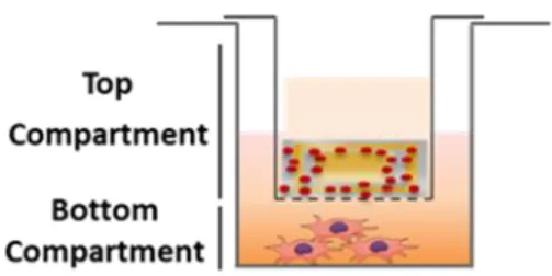
Figure 2.4. Schematic representation of the transwell system used to test MSC differentiation in the presence of different stimuli.. ... 35
Figure 3.1 – Morphology in culture of all tested cell lines.. ... 38
Figure 3.2 - Characterization of cell viability of the three cell lines tested, when cultured in different serum conditions. ... 39
Figure 3.3 – Characterization of EV from KG-1, THP-1 and Jurkat cell lines. ... 41
Figure 3.4. Characterization of EV isolated from KG-1 cells. ... 42
Figure 3.5. (A) B.subtilis culture in Agar. (B) SDS-PAGE to determine the Mw of γ-PGA produced. ... 43
Figure 3.6 - Layer-by-layer system with entrapped EV. ... 44
Figure 3.7 - FTIR spectra of EV-containing LbL systems. ... 45
xiv
Figure 3.9 – MSC recruitment in a transwell migration assay, as influenced by different
stimuli. ... 47
Figure 3.10 - MSC migration profile in time-lapse motility assays. ... 48 Figure 3.11 – Osteogenic and chondrogenic differentiation of MSC in the presence of LbL with
and without EV. ... 49
Figure S1 - Overview of all cutting steps of chitosan scaffolds until the desired size is
achieved. ... 71
Figure S2 – Representative gel image of RNA extracted analyzed by agarose gel
electrophoresis. ... 71
Figure S3 – Representative gel image of RT-qPCR products analyzed by agarose gel
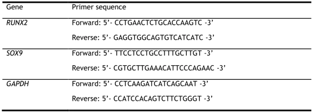
electrophoresis for RUNX2 in MSC stimulated with osteogenesis-inducing media (Osteo), SOX9 in MSC stimulated with chondrogenesis-inducing media (Chondro) and for GAPDH, RUNX2 and SOX9 in MSC stimulated with the LbL delivery system with entrapped EV (from left to right, respectively). ... 72
Figure S4 - Scanning electron microscopy analysis of scaffolds with entrapped EV, as
xv
List of tables
Table 2.1 - Sequence of the primers used in the RT-qPCR analysis performed on MSC, for the
xvii
Abbreviations
AFM Atomic force microscopy
ATR-FTIR Attenuated total reflection-Fourier-transform infrared spectroscopy
BBB Blood brain barrier
BC Bottom compartment
bFGF Basic Fibroblast Growth Factor
CCl4 Carbon tetrachloride
cDNA Complementary DNA
CFSE 5-carboxyfluorescein diacetate succinimidyl ester
Ch Chitosan
Cx43 Connexin 43
DAPI 4′,6-diamidino-2-phenylindole
DC Dendritic cells
DDS Drug delivery system
DLS Dynamic light scattering
DNA Deoxyribonucleic acid
DTH Delayed-type hypersensitivity
ER Endoplasmic reticulum
EV Extracellular vesicle
FBS Fetal bovine serum
FasL Fas ligand
FDA Food and Drug Administration
FTIR Fourier-transform infrared spectroscopy
γ-PGA Poly-gamma-glutamic acid
GM-CSF Granulocyte-macrophage colony-stimulating factor
IL Interleukin
iPS Induced pluripotent stem cell
i.v. Intravenous injection
IFN-γ
Interferon- γ
LB Luria Broth media
LbL Layer-by-layer
LSCM Laser scanning confocal microscopy
MCP-1 Monocyte chemoattractant protein 1
miRNA(s) MicroRNA(s)
xviii
MV Microvesicles
MVB Multivesicular bodies
Mw Molecular weight
MWCO Molecular weight cutoff
NTA Nanoparticle Tracking Analysis
PBS Phosphate-buffered saline
PI Propidium iodide
PGA Poly-glutamic acid
PLGA Poly(D,L-lactide-co-glycolide)
P/S Penicillin/streptomycin
rhBMP-2 Recombinant human bone morphogenetic protein-2
RNA Ribonucleic acid
Rluc Renila luciferase
RS Raman spectroscopy
RT Room temperature
RVG Rabies virus glycoprotein
RT-qPCR Quantitative real time polymerase chain reaction
SDS-PAGE Sodium dodecyl sulfate polyacrylamide gel electrophoresis
siRNA Small interfering RNA
SFM Serum-free media
SMSC Synovium mesenchymal stem cells
SPR Surface plasmon resonance
SN Ultracentrifugation supernatant
TC Top compartment
TEM Transmission electron microscopy
TPP Tripolyphosphate
tRPS Tunable resistive pulse sensing
1
Chapter 1
This chapter was adapted into a manuscript that was submitted for publication, and that is included as an annex (Annex 1) to this thesis.
Alice Pinheiro, Andreia M. Silva, José H. Teixeira, Raquel M. Gonçalves, Maria I. Almeida, Mário A. Barbosa, Susana G. Santos. Extracellular Vesicles: Intelligent delivery strategies
for therapeutic applications. (submitted)
Introduction
The response to tissue injury requires well-orchestrated communication between the cells responsible for its different stages, from the initial inflammation to repair/regeneration. In the last decades, the roles of Extracellular Vesicles (EV), secreted by cells, in these processes have been the subject of intense research [1]. Three different types of EV secreted by cells have been reported and characterized, based on their origin, release mechanism and properties: apoptotic bodies, microvesicles and exosomes. Although the complete distinction of these different vesicle populations is still hindered by methodological limitations, the last two types of vesicles have deserved most attention from the scientific community, due to their biological properties [2]. Nonetheless, due to the persisting difficulties in fully distinguishing these vesicle populations, and in agreement with the recommendations of the International Society of Extracellular Vesicles, in this thesis the designation EV will be preferentially used.
The most intensely studied EV population is that of exosomes, nanometre-sized vesicles formed in the endosomal network and secreted by the majority of cells, including tumor cells [3] [4]. Exosomes are reported to play key roles in intercellular communication, without cell-to-cell contact. They have been reported to participate in different homeostatic biological processes, including antigen presentation, delivery of active biomolecules to target cells, and even cell waste management (initially thought to be their only function) [5]. These natural functions of exosomes have been explored for their application as therapeutic mediators in different fields, from cancer treatment to novel tissue regeneration strategies. However, they may also contribute for the spreading of infecting pathogens and pathologies, namely cancer metastasis [6].
EV have also been greatly explored as biomarkers of disease, because their composition is related to the status of the cell from which they derive. Recent studies have shown that the characterization of exosomes in fluids, particularly in the blood, urine, saliva, breast milk and amniotic fluid can strongly contribute for the diagnosis of various diseases, namely in cancer [7] [8]. This makes the use of EV as biomarkers to be applied in areas such as diagnosis, prognosis, as well as real-time monitoring of therapy of different pathologies by determining their levels and composition in body fluids [7] [9] [10].
On the other hand, EV can be obtained in vitro from different cells, and are reported to be stable when administered in vivo, which are important advantages for their application as a drug, or as drug delivery system. In addition, they can be obtained from the patient own cells, an important advantage regarding their immune compatibility [11]. Indeed, considering their clinical value, EV have been increasingly investigated in the delivery systems field. Many studies have been reporting their use to deliver different molecules such microRNAs (miRNAs), siRNAs, proteins or drugs to target cells [2]. However, despite the increasing research in EV field, there are several difficulties in their isolation and characterization process. Many isolation procedures have been described but in all of them, a very low EV yield is reached. In addition, there is a lack of characterization methods able to efficiently evaluate EV standardized production from different cell culture batches. Both issues constitute a big obstacle to make EV clinical applications succeed. On the other hand, when administered in vivo, exosomes are readily cleared form circulation. Furthermore, the mechanisms controlling the specific delivery of exosomes to a specific target are fairly unknown and need to be studied harder. These limitations are the main motivations underlying this master thesis. Thus, they will be further addressed in the following sections, along with possible strategies to overcome them.
1.1 – Cell-secreted extracellular vesicles
1.1.1 - Extracellular vesicles: characteristics and methods of isolation
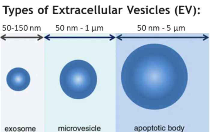
Most cells are capable of releasing different types of EV, which are generally divided in three classes: exosomes, microvesicles, and apoptotic bodies. Each type of EV can be classified based on different criteria, such as origin in the cell, size, and enrichment of specific protein markers. Figure 1.1 establishes a close relationship between the sizes of the three different vesicles. However, this classification is still controversial with different authors reporting different sizes for the same EV population.
1.1 – Cell-secreted extracellular vesicles
3Figure 1.1 - Schematic representation of the sizes of the different types of extracellular vesicles that
can be secreted by cells. Exosomes may range from 50 to 150 nm (are the smallest). Next, microvesicles can range from 50 nm to 1 μm. The largest vesicles are the apoptotic bodies and can reach 5 μm. Adapted from [12].
The microvesicles originate by direct budding from the plasma membrane and their size is quite variable (50 nm - 1 μm) [13]. As for the apoptotic bodies, they are released when the cell is undergoing apoptosis [14], and are an even more heterogeneous population, ranging in size from 50 nm to 5 μm [10]. On the other hand, exosomes are characterized by being the smallest EV, with sizes in the range of 50-150 nm, being derived from the intraluminal bud of multivesicular bodies (MVB), and that are released after fusion of these MVB with the plasma membrane (shown in Figure 1.2) [10]. As a common feature among them, all types of EV are lipidic vesicles that carry a protein content in their core, as well as on their surface. They also carry different types of RNAs, including messenger RNA and miRNAs, DNA, and even different metabolites. Importantly, these biomolecules are functional, even when delivered to target cells. There are three main mechanisms by which EV interact with target/recipient cells: endocytosis; direct fusion with the plasma membrane of the recipient cell; and receptor-mediated endocytosis following receptor–ligand interaction between EV and the recipient cell [15].
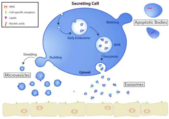
Figure 1.2 - Extracellular vesicles biogenesis, release, and mode of action. Cells secrete different types
of extracellular vesicles, including microvesicles (MV), exosomes and apoptotic bodies. Apoptotic bodies are formed during programmed cell death. MV are released by cell membrane budding of, whereas exosomes have their origin in multivesicular bodies (MVB). Upon release, EV travel body fluids until they reach a target cell. Since EV carry biomolecules such as proteins, messenger RNA, microRNA (miRNA) and DNA, they are able to modulate the phenotype of target cells. Adapted from [16].
Upon secretion, these different EV are present together in body fluids or supernatants of
in vitro cell cultures, and separation methodologies are required to allow the study of each
EV population. Different methods have been developed and optimized in the last years to isolate these EV populations, particularly exosomes, with different degrees of efficiency. These methods include differential centrifugation with a final step of ultracentrifugation, or ultracentrifugation in density gradients, phase separation, size-exclusion chromatography, immunoprecipitation and filtration. Differential (ultra)centrifugation is still the method most commonly reported for the isolation of EV, although EV pellets obtained are generally a mix of exosomes, microvesicles and soluble proteins secreted by cells, since many proteins have sedimentation coefficients similar to EV [14]. In order to isolate pure EV populations, soluble
protein aggregates, denser than EV, may be removed by ultracentrifugation using sucrose
cushions or gradients [14] [17].
Size-exclusion chromatography also allows the separation of EV from contaminant soluble proteins. Importantly, it is perhaps one of the most complete methods for the separation of different EV populations, since apoptotic bodies, microvesicles and exosomes are eluted in different fractions of the eluate [18]. Due to their easy implementation, exosome isolation by phase separation is becoming increasingly popular, being available in commercial kits
1.1 – Cell-secreted extracellular vesicles
5nowadays. However, a major drawback of this method is the common precipitation of microvesicles and soluble proteins alongside exosomes [19].
Up to date, centrifugation in density gradients and size-exclusion chromatography are the preferred techniques for the isolation of pure exosome populations [20]. However, the yield of isolation is usually very low and the isolation protocols are time-consuming, impairing a wider use of these methods.
Considering the difficulties in isolating pure populations of the different types of EV, it is of utmost importance that the vesicles isolated are extensively characterized [17]. However, distinguishing and quantifying the different subtypes of EV remains a difficult task because of several similarities in size and physical properties of the different subtypes, and the lack of specific markers for each one of them [14].
1.1.2 - EV characterization: methods of analysis
In order to characterize the isolated EV several techniques are usually combined. So far, the main techniques used are transmission electron microscopy (TEM), atomic force microscopy (AFM), flow cytometry, western blotting, nanoparticle tracking analysis (NTA), dynamic light scattering (DLS), tunable resistive pulse sensing (tRPS) and surface plasmon resonance (SPR). More recently, Raman spectroscopy has also been applied to the characterization of EV.
A primordial technique used in the field of EV biology, and still the most used for determining EV morphology, and even to visualize their release from cells is transmission electron microscopy (TEM). This technique also allows to determine the size of individual EV with minimal sample processing. Biochemical information about the EV can also be obtained with this method, by using immunogold labelling. However, this technique has limitations related to preparation of the sample, since it must be dehydrated and analysed under vacuum [21]. The AFM is also used for the characterization of EV, however, few studies follow this approach, due to the need of immobilization of the vesicles under study. Moreover, the most suitable EV immobilization method, that could retain the physiological properties of the vesicles, is still being discussed. Besides retrieving information regarding vesicles size and morphology, AFM also allows characterizing their mechanical properties [22] and potentially their interactions, for instance with cells.
For a more detailed biochemical analysis, EV are usually characterized based on the proteins they carry on their surface [7]. Tetraspanins (CD37, CD53, CD63, CD81 and CD9) are highly enriched in EV, and thus are generally accepted as EV markers. CD63 is perhaps the most reported EV marker [23] [24]. However, several positive and negative markers of EV should be tested simultaneously. The specific detection of these markers by antibodies is usually achieved by flow cytometry or western blotting. Flow cytometry allows simultaneous multi-parameter analysis on EV. However, a disadvantage of this technique is that common flow cytometers cannot distinguish light scatter from one single EV, and thus, their analysis by flow cytometry usually requires an additional step of vesicle coupling to carriers of larger size [21]. Multiple EV are often count as a single EV, precluding an accurate analysis of EV concentration and size [25]. Thus, the use of larger beads with EV absorbed, followed by
fluorescent antibody labelling, is still the most common solution to characterize EV by flow cytometry [14]. Protein markers can also be assessed by western blot, the main disadvantage is that the analysis is of a protein pellet, and not individual vesicles. This technique can be associated to immunoprecipitation, using the classical EV markers (CD9, CD63 or CD81), and analysing the immunoprecipitated vesicles directly by western immunoblotting for the detection of additional markers [26].
To analyse changes in the amount of EV produced in different conditions and also to determine EV concentration and size distribution, for analysis of the isolated populations and for standardization of functional assays, the most used methodologies are nanoparticle-tracking analysis (NTA) and dynamic light scattering (DLS). NTA is the most widely used technique to assess EV size and concentration [21] [27]. In general, this technique gives information about EV size distribution and concentration in solution, and allows real-time virtual visualization of the vesicles [28]. In some equipment, fluorescence can be combined with this method, by labelling EV directly with fluorescent dyes or immunolabelling specific molecules with fluorescent-conjugated antibodies [21]. Concerning NTA limitations, it is known that instrument-specific variables affect the characterization of EV, limiting the analysis of very heterogeneous populations [29]. In addition, the use of fluorescently-labelled antibodies for the analysis of EV increases specificity, but may limit the analysis to specific subpopulations, as discussed by Rafal et al. [21]. Similarly to NTA, dynamic light scattering is a technique used for EV size characterization and EV distribution with the variance of scattered light intensities detected in a specific angle and then compared to the wavelength of light [7]. Importantly, this technique allows determining the surface charge of the vesicles, an important parameter for their internalization by target cells. In recent years, a few works were published reporting the use of tunable resistive pulse sensing (tRPS) to characterize EV suspensions. This technique calculates EV absolute size, their concentration in the sample and their surface charge [21]. These values are calculated in comparison with reference particles with known volume and concentration [27]. However, Maas et al. concluded that different operation variables could affect significantly EV quantification [27].
Other tools are being developed to improve accuracy and detail in EV characterization. For instance, surface plasmon resonance (SPR) allows real-time and label-free detection of ligand binding to target receptors located on a sensing film [30]. Im et al. performed EV populations characterization by using a nanohole-based SPR sensor, being 10 000 times more sensitive than western blotting analysis, and allowing a fast characterization of EV [31]. In addition, Raman Spectroscopy (RS) analyses chemical vibrational modes of the samples due to wavelength shifts [32]. RS generates a spectrum with qualitative and quantitative information to describe chemical composition of EV. With this, the use of specific protein biomarkers is not required. Unlike all the previously described techniques, RS provides label-free determination of chemical composition of even a single EV in a short time [33]. Due to these advantages, this technique is being increasingly investigated as a fundamental analytical tool in EV characterization field [34].
Despite all the technical advances achieved in the last years, basic EV characterization techniques are far from being used in diagnostics. In addition, they are still very limited for the characterization of EV to be administered in in vivo models, and to quantify their distribution in the body upon administration. The important role of EV, in particular as potential therapeutic tools, makes it necessary to further improve the techniques of
1.1 – Cell-secreted extracellular vesicles
7quantification and characterization of these vesicles. To reach clinical practice, a fast sample characterization, based on a fingerprint characterization of EV populations, must be achieved, and improved experimental settings and routines of analysis must be implemented [27] [35].
1.1.3 - Therapeutic potential of EV
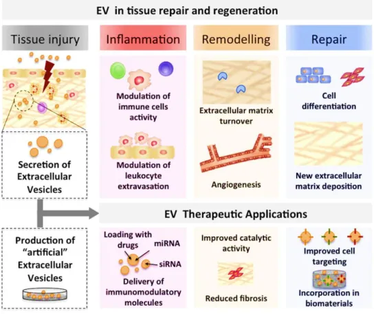
It is nowadays recognized that EV play important roles in intercellular communication, which renders them a natural mechanism for transferring information between cells. In particular taking advantage of their nanosize, EV are being explored as nanodevices for therapeutic applications [36]. Our group recently reviewed the potential contributions of EV to the different stages of tissue repair and regeneration, as illustrated in Figure 1.3.
Figure 1.3 - Extracellular Vesicles are natural and tunable delivery vehicles with clinical potential in
tissue repair and regeneration. EV are produced by virtually all cell types in the body and their content varies with physiological conditions, playing crucial roles during the inflammatory, remodelling and repair stages of tissue regeneration. Furthermore, natural EV can be modified to carry miRNAs, mRNAs, proteins and drugs of interest, functioning as improved therapeutic vehicles. Adapted from [37].
Naturally, EV carry active biomolecules that are loaded into their core or incorporated in their membrane through cell endogenous mechanisms [10] [38] [39]. The content of EV constitutively secreted by a cell reflects its physiological condition, and will impact on their
effect on target cells. Different roles have been described for EV, that may contribute to the different stages of tissue repair/regeneration. For instance, researchers previously showed that the RNA cargo of MSC-derived EV is different according to the MSC tissue source [40]. Also, EV secreted by immune cells in different stages of activation may elicit either immunostimulatory or immunosuppressive responses on target cells [41]. In addition, EV have been described for many years as having a role in bone matrix mineralization [42].
In order to improve the therapeutic potential and to standardize the effects of EV, the investigation into modified and artificial EV has been increasing. Much like with synthetic nanoparticles, different types of molecules such miRNAs, messenger RNAs, siRNAs, proteins, and even drugs, can be specifically incorporated into EV, which become the transporters of these molecules to target cells [43]. EV may be loaded with different molecules either exogenously or endogenously. In the first approach, the molecule of interest is included into EV after their isolation and purification, while the endogenous approach is based on active encapsulation during EV biogenesis, via transforming the EV-producing cells with the selected molecule [43].
EV have been most explored in the cancer field, where their potential roles and applications are more intensely researched. Their use in immunomodulatory therapies or directly anti-tumor therapies has been recently reviewed [17]. Recently, the anti-tumor therapies based on anti-cancer drug loading on EV have been the most studied. Saari et al. studied the cytotoxic effect of EV isolated from prostate cancer lines cultures, and loaded with the antimitotic cancer drug Paclitaxel, upon the same type of cancer cell [44]. They showed that loaded EV decreased cell viability, and generated better cytotoxic effects than the free drug, which was mediated by EV internalization by cancer cells. Also, using EV loaded with high dosage of Paclitaxel, Pascucci et al. showed great results in vitro in pancreatic cancer therapy [45]. This study introduced the ability of EV secreted by MSCs, which were previously loaded with Paclitaxel, to incorporate the drug during vesicle biogenesis. Interestingly, these EV retained anti-tumor activity in vitro. Many other studies reported the use of chemotherapeutics loaded in EV, supporting their use as drug vehicles as a good alternative to traditional cancer therapy. This is mainly due to a great reduction in toxic side effects, since EV biodistribution to target tissues may be increased by choosing appropriate parental cells for EV production [46] [47]. However, many different ways to further enhance effects and tailored targeting of these EV are being tested, so that EV therapies become a reality in clinical applications.
EV have also been studied for the development of novel therapies for neurodegenerative disorders, which affect increasing proportions of the world population. Haney et al. took advantage of the ability of EV to cross the blood-brain barrier (BBB) and incorporated catalase in the vesicles using different methods, in order to restore the reduced levels of redox enzymes, commonly observed in the brains of Parkinson’s patients. A significant amount of catalase-loaded EV was detected in the brain of mice tested after intranasal administration, particularly in their neurons and microglial cells, which resulted in a neuroprotective effect [48]. Similarly, EV were suggested as therapeutic vehicles in Alzheimer's disease. Alvarez-Erviti et al. have shown the ability of engineered EV (from self-derived dendritic cells, DC), loaded with siRNA against BACE1 protein, to cross the BBB in mice, working as a treatment for Alzheimer's disease. Importantly, these EV were injected intravenously but non-specific effects in other tissues were not detected [49]. This work, along with other studies in the
1.1 – Cell-secreted extracellular vesicles
9field, is very promising for the EV-based treatment of different neurodegenerative diseases. However, some issues need to be overcome to achieve clinical trials. For example, the fact of the RVG peptide is exogenously expressed in EV surface in many studies targeting the brain, and the fact that the receptor for this peptide is often downregulated in Alzheimer’s disease brains, makes this practice unable to proceed to the next investigation step before further in
vivo studies [50].
Although cancer and neurodegenerative disorders are the fields where EV research is most intense, using EV as delivery systems in other areas, such as tissue repair and regeneration, has also gained increasing attention, particularly in models of hepatic failure, renal and myocardial injury. Interestingly, Wang and colleagues used EV derived from human umbilical cord-derived MSCs to study their capacity in preventive prophylaxis of acute graft-versus-host disease, in a mouse model of allogeneic hematopoietic stem cell transplantation. EV were intravenously administrated, and results demonstrated a decrease in in vivo manifestations of acute graft-versus-host disease along with decrease of mortality [51].
In the field of tissue repair, EV secreted by MSC have been the most studied for their regenerative capacity. Many studies have been demonstrating their positive effects on the recovery of homeostasis, as well as on the regeneration and repair of damaged tissues. This suggests a therapeutic effect of MSC-derived EV that can be used as an aid to various therapies [52] [53].
Different injury models have been used to test the potential of MSC-derived EV as therapeutics. For instance, Li et al. studied the effect of EV derived from umbilical cord-MSCs
in a carbon tetrachloride (CCl4)-induced mouse liver fibrosis. This treatment inhibited hepatic
lobule destruction, hepatocyte apoptosis and epithelial-to-mesenchymal transition, and ameliorated fibrosis [54]. Many other studies used EV of different origin as delivery systems
for the treatment of the same model of CCl4-induced liver fibrosis in mouse, reporting an
amelioration of the condition [4] [55] [56]. Using a renal injury model, Gatti et al. demonstrated significant effects in reduction of epithelial tubular cell damage in an ischemia rat model, by using MSC-derived EV [57]. However, this knowledge has not yet been translated to human clinical trials [58]. EV derived from MSC were also shown to reduce infarct size in a mouse model of myocardial ischemia/reperfusion injury [59]. In a similar approach, Ibrahim et al. used EV from human cardiosphere-derived cells in a chronic myocardial infarction murine model. Exosome administration inhibited apoptosis of cardiomyocytes and interestingly, promoted their proliferation, helped by an increase of angiogenesis [60]. The regenerative effects of EV from MSCs were also reported in models with osteochondral defects [40]. In the study from Zhang et al., MSC-derived EV were intra-articular administrated in an osteochondral defect created on distal femurs of adult rats. After 12 weeks, a full reparative response of the femurs was observed in rats exosome-treated. In addition, only fibrous repair tissues were found in the control rats, PBS-exosome-treated. This was the first study reported the positive effects of EV derived from MSC in cartilage repair [61].
In our group, we have been focusing on the crosstalk between immune cells and MSC, particularly on the way immune cells can influence MSC behaviour. Our previous work showed that DC are able to recruit MSC [62], and that secreted EV are main effectors of DC-mediated recruitment [63].
In the different studies published so far exploring the therapeutic potential of EV in vivo and in clinical trials, the strategy for vesicle administration is highly variable, using different routes and in different dosages. Furthermore, several EV-based therapies have been promoted for commercialization by several companies, for which EV dose, administration route, and timeplan of administration are still undisclosed and not well defined [64].
1.2 – Strategies for EV delivery
1.2.1 – Systemic delivery of EV
To reach the target tissue or organ, EV can be administered via different routes, such as intravenous, intraperitoneal, oral, intranasal, and subcutaneous (Figure 1.4), that may lead to their systemic distribution.
Figure 1.4 - Routes of administration of Extracellular Vesicles. EV may be used in vivo as a drug. They
can be administered either as free vesicle suspensions or loaded in drug delivery systems that allow their controlled release along time. Depending on the clinical application intended, they can be administered in different body locations, which will affect their time of retention in circulation and in target organs, as well as their body biodistribution.
1.2.1.1 - EV intravenous administration
The most widely used strategy for EV in vivo delivery is the intravenous injection (i.v.), considered the fastest way to deliver drugs and also EV, due to their direct delivery to
1.2 - Strategies for EV delivery 11
systemic circulation. In one of the earliest studies dedicated to determine EV biodistribution, reporter EV derived from HEK 293T cells were injected subcutaneously in athymic mice and their biodistribution followed by in vivo imaging [65]. These EV were coupled to Gaussia luciferase and biotin, allowing conjugation with labeled streptavidin for non-invasive in vivo fluorescence imaging, and also analysis of EV distribution by bioluminescence, including in biofluids such as blood and urine, upon the addition of luciferase substrate. As early as 30 min after administration, EV could be detected in liver and spleen of mice, being detected in low amounts in the brain, heart and muscle at all timepoints analyzed. In accordance, EV were detected at the highest concentration in blood at 30 min after administration, peaking in the urine at 60 min post-injection [65]. In a later work, Manca et al. tested the biodistribution of fluorophore-labeled milk-derived EV upon i.v. and oral administration. After 24h, EV administered i.v. accumulated in liver and spleen, while those administered by oral route accumulated only in small intestine [66]. The i.v. route has been used in the majority of the in vivo studies investigating EV as new anti-cancer therapies. For instance, i.v.-injected EV were already demonstrated to work as potent vaccines for induction of antitumor immunity, in the mouse [67]. In one of the first studies performing EV administration i.v., EV ameliorated functional recovery in a mouse model of acute kidney injury [68]. Several other studies performing systemic i.v. injection have been successful in ameliorating different brain conditions, implicating that EV in circulation are capable of crossing the BBB [69] [70]. Indeed, in one of the first studies using siRNA-loaded EV, discussed above, EV injected intravenously, could reach the mouse brain, resulting in specific gene knockdown [49]. In further studies, MSC-derived EV injected in the tail vein were capable of improving animal functional recovery in traumatic brain injury [71] and cerebrovascular accident [72]. This capacity of EV to readily reach the brain is a great advantage comparing to most of the synthetic delivery systems under development, that usually need to be functionalized in order to improve their penetrance across the BBB. The i.v. administration of EV has also been explored in the cardiovascular field, using vesicles of different cellular origin. In the study by Vandergriff et al. the authors show that EV isolated from cardiosphere stem cells cultures, when injected in the tail vain of mice with induced dilated cardiomyopathy, promoted heart function recovery by reducing apoptosis and fibrosis [73]. Furthermore, i.v. administration of EV derived from MSC were shown to reduce infarct size in a mouse model of myocardial ischemia/reperfusion injury [59]. The systemic administration of EV was also previously shown to improve local cutaneous wound healing. The recent work from Wang and colleagues suggested that EV derived from human adipose MSC decreased the size of incisional dorsal wounds in mice, upon intravenous injection [74].
Intravenous administration allows, with a minimally invasive procedure, that EV reach their target anywhere in the body, even locations distant from the place of injection, like internal organs. However, the short half-life index of EV in circulation after injection is one of the major limitations of this route of administration [75]. Indeed, as demonstrated by several
in vivo studies, EV are readily cleared from circulation, with some works reporting a
calculated half-life close to only 2 minutes [75]. Most commonly, EV are cleared to liver and lungs [75]. In a detailed study by Morishita et al., EV from B16BL6 melanoma cells were radioactively labeled and injected intravenously. By collecting blood and organs at different timepoints, and measuring radioactivity, the authors showed that, 30 min after injection, only 1 % of injected EV were still in blood. At the same timepoint, almost 40 % of the injected dose could be detected in the liver, and 10 % in the lungs, a distribution pattern that was
specific for those vesicles [76]. In a follow-up study, the same group indicated that the rapid clearance of EV from circulation after i.v. injection was mainly driven by macrophages, reason why the injected EV usually accumulate in the liver [77].
Although EV have a certain degree of specific tissue targeting, which is usually considered better than synthetic nanoparticles [78] [79], it is important to understand that EV clearance from circulation into different off-target organs might be an important drawback, for instance if vesicles are being used for the delivery of cytotoxic drugs. Furthermore, it is necessary to take into account that there are consequences of maladministration, which may be irreversible [10]. Indeed, serious consequences may occur when over-dosing EV is reached. For instance, Smith et al. reported a mouse died by asphyxiation with accumulation of EV in lungs, despite all the positive effects reported in regeneration of many organs [80].
1.2.1.2 - EV intraperitoneal administration
Another common method of EV administration is via the intraperitoneal route. Studies like the one by Liu et al. performed intraperitoneal administration of EV to modulate the immune response to a vein graft performed distantly, in the carotid of mice [81]. In this work, EV isolated from cultures of adipose tissue-derived MSC, and labeled with the PKH26 dye, were administered intraperitoneally and could be found after 30 min in the aortic endothelium, supporting the systemic distribution of the EV administered by this route. More interestingly, mice groups receiving the EV showed thinner neointima in the grafted carotid, which was correlated with a decreased macrophage infiltration and decreased expression of IL-6 and MCP-1 in the graft [82]. Of note, the intraperitoneal administration route allow the loading of larger EV doses, which may represent an advantage compared to other routes of systemic administration [3]. However, injected EV are also rapidly diluted to unwanted sites due to the big area of the peritoneal cavity [83].
1.2.1.3 - EV oral administration
Oral administration is the most convenient route for pharmaceutical products intake, due to ease of administration, facilitating patients’ compliance to treatment. However, in this route EV face important obstacles. The intestinal barrier, changes in pH along the gastrointestinal tract and the characteristics of the intestinal microflora are further aspects that EV need to overcome to reach their target tissue or organ. Rezaie et al. suggested that this type of administration is mostly successful for delivery in luminal epithelial surface of the gastrointestinal tissues than in other target tissues [83]. Comparatively to other routes of administration, few studies were published so far reporting EV administration by oral route. Moreover, the works published mainly report the administration of milk-derived EV. In 2015, Arntz et al. reported the first oral delivery of bovine milk-derived EV with effects in arthritis [84]. Researchers used two different mouse models of arthritis and tested the capacity of these EV to delay the appearance of the disease and the evolution of the inflammatory phenotype. After EV administration, by oral gavage or in drinking water, histology analysis confirmed the delay of the arthritis as well as a decrease of cartilage and bone marrow inflammation. Serum levels of pro-inflammatory cytokines were also reduced [84]. Agrawal et
1.2 - Strategies for EV delivery 13
al. used EV from bovine milk for oral delivery of the chemotherapeutic drug Paclitaxel. To
demonstrate the therapeutic effect of these vesicles, they used a female athymic nude mice model with lung tumor xenografts. Results showed a higher effect of drug-loaded EV on the inhibition of tumor growth, compared to control groups treated with PBS, the free drug or EV alone. Importantly, they observed a reduced systemic toxicity and inflammation when loaded EV were administered orally, comparing to their intravenous administration [85]. Thus, from this study, oral administration appears as a good alternative to decrease toxic side effects of EV systemic administration and to make EV-based therapies more efficient. The same group reported similar observations for milk EV loaded with the anti-cancer drug curcumin [86]. Upon oral administration of these loaded EV in mice bearing cervical tumor xenografts, tumors had lower volumes compared to control animals. However, curcumin could be found in lungs, liver and brain, in a dose dependent on EV dose administered [86].
1.2.1.4. EV intranasal administration
The intranasal route is also used for EV administration, with the first studies reporting this approach for the induction of a systemic immune response against air-borne pathogens using EV from infected macrophages [87], and to promote tolerogenic immune responses to air-borne allergens using bronchoalveolar lavage fluid-derived EV [88]. However, this route is also particularly interesting for the delivery of pharmaceutical products to the brain, either by entering the nearby circulation followed by crossing of the BBB, or directly through olfactory and trigeminal nerve cells [89]. Moreover, EV passage to circulation via the nasal route diminishes EV loss, because it avoids the intestinal and hepatic metabolism [90]. Indeed, this kind of administration showed great results in studies for Parkinson’s disease therapy in mouse models of this condition, having neuroprotective effects [48]. Furthermore, the EV intranasal administration was reported to be more effective in retaining the vesicles in brain tissue than their systemic administration via intravenous injection in the tail vein [91].
Comparing different routes of administration, Yan et al. used EV from human umbilical
cord MSCs in a murine model of hepatic oxidant injury, the CCl4-induced acute liver failure.
EV administration was tested orally and by injection in the tail vein, with different doses being tested. Both investigated routes showed similar effects, which gives advantage to the oral route due to the ease of administration. The results demonstrated significant antioxidant
effects in CCl4- and H2O2-injured cells. In addition, cell viability increased after EV
administration. However, the mechanism underlying EV effect in hepatic oxidative stress remained unclear [92].
It is important to note that the classification of systemic and local routes of administration is not entirely linear. There are routes of administration, like the intraperitoneal, the intranasal or the subcutaneous, that are most commonly considered routes of local delivery. However, they can also be considered routes of systemic delivery, depending on the situation. In the case of subcutaneous injection, it can be used for local administration or be considered systemic administration (as detailed in the next section), when intended to act far from the site of administration, achieving systemic circulation. Intraperitoneal administration is considered systemic, but in fact it is mainly used to reach the gut, without necessarily going through the systemic circulation. Also, intranasal
administration can be used to reach the systemic circulation, but it is mainly exploited for its proximity to the brain.
Altogether, the different routes of systemic administration have some common limitations. The distance and barriers that EV have to travel until reaching their target cells, implies that most vesicles do not reach the target, and instead accumulate in other organs. Common constraints influencing EV biodistribution include passage through physical barriers such as capillary endothelium, gastrointestinal epithelium and the BBB, uptake by non-target tissues, including entrapment in their extracellular matrix, phagocytosis by immune cells, particularly macrophages, and clearance in urine. Ultimately, this implies that higher EV doses have to be administered in order to achieve the intended biological effect at the target tissue. One possible strategy to overcome these problems is to increase EV tropism to the tissue of interest. This may be achieved by EV functionalization with surface proteins binding ligands on target tissues. In fact, Tominaga and colleagues demonstrated that the transfection of surface proteins from donor cells in EV-producing cells functioned as strategy to harness EV with special tropism to the brain [93]. Thus, it remains a challenge in the EV delivery field to find the responsible proteins for different targets, in order to achieve natural organ tropism for EV. Another possible strategy to increase the chance of EV reaching their target tissue is their modification with “don’t eat me” signals, such as CD47, which allows escaping the engulfment by immune cells [94]. Alternatively, to minimize the adverse effects of EV systemic administration, they can also be delivery directly to the target tissue.
1.2.2 - Local delivery of EV
Local drug administration is generally associated with reduced side effects [10], which will likely be the case also for EV. Furthermore, local administration of EV may overcome the often-reported low retention rates of the vesicles at specific target sites, compared to non-specific organs.
One of the first studies reporting the local effect of EV dates back over a decade, to 2006, when Kim et al. demonstrated that EV derived from genetically modified murine bone marrow DC (overexpressing FasL), when administered locally, could resolve inflammation in a murine model of delayed-type hypersensitivity (DTH), and suppress the signals of arthritis in a collagen-induced arthritis mouse model. They administered EV via local injection in the footpad and via systemic intravenous injection, and concluded that both were effective in suppression of the DTH response and in the treatment of collagen-induced arthritis. However, EV injected locally were not detected in the animals liver or spleen, suggesting the effect of EV was not mediated systemically [95].
In the cancer field, intratumoral injection of EV as an anti-cancer treatment strategy has been greatly explored. Indeed, some biodistribution studies reported unmodified EV to be unsuccessful in tumor-specific delivery upon i.v. administration [96] [97]. Alternatively, direct injection of EV into tumors has been shown to be a much more effective anti-tumor therapy. Indeed, the work of Smyth et al. suggests that the intratumoral injection of DIR-labeled EV derived from 4T1 cells allows much higher retention of the vesicles inside the tumor, than their i.v injection in the tail [11]. Furthermore, intratumoral EV injection has been reported as efficacious in tumor growth inhibition. In the work of Martins-Marques et al., EV loaded
1.2 - Strategies for EV delivery 15
with luciferin and the chemotherapeutic agent doxorubicin, were injected in mouse tumors. Tumor growth was then monitored by bioluminescence imaging and a cytotoxic effect specifically in tumor cells was demonstrated. Interestingly, they concluded the presence of the gap junction protein connexin43 (Cx43) embedded in EV membrane improved the release of EV content into target cells, contributing to the reduction of cardiotoxicity of free doxorubicin injected systemically [98]. Intratumoral injection may be generally associated with reduced toxicity in non-target tissues. However, similar results using local or intravenous administration have also been reported [46].
Although intratumoral EV injection is apparently more effective in tumor eradication, this type of administration is much more invasive than systemic administration, and thus unlikely to be as well accepted by patients [83]. In addition, it is not easily applicable for the treatment of tumors in deep body locations. However, injectable systems are also being developed that may facilitate delivery by minimally invasive surgery, and when surgery is required, for example for removal of the tumor, a delivery system could be implanted for the removal of cancer cells potentially remaining at the site. Indeed, a recent study by Palamà et
al. leads us to speculate that an approach similar to the one they followed may be applicable
for EV delivery. The authors reported the use of a poly-ε-caprolactone scaffold loaded with the anti-inflammatory drug dexamethasone in the treatment of osteosarcoma based on pre-and-post-operative surgery [99]. Results demonstrated a sustained drug delivery and thus, a good strategy of localized tumor therapy.
Another route of EV local administration often used is the subcutaneous injection. Investigations have shown that administration of EV by subcutaneous injection may be an effective anti-cancer treatment. An interesting study suggested the use of EV derived from M1-polarized macrophages as a cancer subcutaneous vaccine adjuvant [100]. A melanoma mouse model was used, and EV were injected subcutaneously in their right flank. To verify the EV distribution after administration, they were labeled with the lipophilic fluorescent dye Dil. Using an in vivo imaging system after animal’s sacrifice at 7 days after injection, EV were localized in inguinal and axillary lymph nodes near the injection local. This observation suggests that EV did not achieve systemic circulation in the lymphatics but mainly accumulated in lymph nodes near the place of injection, where they promoted an increase in the secretion of proinflammatory cytokines [100].
Since EV efficiency is dependent on their administration route, Hao et al. compared effects of EV administered via subcutaneous injection and via intradermal injection in antitumor immunity. This was performed in a mouse model with induced tumors in the tights. To monitor their distribution upon administration, EV were labeled and, despite the proximity of both injection routes to the tumor, by the intradermal route, labeled EV were detected in 3.8 % of lymph nodes cells, against only 0.9 % of cells when subcutaneous route was used. These results are in agreement with the mice survival rate upon the two different administration routes: intradermal route demonstrated to be more successful in antitumor immunity than subcutaneous administration [101]. Interestingly, the first clinical trials (phase I) using EV performed repeated injections of EV, derived from autologous dendritic cells (DC), into melanoma and non-small cell lung cancer patients using the intradermal (10 % of the dose) and subcutaneous routes (90 % of the dose). Nonetheless, only modest antitumoral effects were achieved [102]. More recently, a phase II clinical trial for the treatment of non-small cell lung cancer used conventional chemotherapy, combined with DC-derived EV for