online | memorias.ioc.fiocruz.br
Prognostic value of serum brain-derived neurotrophic factor levels
in patients with Chagas cardiomyopathy
Henrique S Costa1/+, Marcia Maria O Lima2, Pedro Henrique S Figueiredo2,
Patrícia M Martinelli3, Elizabeth RS Camargos3, Ana Thereza Chaves1,
Maria Carmo Pereira Nunes1, Manoel Otavio C Rocha1
1Universidade Federal de Minas Gerais, Departamento de Clínica Médica, Curso de Pós-Graduação em Infectologia e Medicina Tropical,
Belo Horizonte, MG, Brasil
2Universidade Federal dos Vales do Jequitinhonha e Mucuri, Faculdade de Ciências Biológicas e da Saúde, Departamento de Fisioterapia,
Diamantina, MG, Brasil
3Universidade Federal de Minas Gerais, Instituto de Ciências Biológicas, Departamento de Morfologia, Belo Horizonte, MG, Brasil
BACKGROUND Serum brain-derived neurotrophic factor (BDNF) levels have been shown to be lower in patients with Chagas cardiomyopathy (ChC) than in patients with non-dilated chagasic cardiomyopathy. However, its prognostic value was not established in patients with ChC.
METHODS Forty-nine patients with ChC [50 ± 7 years, New York Heart Association (NYHA) I-III] were evaluated by echocardi-ography, exercise testing, and blood analysis. Serum BDNF levels were determined using enzyme-linked immunosorbent assay sandwich. Patients were followed-up, and cardiac death was considered the end-point. The survival analyses were performed using Kaplan-Meier and Cox regression.
RESULTS After 39 ± 14 months of follow-up, 12 patients (25%) died. The concentration of 2.5 ng/mL was the optimal cut-off value
to predict survival with significant difference between the groups with low (≤ 2.5 ng/mL) and high (> 2.5 ng/mL) BDNF levels
(p = 0.006). Lower serum BDNF levels (hazards ratio (HR) 1.1, 95% confidence interval (CI) 1.1-1.4; p = 0.001), peak oxygen uptake (HR 1.2, 95% CI 1.0-1.3; p = 0.009), and left ventricular ejection fraction (HR 0.8, 95% CI 0.7-0.9; p = 0.001) were the independent predictors of survival. The combination of low serum BDNF levels and reduced left ventricular ejection fraction were highly predictive of death (HR 5.6, 95% CI: 1.2-9.7; p = 0.026).
CONCLUSION In patients with ChC, reduced serum BDNF levels, especially if associated with systolic function, may provide useful prognostic information.
Key words: brain-derived neurotrophic factor - Chagas cardiomyopathy - echocardiogram - exercise testing - prognosis
Chagas disease still remains an important public health problem in Latin America.(1) Due to immigration and globalisation, its prevalence has increased in the last decades both, in Europe(2) and the United States.(3) Dysautonomia(4), inflammatory cytokine expression(5), and functional impairment(6) are important clinical fea-tures in all stages of infection, especially in Chagas cardiomyopathy (ChC), the most severe clinical mani-festation of the disease.
Brain-derived neurotrophic factor (BDNF) is a neu-rotrophin widely distributed in the central nervous sys-tem(7) that regulates various neurotrophic functions(8) and is involved in metabolic(9) and inflammatory(10) pro-cesses. In patients with heart diseases, BDNF has proved to be a valuable cardioprotective factor against ischemic injury after myocardial infarction,(11) and low BDNF levels were associated with worse prognosis in patients with heart failure and angina pectoris.(12)
doi: 10.1590/0074-02760180224
+ Corresponding author: henriquesilveira@yahoo.com.br Received 1 May 2018
Accepted 3 August 2018
A previous study(13) showed that serum BDNF levels are lower in patients with ChC than in those with non-dilated chagasic cardiomyopathy (p < 0.05) by intense fibrosis. Furthermore, other studies have reported the positive effect of moderate aerobic exercise after a sin-gle exercise session(14) and after exercise training(15) on serum BDNF levels. However, the role of serum BDNF levels on the survival of patients with ChC remained un-known. The present study aimed to verify the prognostic value of serum BDNF levels in patients with ChC.
SUBJECTS AND METHODS
A prospective study with clinically stable patients with ChC was conducted at scheduled clinic visits from an Outpatient Reference Centre for Chagas Disease in the state of Minas Gerais, Brazil. The research was approved by the Institutional Ethics Committee, and all patients gave their written informed consent before participating.
blood transfusion within six months, use of antidepres-sant medication, and inability to perform exercise test.
At baseline, the blood sample was collected, and previous patients underwent echocardiogram and symp-tom-limited exercise testing.
Blood sample and serum BDNF analysis - A 5-mL
sample of blood was collected by venepuncture using a sterile Vacuntainer flask without anticoagulant, after patients were rested for 30 min. Serum samples were stored at -80ºC, and BDNF levels were determined by enzyme-linked immunosorbent assay (ELISA),(17) ac-cording to the R&D Systems protocol (Minneapolis, MN, USA). The biochemical analysis was performed by two different researchers.
Echocardiography evaluation - Left ventricular ejec-tion fracejec-tion (LVEF) was obtained using modified Simp-son’s rule. Diastolic function was assessed using pulsed-wave Doppler examination of mitral inflow and tissue Doppler imaging (TDI). Early diastolic velocity (e’) at the medial border of the mitral annulus was obtained, and the ratio between peak mitral E and e’ (E/e’) was calculated.
Treadmill exercise testing - A symptom-limited
ex-ercise test was performed on a treadmill (Digistress Pul-sar, Micromed, Brazil) using a standard Bruce protocol. On the day of the treadmill test, patients received their usual cardiac medications and were requested to abstain from eating, drinking, smoking, and performing rigor-ous physical activity for at least 3 h before the test. A 12-lead electrocardiogram was continuously monitored and recorded every 1 min. The maximal exercise capac-ity was verified by VO2peak, expressed in mL/kg/min and calculated indirectly using the formula VO2peak = 2.33 (time in min) + 9.48.(18)
Follow-up period - Follow-up started after the base-line evaluations and was conducted through scripted telephone interviews every four months for five years. The end-point was defined as cardiac death.
Statistical analysis - The data distribution was veri-fied by the Kolmogorov-Smirnov test. The descriptive analysis was expressed as the mean with standard devia-tion or median and interquartile range, as appropriate. Categorical variables are presented as absolute number (percentage). Independent T-test, chi-square, and Mann-Whitney were performed for data analysis, with signifi-cance levels at 0.05.
The prognostic value of BDNF levels was verified with uni- and multivariate Cox regression analysis. In the Cox regression model, sex, New York Heart Associa-tion (NYHA) funcAssocia-tional class, and BDNF levels (cut-off value) were used as categorical variable. The other vari-ables were continuous.
A receiver operating curve was obtained to deter-mine the cut-off value of the serum BDNF levels and variables that remained as independent predictors of car-diac death in the multivariate Cox analysis. The optimal cut-off considered was the value with the best combina-tion of sensitivity and specificity to predict cardiac death. The cut-off value was used in the Kaplan-Meier curve.
Data were analysed with SPSS software, version 20.0 (Chicago, Illinois).
RESULTS
A total of 49 patients with ChC were evaluated. The median serum BDNF concentration was 6.2 (1.9-8.5) ng/ mL. Serum BDNF levels, demographic data, functional status, and echocardiographic features are listed in Table I.
By the final follow-up (39 ± 14 months), 12 patients (25%) had died. Non-survivors had lower serum BDNF levels (p = 0.030) and lower VO2peak compared to survi-vors. Inter-group differences are shown in Table II.
The area under the receiver operating characteristic (ROC) curve to identify the risk of cardiac death accord-ing to serum BDNF levels in patients with ChC was 0.74 (95% CI: 0.56-0.93) (Fig. 1) and the concentration of 2.5 ng/mL was the optimal cut point value, with 75% sensi-tivity and 70% specificity. Based on this cut-off point, the groups were stratified into low-BDNF level group (17 patients with serum BDNF levels of ≤ 2.5 ng/mL) and high-BDNF level group (32 patients with serum BDNF levels of > 2.5 ng/mL).
The frequency of cardiac death was higher in the low-BDNF level group (≤ 2.5 ng/mL) than in the high-low-BDNF level group (> 2.5 ng/mL; 47% versus 12%; p = 0.011).
TABLE I
Baseline characteristics of the sample
Variables All patients (n = 49)
Serum BDNF (ng/mL) 6.2 (1.9 - 8.5)
Age (years) 50 ± 7
Male sex (%) 28 (57)
BMI (kg/m2) 24.8 ± 4.0
NYHA class (%)
I 28 (57)
II 16 (33)
III 5 (10)
Medication, n (%)
Amiodarone 29 (59)
β-blockers 15 (31)
ACE-inhibitor 34 (71)
Diuretics 35 (71)
Digitalis 12 (24)
Anticoagulants 06 (12)
Exercise testing
VO2peak (mL.kg.min) 27.2 ± 7.4 Echocardiography
LVEF (%) 36.0 (31.0 - 41.0)
LVDd (mm) 63.5 ± 6.3
E/e’ ratio 10.9 ± 5.0
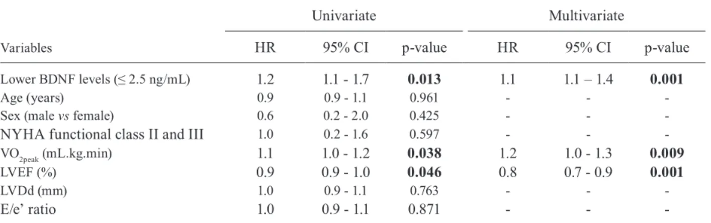
The univariate Cox analysis showed that lower se-rum BDNF levels, reduced VO2peak, and lower LVEF were associated with poor prognosis at the end of fol-low-up (Table III). In the final multivariate model, lower BDNF levels [hazards ratio (HR) 1.1, 95% confidence interval (CI): 1.1-1.4; p = 0.001], LVEF (HR 0.8, 95% CI: 0.7-0.9; p = 0.001), and VO2peak (HR 1.2, 95% CI: 1.0-1.3; p = 0.009) remained as independent predictors of cardiac death in patients with ChC.
When comparing the variables as independent pre-dictors of cardiac death using the Cox regression analy-sis, the serum BDNF level showed a prognostic value similar to that of LVEF, based on the area under the ROC curve and negative and positive predictive values (Table IV). The prognostic value of VO2peak was lower than that of serum BDNF and LVEF.
In the Kaplan-Meier analysis using the cut-off points obtained using the ROC curve, only VO2peak showed no significant difference (log rank = 0.361) between the groups with values below and above the established point, that is, 25 mL/kg/min (Fig. 2).
In Cox survival analysis using the cut-off points obtained with the ROC curve, the combination of low BDNF values (≤ 2.5 ng/mL) and LVEF (≤ 31.5%) were highly predictive of cardiac death (HR 5.6, 95% CI: 1.2-9.7; p = 0.026).
TABLE II
Differences in serum brain-derived neurotrophic factor (BDNF), demographic data, functional status and echocardiographic parameters between survivors and
non-survivors patients
Variables
Survivors (n = 37)
Non-survivors (n = 12) p-value
Serum BDNF (ng/mL) 4.4 (2.4 - 11.9)
2.0 (1.4 - 5.2)
0.041
Age (years) 50 ± 7 49 ± 9 0.926
Sex (male/female) 20/17 8/4 0.336 NYHA class I/II/III 23/12/2 5/4/3 0.391
Exercise testing
VO2peak (mL.kg.min) 30.7 ± 7.9 26.1 ± 6.9 0.048
Echocardiography
LVEF (%) 37.0
(32.5 - 41.0)
32.0 (24.2 - 40.5)
0.169
LVDd (mm) 65.2 ± 6.5 65.8 ± 5.8 0.449 E/e’ ratio 10.5 ± 5.5 11.0 ± 4.9 0.651
Data presented as mean and standard deviation (mean ± SD), median (MD) and interquartile range (25-75%) or absolute number. p-values highlighted in bold are statistically signif-icant (p < 0.05). BMI: body mass index; E/e’ ratio: ratio of the early diastolic transmitral flow velocity to early diastolic mitral annular velocity; LVDd: left ventricular end-diastolic diameter; LVEF: left ventricular ejection fraction; ng/mL: nanograms per milliliters; NYHA: New York Heart Associa-tion funcAssocia-tional class; VO2peak: peak oxygen uptake.
Fig. 1: accuracy of serum brain-derived neurotrophic factor (BDNF) levels in predicting cardiac death based on the receiver operating char-acteristic (ROC) curve in patients with Chagas cardiomyopathy (ChC).
DISCUSSION
The present study showed, for the first time, the prog-nostic value of serum BDNF levels in patients with ChC. The main findings of the present study were as follows: (1) serum BDNF levels and VO2peak were lower in non-sur-vivor patients with ChC than in the surnon-sur-vivors; (2) serum BDNF concentration of 2.5 ng/mL was the optimal cut-off value to identify patients at risk of adverse outcome; (3) low serum BDNF levels, LVEF, and VO2peak were indepen-dent predictors of cardiac death, and (4) the combination of low serum BDNF levels and LVEF are highly predictive of cardiac death in ChC after 39 ± 14 months of follow-up. Our results suggest that serum BDNF levels, especially when associated with systolic function, may provide use-ful prognostic information in patients with ChC.
Takashio et al.(21) reported that plasma concentration of BDNF in patients with heart failure (n = 242, 71 ± 12 years, NYHA I-III) was significantly lower than in healthy individuals (p < 0.001). At the end of follow-up, 14% of patients died of any cause. Patients with low plasma BDNF concentration (≤ 3.7 ng/mL) had higher mortality rate than those with higher plasma BDNF lev-els (HR 2.22, 95% CI: 1.03-4.82, p = 0.04).
In the present study, lower BDNF level was an inde-pendent predictor of death in patients with ChC. Abnor-malities in the skeletal muscles, autonomic denervation, and reduction of cardiomyocytes by fibrosis, the main pathological signs of an infected heart, are common clini-cal findings with the progression of Chagas disease. Both skeletal(22) and cardiac muscles(23) are important sources of BDNF. In addition, we believe that BDNF, being a neu-rotrophic factor, may reflect the autonomic dysfunction presented by patients with ChC. Thus, muscle atrophy, myocardial fibrosis, and autonomic denervation, the signs of disease progression, may contribute to the reduction of circulating levels of this neurotrophic factor.
The association between BDNF levels and predictors of poor prognosis in ChC has also been reported in
an-other study(13), which found a positive correlation between serum BDNF and LVEF levels (r = 0.3137, p = 0.0431) and a negative correlation with ventricular dilatation index (r = -0.3146, p = 0.0424). In patients with heart failure, a pre-vious study(19) reported that compared to BNP, the first-line biomarker in the prognosis of this disease, the BDNF was similarly effective in predicting adverse events in this population (AUC 0.827 and 0.798, respectively).
However, compared to the aforementioned studies, the present study showed different HR values. A few hypotheses are proposed. Firstly, Kadowaki et al.(20) and Fukushima et al.(19) considered cardiac death and hospi-talisations due to heart failure as end-points, while the present study considered only cardiac death as the end-point. Secondly, studies by Kadowaki et al.(20) and Fuku-shima et al.(19) selected older patients (71 ± 13 and 59.2 ± 13.7 years, respectively) than those enrolled in the pres-ent study (50 ± 7 years). Furthermore, both studies used a sample set of patients with heart failure and comor-bidities, such as diabetes, dyslipidaemia, and hyperten-sion, which have been shown to reduce patient survival. Finally, the study by Kadowaki et al.(20) did not report the use of antidepressant medication as an exclusion
cri-TABLE III
Uni and multivariate Cox analysis for cardiac death in Chagas cardiomyopathy (ChC) patients
Variables
Univariate Multivariate
HR 95% CI p-value HR 95% CI p-value
Lower BDNF levels (≤ 2.5 ng/mL) 1.2 1.1 - 1.7 0.013 1.1 1.1 – 1.4 0.001
Age (years) 0.9 0.9 - 1.1 0.961 - -
-Sex (male vs female) 0.6 0.2 - 2.0 0.425 - -
-NYHA functional class II and III 1.0 0.2 - 1.6 0.597 - -
-VO2peak (mL.kg.min) 1.1 1.0 - 1.2 0.038 1.2 1.0 - 1.3 0.009
LVEF (%) 0.9 0.9 - 1.0 0.046 0.8 0.7 - 0.9 0.001
LVDd (mm) 1.0 0.9 - 1.1 0.763 - -
-E/e’ ratio 1.0 0.9 - 1.1 0.871 - -
-BDNF: brain-derived neurotrophic factor; E/e’ ratio: ratio of the early diastolic transmitral flow velocity to early diastolic mitral annular velocity; HR: hazard ratio; LVDd: left ventricular end-diastolic diameter; LVEF: left ventricular ejection fraction; ng/mL: nanograms per milliliters; NYHA: New York Heart Association functional class; VO2peak: peak oxygen uptake; 95% CI: 95% con-fidence interval.
TABLE IV
Cutoff values, area under the receiver operating characteristic (ROC) curve, sensitivity, specificity and positive and negative predictive values of independent predictors of cardiac death
Predictors Cutoff value AUC (95% CI) Sensitivity Specificity Negative predictive values
Positive predictive values
Serum BDNF 2.5 ng/mL 0.74 (0.56-0.93) 75% 70% 0.31 0.76
VO2peak 25 mL.kg.min 0.69 (0.50-0.90) 75% 60% 0.17 0.59
LVEF 31.5% 0.76 (0.59-0.95) 80% 73% 0.43 0.78
terion; such medication could alter BDNF levels, reduce potential depressive symptoms, increase patient’s qual-ity of life, and increase adherence to treatment.
The cut-off point of serum BDNF in the prediction of cardiac death was also found to be different in the present study compared to those in the studies by Fu-kushima et al.(19) and Kadowaki et al.(20). ChC evolves with incessant and progressive fibrosis. Moreover, fi-brosis causes destruction of cardiomyocytes, which are important sources of BDNF, as justified by Martinelli et al.(13). Thus, the baseline concentration of serum BDNF and the cut-off point in predicting cardiac death were expected to be lower in patients with heart failure due to ChC than in other aetiologies. Corroborating with this hypothesis, BDNF levels at baseline were lower in the present study [6.2 (1.9-8.5) ng/mL] when compared to the study by Fukushima et al.(19) (19.0 ± 5.6) and Kad-owaki et al.(20) (14.7 ± 8.4 ng/mL).
Although low concentrations of serum BDNF are a statistically significant prognostic marker in ChC, it demonstrates a borderline HR when analysed alone. However, when combined with reduced LVEF, a well-established independent predictor of death in ChC, HR has been demonstrated to become clinically relevant. Patients with BDNF concentration of below 2.5 ng/mL and LVEF of < 31.5% had HR of 5.6 (95% CI: 1.2-9.7; p = 0.026) for cardiac death. Therefore, the association between reduced serum BDNF levels and LVEF had a higher prognostic value (HR 5.6, 95% CI: 1.2-9.7) than LVEF alone (HR 0.8, 95% CI: 0.7-0.9), which may have an impact on the assessment of prognosis and risk strati-fication of the patient. We believe that patients with se-rum BDNF level of below 2.5 ng/mL and LVEF of below 31.5% are high risk and should be monitored more fre-quently in the clinical management. In addition, patients with low serum BDNF and reduced LVEF levels should be given priority in exercise-based treatment, as physical training in ChC appears to increase the BDNF levels.(15)
In the multivariate Cox analysis, LVEF and VO2peak, as well as the low concentration of serum BDNF level, remained as independent predictors of death. The LVEF is a well-established independent predictor of survival in patients with ChC(24,25) and serum BDNF showed similar predictive values. However, the role of VO2peak in predict-ing adverse events in this population remains poorly un-derstood. Mady et al.(26) conducted one of the first stud-ies that aimed to verify the prediction of adverse events in male patients with ChC (n = 104, 40.3 ± 9 years) based on VO2peak. Under Cox multivariate regression, VO2peak (p < 0.001) and LVEF (p < 0.001) were highly associated with survival time. Functional impairment has also been shown to suggest subclinical myocardial injury, preced-ing even the reduction of LVEF.(6) Additionally, Ritt et al.(27) found that VO
2peak was associated with poor prog-nosis in patients with ChC (n = 55, LVEF < 45%), but was not considered as an independent predictor of death (HR: 0.97, 95% CI: 0.91-1.04, p = 0.44). In the present study, VO2peak was observed to demonstrate a significant predictor of survival, but failed to determine the cut-off point for the stratification of patients at higher risk of cardiac death. We believe that functional capacity is
sociated with patient’s clinical condition; however, the establishment of the role of VO2peak on survival should be better investigated in studies with larger sample size.
Study limitations include small sample size and the performance of the stress test using conventional maxi-mal exercise testing, without direct gas analysis. How-ever, our sample size is in agreement with a prognostic study of BDNF in patients with heart failure.(19) Regarding exercise testing, the indirect assessment of the VO2peak has been established to be highly correlated with the direct measurement,(28) which does not compromise our results.
In conclusion, low serum BDNF level, especially in association with systolic function and functional capacity, was an independent predictor of survival in patients with ChC and may aid in the risk stratification of these patients.
REFERENCES
1. Rocha MO, Teixeira MM, Ribeiro AL. An update on the manage-ment of Chagas cardiomyopathy. Expert Rev Anti Infect Ther. 2007; 5(4): 727-43.
2. Requena-Mendez A, Aldasoro E, de Lazzari E, Sicuri E, Brown M, Moore DA, et al. Prevalence of Chagas disease in Latin-American migrants living in Europe: a systematic review and meta-analysis. PLoS Negl Trop Dis. 2015; 9(2): e0003540.
3. Gascon J, Bern C, Pinazo MJ. Chagas disease in Spain, the Unit-ed States and other non-endemic countries. Acta Trop. 2010; 115(1-2): 22-7.
4. Punukollu G, Gowda RM, Khan IA, Navarro VS, Vasavada BC. Clinical aspects of the Chagas’ heart disease. Int J Cardiol. 2007; 115(3): 279-83.
5. Sousa GR, Gomes JA, Fares RC, Damasio MP, Chaves AT, Ferreira KS, et al. Plasma cytokine expression is associated with cardiac morbidity in chagas disease. PLoS One. 2014; 9(3): e87082.
6. Mady C, Ianni BM, Arteaga E, Salemi VM, Frimm CC. Maximal functional capacity in patients with Chagas’ cardiomyopathy without congestive heart failure. J Card Fail. 2000; 6(3): 220-4.
7. Teixeira AL, Barbosa IG, Diniz BS, Kummer A. Circulating lev-els of brain-derived neurotrophic factor: correlation with mood, cognition and motor function. Biomark Med. 2010; 4(6): 871-87.
8. Mattson MP, Maudsley S, Martin B. BDNF and 5-HT: a dynamic duo in age-related neuronal plasticity and neurodegenerative dis-orders. Trends Neurosci. 2004; 27(10): 589-94.
9. Gómez-Pinilla F, Vaynman S, Ying Z. Brain-derived neurotrophic factor functions as a metabotrophin to mediate the effects of exer-cise on cognition. Eur J Neurosci. 2008; 28(11): 2278-87.
10. Qiao LY, Shen S, Liu M, Xia C, Kay JC, Zhang QL. Inflammation and activity augment brain-derived neurotrophic factor peripheral release. Neuroscience. 2016; 318: 114-21.
11. Okada S, Yokoyama M, Toko H, Tateno K, Moriya J, Shimizu I, et al. Brain-derived neurotrophic factor protects against car-diac dysfunction after myocardial infarction via a central nervous system-mediated pathway. Arterioscler Thromb Vasc Biol. 2012; 32(8): 1902-9.
12. Jiang H, Liu Y, Zhang Y, Chen ZY. Association of plasma brain-derived neurotrophic factor and cardiovascular risk factors and prognosis in angina pectoris. Biochem Biophys Res Commun. 2011; 415(1): 99-103.
13. Martinelli PM, Rocha MOC, Teixeira AL, Nunes MCP, Camargos ERS. Brain-derived neurotrophic factor is up regulated in chronic Chagas disease. Int J Cardiol. 2011; 149(2): 277-8.
14. Costa HS, Lima MM, Silva MG, Alencar MC, Nunes MC, Ca-margos ER, et al. Effect of acute aerobic exercise on serum BDNF levels in patients with Chagas heart disease. Int J Cardiol. 2014; 174(3): 828-30.
15. Lima MM, Nunes MC, Nascimento B, Costa HS, Sousa LA, Teixeira AL, et al. Improvement of the functional capacity is as-sociated with BDNF and autonomic modulation in Chagas dis-ease. Int J Cardiol. 2013; 167(5): 2363-6.
16. Andrade JP, Marin Neto JA, Paola AA, Vilas-Boas F, Oliveira GM, Bacal F, et al. I Latin American Guidelines for the diagnosis and treatment of Chagas’ heart disease: executive summary. Arq Bras Cardiol. 2011; 96(6): 434-42.
17. Trajkovska V, Marcussen AB, Vinberg M, Hartvig P, Aznar S, Knudsen GM. Measurements of brain-derived neurotrophic fac-tor: methodological aspects and demographical data. Brain Res Bull. 2007; 73(1-3): 143-9.
18. ACSM. ACSM’s Guidelines for Exercise Testing and Exer-cise Prescription. 7th ed. Philadelphia: Lippincott, Williams & Wilkins; 2006.
19. Fukushima A, Kinugawa S, Homma T, Masaki Y, Furihata T, Yo-kota T, et al. Serum brain-derived neurotropic factor level predicts adverse clinical outcomes in patients with heart failure. J Card Fail. 2015; 21(4): 300-6.
20. Kadowaki S, Shishido T, Honda Y, Narumi T, Otaki Y, Kinoshi-ta D, et al. Additive clinical value of serum brain-derived neu-rotrophic factor for prediction of chronic heart failure outcome. Heart Vessels. 2015; 31(4): 535-44.
21. Takashio S, Sugiyama S, Yamamuro M, Takahama H, Hayashi T, Sugano Y, et al. Significance of low plasma levels of brain-derived neurotrophic factor in patients with heart failure. Am J Cardiol. 2015; 116(2): 243-9.
22. Matthews VB, Astrom MB, Chan MH, Bruce CR, Krabbe KS, Prelovsek O, et al. Brain-derived neurotrophic factor is produced by skeletal muscle cells in response to contraction and enhances fat oxidation via activation of AMP-activated protein kinase. Dia-betologia. 2009; 52(7): 1409-18.
23. Hiltunen JO, Laurikainen A, Vakeva A, Meri S, Saarma M. Nerve growth factor and brain-derived neurotrophic factor mRNAs are regulated in distinct cell populations of rat heart after ischaemia and reperfusion. J Pathol. 2001; 194(2): 247-53.
24. Nunes MC, Carmo AA, Rocha MO, Ribeiro AL. Mortality predic-tion in Chagas heart disease. Expert Rev Cardiovasc Ther. 2012; 10(9): 1173-84.
25. Rassi Jr A, Rassi A, Rassi SG. Predictors of mortality in chronic Chagas disease: a systematic review of observational studies. Cir-culation. 2007; 115(9): 1101-8.
26. Mady C, Cardoso RH, Barretto AC, da Luz PL, Bellotti G, Pileggi F. Survival and predictors of survival in patients with congestive heart failure due to Chagas’ cardiomyopathy. Circulation. 1994; 90(6): 3098-102.
27. Ritt LE, Carvalho AC, Feitosa GS, Pinho-Filho JA, Andrade MV, Feitosa-Filho GS, et al. Cardiopulmonary exercise and 6-min walk tests as predictors of quality of life and long-term mortality among patients with heart failure due to Chagas disease. Int J Cardiol. 2013; 168(4): 4584-5.