DOI: 10.5935/2359-4802.20180075
ORIGINAL ARTICLE
Mailing Address: Marta Madeira
Serviço de Cardiologia, Centro Hospitalar e Universitário de Coimbra - Quinta dos Vales, S. Martinho do Bispo. Postal Code: 3045-043, Coimbra - Portugal E-mail: [email protected]
Does Percutaneous Left Atrial Appendage Closure Affect Left Atrial Performance?
Marta Madeira,*1,2 Rogério Teixeira,*1,2 Liliana Reis,1 Paulo Dinis,1 Luís Paiva,1,2 Ana Botelho,1 Marco Costa,1 Lino Gonçalves1,2
Serviço de Cardiologia, Centro Hospitalar e Universitário de Coimbra - Hospital Geral,1 Coimbra - Portugal
Faculdade de Medicina da Universidade de Coimbra,2 Coimbra - Portugal
* Both authors contributed equally to the paper
Manuscript received September 09, 2017, revised manuscript April 11, 2018, accepted July 02, 2018.
Abstract
Background: Percutaneous left atrial appendage (LAA) occlusion may be an alternative therapy for atrial fibrillation (AF) patients with contraindication for anti-coagulation therapy. However, the influence of LAA occlusion on left atrial (LA) performance has not been studied.
Objective: Our aim was to evaluate the influence of percutaneous LAA occlusion device on LA function by transthoracic echocardiography plus speckle-tracking echocardiography (STE).
Methods: We included 16 patients undergoing percutaneous LAA closure with adequate echocardiographic window for the study of LA mechanics. Transthoracic echocardiography was performed before and after the procedure. LA volumes were calculated using the biplane method, and LA mechanics were assessed using STE. The analysis focused on the LA reservoir phase strain and strain rate.
Results: Seventy-five percent of patients had permanent atrial fibrillation. Embolic and bleeding risk scores used were CHA2DS2-VASc [median of 4-5] and HAS-BLED [median of 2-3]. Major bleeding (62%) was the most common indication for the procedure. Percutaneous LAA closure was performed successfully in all patients, without major complications. No differences were found in maximum LA volume (44 ± 11 vs. 46 ± 13 mL/m2; p = 0.54), minimum
LA volume (32 ± 8 vs. 37 ± 14 mL/m2; p = 0.09) or LA emptying fraction (26 ± 17 vs. 21 ± 14%; p = 0.33) before and
after the procedure. Similarly, no differences were noted in left atrial strain (13.7 ± 11.1 vs. 13.0 ± 8.8%; p = 0.63) or strain rate (1.06 ± 0.26 vs. 1.13 ± 0.34 s-1; p = 0.38) in the reservoir phase.
Conclusions: Our data suggest that percutaneous LAA closure does not affect LA reservoir function. (Int J Cardiovasc Sci. 2018;31(6)569-577)
Keywords: Atrial Fibrillation; Atrial Appendage; Heart Atria; Echocardiography, Transthoracic.
Introduction
Atrial fibrillation is the most common sustained cardiac
arrhythmia,1 with a current estimated prevalence of 1.5%
to 2%.2 It is considered a major cause of systemic embolism,
increasing the risk for ischemic stroke by 5 times.2,3 Oral
anticoagulation has been shown to effectively reduce the risk for stroke in patients with atrial fibrillation and
is one of the cornerstones of management.2 However,
a significant proportion (30% - 50%) of eligible patients do not receive oral anticoagulation due to the presence of absolute contraindications or a perceived high risk of
bleeding.3 Several studies have shown that, in patients
with nonvalvular atrial fibrillation, 90% of thrombus
formation occurs in the left atrial appendage (LAA).4,5
Therefore, devices for LAA closure have been developed as an alternative to oral anticoagulation in patients at high risk for stroke with contraindications to anticoagulation
therapy.6 Recently, the non-inferiority of LAA exclusion
over warfarin for stroke prevention was demonstrated in
patients with nonvalvular atrial fibrillation.7
hemodynamics.8 The appendage is more compliant
than the left atrium, acting as a reservoir to attenuate the rise in intra-atrial pressure in response to various
hemodynamic factors.9 Surgical clamping or removal of
the LAA has been shown to cause an immediate increase in left atrial pressure, left atrial size, and pulmonary- and
mitral-inflow velocities.10 However, both the relative
contribution of appendage distensibility to the passive elastic-chamber properties of the left atrium and the physiological and hemodynamic importance of the LAA are currently uncertain. Furthermore, the influence of the LAA occlusion device on left atrial performance has not yet been defined.
Two-dimensional speckle-tracking echocardiography (2D-STE) is a recently developed, angle-independent, semiautomated technique used to evaluate the
myocardium.11 It uses standard B-mode images to track
blocks of speckles from frame to frame, and measures myocardial lengthening and shortening relative to the baseline – the Lagrangian method. 2D-STE provides local myocardial information from which displacement, velocity, strain, and strain rate can be derived, allowing an accurate assessment of longitudinal, radial, and
circumferential myocardial mechanics.11 In recent years,
left atrial mechanics have been used as a surrogate for left atrial performance, which is influenced by the left atrial wall properties, left atrial volume, and left atrial pressure, and also by the left ventricular longitudinal
systolic function.12 Measurements of left atrial strain
(ƐR) and strain rate (SRR) during the reservoir phase can
be used to describe atrial function physiology and are sensitive to detect early functional remodeling before
anatomical changes occur.12,13
We hypothesized that left atrial function, assessed by echocardiographic parameters and 2D-STE, would decrease after percutaneous LAA closure. Therefore, our aim was to evaluate the influence of the LAA closure device on left atrial physiology.
Methods
Patients
Twenty-five patients with non-valvular atrial
fibrillation and a high risk for stroke with a CHA2DS2
-VASc Score of ≥ 1 admitted to our centre for percutaneous LAA closure between August of 2010 and August of 2015 were enrolled in this retrospective study. Nine patients were excluded due to lack of adequate echocardiographic evaluation before or after the procedure or poor
echocardiographic window for the evaluation of left atrial mechanics.
Referral indications for percutaneous LAA closure were contra-indication for long-term oral anticoagulation, bleeding events during oral anticoagulation, labile international normalized ratio (INR) or embolic events despite proper anticoagulation.
Clinical data included past medical history, current
medication, the CHA2DS2-VASc and HAS-BLED scores,
and diagnostic evaluation by routine laboratory testing, electrocardiography, and echocardiography.
Sixteen patients with good echocardiographic window for assessment of the left atrial mechanics were included in our study.
The study was approved by the ethics committee of our institution.
Echocardiographic evaluation
Echocardiography was performed on the day before and 3 months after percutaneous closure of the LAA.
Echocardiographic examinations were performed
using an ultrasound system (Vivid 7, General Electric®,
Horten, Norway) and tissue harmonic imaging at 1.7/3.4 MHz. A complete echocardiographic study was performed using standard views according to
current guidelines.14 Three consecutive heart cycles
were acquired for quantification of the left atrial size and 2D-STE analysis for sinus rhythm patients, and five consecutive heart cycles were obtained for atrial fibrillation patients.
Left atrial volume was assessed by the biplane method of disks from the apical 4- and 2-chamber views and the measurements were indexed to the body surface area
according to established recommendations.15 Minimum
left atrial volume was measured at left ventricular end-diastolic volume, and maximum left atrial volume at end-systole. Left atrial emptying fraction was calculated as (maximum left atrial volume - maximum left atrial
volume)/ maximum left atrial volume.15
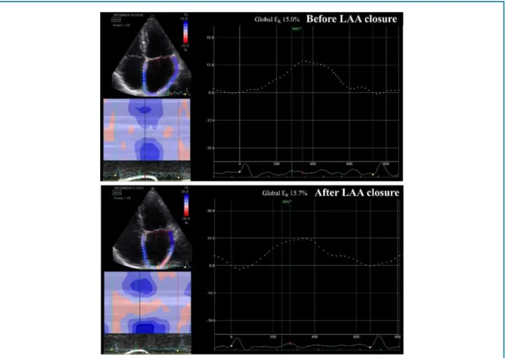
The 2D-STE method was used to calculate regional and
global longitudinal ƐR and SRR (Figure 1). A minimum
frame rate of 60 frames/sec was required for a reliable operation of the program. The recordings were processed using an acoustic-tracking dedicated software (EchoPAQ
9.0, GE Healthcare®, Horten, Norway), which allowed
Figure 1 - Global left atrial strain during reservoir phase (ƐR) before and after percutaneous closure of the left atrial appendage (LAA). Differences between baseline and post-LAA occlusion device implantation data were analysed by paired sample t-test.
traced in end-systole in both four- and two-chamber views by a point-and-click approach. An epicardial surface tracing was then automatically generated by the system, generating the region of interest (ROI). For definition of the ROI at the discontinuity of the left atrial wall (corresponding to pulmonary veins and left atrial appendage), the limit of left atrial endocardial and epicardial surfaces at the junction of these structures was extrapolated. After manual adjustment of ROI width and shape to ensure optimal tracking, the software divided the ROI into six segments (basal, middle and apical segments of the atrial septum and lateral wall), and the tracking quality of each segment was automatically scored as either acceptable or non-acceptable, with possible further manual correction. Segments from which good quality images could not be obtained were rejected by the software and excluded from the analysis. In subjects with good quality images, a total of twelve segments were analyzed. The software displayed peak
longitudinal ƐR and strain rate for each of the twelve
segments and the average global strain. Peak ƐR were
expressed in percentages and SRR in s-1. Since left atrial
wall strain is reliably imaged and is not constrained by other cardiac chambers, recent consensus of imaging for evaluation of atrial fibrillation patients recommend
the evaluation of this parameter rather than global ƐR.16
Therefore, we also performed a comparison between
left atrial lateral wall strain and SRR at baseline and after
device implantation. Since we included patients with atrial fibrillation and sinus rhythm, we used the first left ventricular systolic frame as the frame of interest – QRS timed analysis.
LAA closure procedure
LAA closure device was implanted in the catheterization
laboratory. The device used was an Amplatzer® (St. Jude
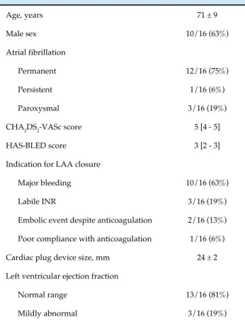
Table 1 - General characteristics of the study group (n = 16)
Age, years 71 ± 9
Male sex 10/16 (63%)
Atrial fibrillation
Permanent 12/16 (75%)
Persistent 1/16 (6%)
Paroxysmal 3/16 (19%)
CHA2DS2-VASc score 5 [4 - 5]
HAS-BLED score 3 [2 - 3]
Indication for LAA closure
Major bleeding 10/16 (63%)
Labile INR 3/16 (19%)
Embolic event despite anticoagulation 2/16 (13%)
Poor compliance with anticoagulation 1/16 (6%)
Cardiac plug device size, mm 24 ± 2
Left ventricular ejection fraction
Normal range 13/16 (81%)
Mildly abnormal 3/16 (19%)
LAA: left atrial appendage; INR: international normalized ratio. Data expressed as mean and standard deviation, percentage or median and interquartile range.
through an appropriate sheath depending on the size of the selected occluder through a puncture in the femoral vein. Deployment and position of the device were controlled by fluoroscopy, and by periprocedural transesophageal or intracardiac echocardiography. LAA was reached through a transseptal puncture. Decision on device size was made upon anatomical morphology, and measurements in echocardiography and fluoroscopy. Oral anticoagulation, if present, was discontinued 48 hours prior to the procedure. During procedure, heparin was administered with an activated clotting time of 250s. Dual antiplatelet therapy with aspirin 100 mg and clopidogrel 75 mg was recommended for 1 month, followed by long-term antiplatelet therapy with aspirin 100 mg daily. No oral anticoagulation was recommended after device implantation.
Statistical analysis
The Kolmogorov-Smirnov test was used to evaluate the distribution of the continuous variables. In the overall sample, all variables were normally distributed,
except for ƐR and follow-up time; and when patients
were separated by group (no change and decrease of ƐR
and SRR), the variables were not normally distributed.
According to distribution normality, continuous data were presented as mean and standard deviation or as median and interquartile range. Quantitative variables with normal distribution were compared by the t-test and quantitative variables without normal distribution by the Mann-Whitney test. Qualitative variables were compared using the chi-square test. Differences between baseline and post-implantation of the LAA occlusion device were analysed by the paired sample t-test.
Statistical analysis was carried out with SPSS®15 and
GraphPad Prism® 6.05. A two-tailed p value < 0.05 was
considered statistically significant.
Results
Population characteristics
Mean age of our sample was 71 ± 9 years, with male predominance (63%). Seventy-five percent of patients had permanent atrial fibrillation. There was no history of percutaneous atrial fibrillation ablation attempt or surgical Maze procedure. Our population had a high embolic and bleeding risk, expressed by a median
CHA2DS2-VASc score of 5 [4-5] and HAS-BLED score
of 3 [2-3].
Major bleeding (62%) was the most common indication for the procedure, followed by labile INR (19%), embolic events despite anticoagulation (13%), and poor compliance with anticoagulation medication (6%). Percutaneous LAA closure was performed successfully in all patients using the cardiac plug device (size, 24 ± 2 mm), without any major complications during or after the procedure.
Characteristics of the study population are summarized in Table 1.
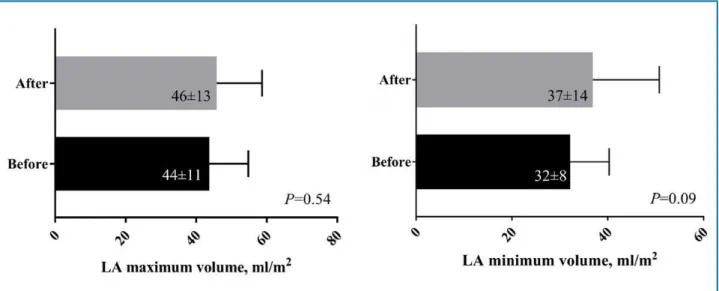
LA volume and emptying fraction
Maximum and minimum values of left atrial volume and the left atrial emptying fraction before and after the procedure are represented in Figure 2 and Figure 3, respectively. No differences were found in maximum
left atrial volume (44 ± 11 vs. 46 ± 13 mL/m2; p = 0.54),
minimum left atrial volume (32 ± 8 vs. 37 ± 14 mL/m2;
Figure 2 - Maximum and minimum volume of the left atrium before and after percutaneous closure of the left atrial appendage (LAA). Differences between baseline and post-LAA occlusion device implantation data were analysed by paired sample t-test.
Figure 3 - Left atrial emptying fraction before and after percutaneous closure of the left atrial appendage (LAA). Differences between baseline and post-LAA occlusion device implantation data were analysed by paired sample t-test.
vs. 21 ± 14%; p = 0.33) after the intervention compared with the baseline values.
Left atrium reservoir ƐR and SRR
Global and regional peak ƐR and SRR of the 12
segments before and months after percutaneous closure
of the LAA are listed in Table 2. Similar values of ƐR (10.1
[8.1–14.7] vs. 12.7 [5.4–16.5]%; p = 0.81) and SRR (1.06 ±
0.26 vs. 1.13 ± 0.34 s-1; p = 0.38) were observed before and
after the procedure (Figure 4).
Assessment of left atrial lateral wall revealed similar
ƐR (11.0 [6.5–19.8]% vs. 8.2 [2.7–15.9]%; p = 0.60) and SRR
(1.01 [0.78–1.54] vs. 1.02 [0.85–1.56] s-1; p = 0.75) before
and after the procedure.
In 44% of patients, there was a decrease in ƐR. There
were no differences regarding patient age, baseline left
atrial volume, left atrial emptying fraction, ƐR, SRR,
CHA2DS2-VASc or HAS-BLED scores, cardiac-plug
device size, or incidence of cardiovascular adverse events during follow-up between patients with decreased and
Table 2 - Global and regional peak left atrial strain (ƐR) and strain rate (SRR) during reservoir phase of the 12
segments before and after percutaneous closure of the left atrial appendage (LAA)
Basal After LAA
occlusion p
Global ƐR, % 10.1 [8.1 - 14.7] 12.7 [5.4 - 16.5] 0.81
Lateral basal ƐR, % 18.6 [10.5 - 28.8] 17.6 [11.0 - 22.3] 0.49
Lateral mid ƐR, % 10.1 [6.0 - 18.8] 9.0 [4.4 - 16.4] 0.40
Lateral apical ƐR, % 7.1 [3.9 - 13.6] 5.9 [2.0 - 15.7] 0.84
Septal apical ƐR, % 9.1 [4.9 - 18.4] 8.5 [3.9 - 16.8] 0.38
Septal mid ƐR, % 12.0 [8.1 - 17.5] 9.8 [3.2 - 22.5] 0.86
Septal basal ƐR, % 13.2 [5.2 - 21.8] 12.2 [2.7 - 23.6] 0.84
Global SRR, s-1 1.06 ± 0.26 1.13 ± 0.34 0.38
Lateral basal SRR, s-1 1.14 ± 0.49 1.20 ± 0.62 0.60
Lateral mid SRR, s-1 1.04 ± 0.42 1.10 ± 0.56 0.61
Lateral apical SRR, s-1 1.18 ± 0.50 1.05 ± 0.48 0.41
Septal apical SRR, s-1 1.00 ± 0.54 1.10 ± 0.45 0.50
Septal mid SRR, s-1 0.94 ± 0.45 1.01 ± 0.29 0.45
Septal basal SRR, s-1 1.39 ± 0.53 1.35 ± 0.62 0.86
ƐR: left atrial strain during the reservoir phase; LAA: left atrial appendage; SRR: left atrial strain rate in the reservoir phase. ƐR values compared by the Mann-Whitney test and SRR values by the t-test.
Figure 4 - Global left atrial strain (ƐR) and strain rate during reservoir phase (SRR) before and after percutaneous closure of the left atrial
appendage. Differences between baseline and post-LAA occlusion device implantation data were analysed by paired sample t-test.
Discussion
Our investigation demonstrates that changes in left atrial fraction volume are minimal after LAA percutaneous closure, and mechanics of the left atrial reservoir phase assessed by 2D-STE are not significantly different before and after the procedure.
Structural and functional remodelling of the left atrium has been proposed as a surrogate for diastolic dysfunction and a predictor of cardiovascular outcomes such as new-onset atrial fibrillation, stroke, heart failure, mortality after myocardial infarction, severity of diastolic
dysfunction, and cardiovascular death.12 2D-STE is a
novel method for quantitative real-time assessment of regional myocardial deformation. The technology tracks acoustic speckles or kernels rather than using Doppler
myocardial velocities.17 Considering the limitations of the
classical indices of left atrial function, assessment of ƐR
by 2D-STE may represent a relatively rapid and easy-to-perform technique for assessing left atrial function, due to its semiautomated nature and off-line processing. In fact, in contrast to Doppler-derived parameters, 2D-STE has the advantage of being angle-independent, and less affected by reverberation, side lobe and drop-out
artefacts.18 Furthermore, recent studies have shown
that 2D-STE is feasible and reproducible.18-20 It has been
suggested that ƐR allows an excellent assessment of the
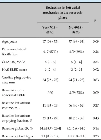
Table 3 - Patients with reduction in left atrial mechanics in the reservoir phase
Reduction in left atrial mechanics in the reservoir
phase
p
Yes (7/16 - 44%)
No (9/16 - 56%)
Age, years 67 [66 - 73] 77 [69 - 81] 0.09
Permanent atrial
fibrillation 4/7 (57%) 8/9 (89%) 0.26
CHA2DS2-VASc 5 [3 - 5] 5 [4 - 6] 0.35
HAS-BLED score 3 [2 - 4] 3 [2 - 3] 0.92
Cardiac plug device
size, mm 24 [22 - 25] 24 [21 - 25] 0.83
Baseline mildly
abnormal LVEF 0/0 3/9 (33%) 0.09
Baseline left atrium
volume, mL 41 [33 - 45] 46 [40 - 62] 0.27
Baseline left atrium
emptying fraction, % 25 [13 - 49] 18 [15 - 39] 0.43
Baseline global ƐR, % 14.4 [8.7 - 26.4] 9.2 [5.6 - 14.0] 0.14
Baseline global SRR, s-1 1.1 [0.9 - 1.2] 1.0 [0.8 - 1.1] 0.25
LVEF: left ventricle ejection fraction; ƐR: left atrium strain during the reservoir phase; SRR: left atrium strain rate reservoir phase. Comparison of variables was performed with a Mann-Whitney test and SRR values by the t-test. Data expressed as mean and standard deviation or median and interquartile range.
closely following left atrial physiology, and can be used to
evaluate dynamic left atrial function.18,19 It has also been
demonstrated that the left atrial reservoir ƐR is associated
with fibrosis and can thus represent left atrial stiffness.21
Contrary to earlier belief, LAA is now thought to play an important role in normal cardiac hemodynamics, acting as an adaptive chamber in conditions of volume
overload to attenuate the rise in intra-atrial pressure.22,23
Furthermore, the highest density of atrial natriuretic-peptide granules of the heart is found in LLA, and the release of atrial natriuretic peptide with consequent diuresis is an important compensatory mechanism
involved in the maintenance of normal fluid homeostasis.24
Hondo et al.,23 in a study performed in 10 open-chest
dogs, reported that the LAA is more compliant than the left atrial main chamber. They also found a higher dimensional increase in the LAA than the left atrial main
chamber during left atrial volume overload. Davis et al.,25
reported, in a study using 6 isolated canine left atria, that the LAA may enable the entire left atrium to better adapt reservoir function to physiologic conditions by protecting the pulmonary capillary system from encountering a rise in pressure.
In a study conducted by Kamohara et al.,26 to
investigate the short-term and midterm effects of LAA exclusion on left atrial function, involving 19 dogs with 90 days of follow-up, the authors showed no significant difference in the transmitral flow tissue Doppler imaging measurements, left atrial pressure, left ventricular
volume, or stroke volume. Tabata et al.,27 evaluated the
role of LAA in left atrial reservoir function by assessing changes in left atrial flow dynamics after LAA clamping during cardiac surgery. The subjects of the study were 8 patients who had undergone coronary artery bypass grafting and 7 who had undergone valvular surgery for mitral regurgitation; all patients were in sinus rhythm. They demonstrated that, in both groups, mean left atrial pressure and maximum left atrial dimension significantly increased during LAA clamping. The authors concluded that the LAA is more compliant than the left atrial main chamber and plays an important role in left atrial
reservoir function. Johansson et al.,28 explored the
effects on atrial and ventricular function of restoring sinus rhythm after epicardial cryoablation and closure of the LAA in 65 patients with mitral valve disease and atrial fibrillation. In patients who were in sinus rhythm, peak velocity during atrial contraction and the reservoir function were lower in patients that underwent LAA closure than in the control group at 6 months of follow-up. In summary, it seems that in patients who are in sinus rhythm, LAA occlusion might negatively influence left atrial reservoir function. In fact, our patients in sinus
rhythm had a decrease in ƐR and SRR after the procedure.
However, in patients with atrial fibrillation, closure of the LAA does not seem to have an impact on left atrial
reservoir function. Hanna et al.,29 conducted a study
increased left atrial volumes and decreased left atrial
emptying fractions,15ƐR, and SR
R.
12 Furthermore, Sasaki
et al.,30 demonstrated that left atrial peak systolic ƐR
is independently associated with LAA dysfunction in patients with atrial fibrillation. Hence, in our population of patients with left atrial chamber dysfunction at baseline, a reduced LAA function might also be present. Our
results, along with those of Hanna et al.,29 suggest that
the exclusion of the LAA does not seem to have a further impact on compromised left atrial physiology.
Nevertheless, a recent study with 33 patients (20 patients with atrial fibrillation) demonstrated that LAA closure was associated with an improvement in left atrial mechanical function in a 45-day follow-up, and these changes appeared to be related to changes
in loading conditions (Frank-Starling effect).31 Despite
favourable short-term outcomes, the long-term effects of an increase in left atrial volume might lead to deleterious effects, mainly in patients with sinus rhythm. We must also highlight that although there was an increase in peak atrial longitudinal strain at discharge compared to baseline, peak atrial longitudinal strain tended to be lower 45 days as compared with discharge (p = 0.08). Therefore, with a longer follow-up, peak atrial longitudinal strain might return to baseline levels as observed in our study.
Most previous studies were performed in patients with sinus rhythm and evaluated left atrial function immediately after LAA closure; the long-term hemodynamic effects of this procedure in patients who are in sinus rhythm are currently not known. Although we did not find any statistically significant difference between patients with decreased and with similar left atrial reservoir function after the procedure, the former group might have better left atrial function at baseline since a lower number of patients had lower left atrial volumes, higher left atrial
emptying fractions, and higher ƐR values, consistent with
the results of the studies mentioned above.
Limitations
Our study has several limitations. First, this was a single-center study with a relatively small sample size. However, there is a paucity of data regarding the effects of percutaneous LAA closure on left atrial function in the literature. Second, the retrospective nature of the study limited the evaluation of additional clinical and analytical parameters. Third, although we analyzed the impact of LAA percutaneous closure on left atrial mechanics, the design of our study made the assessment of clinical
outcomes impossible. Finally, most of our patients had atrial fibrillation and left atrial dysfunction at baseline. It would therefore be important to study the influence of LAA percutaneous closure on left atrial function in patients who are in sinus rhythm with normal or slightly altered left atrial function.
Further studies including large populations of patients in sinus rhythm and in atrial fibrillation are needed to provide definitive evidence of the impact of LAA occlusion not only on left atrial physiology at long term, assessed by 2D-STE, but also on clinical outcomes.
Conclusion
We have demonstrated that in patients with atrial fibrillation and contraindication to oral anticoagulation, percutaneous LAA closure does not have a negative effect on left atrial reservoir function in patients with permanent atrial fibrillation. Further studies with a larger population of patients are warranted to confirm this finding.
Author contributions
Conception and design of the research: Madeira M, Teixeira R, Costa M. Acquisition of data: Madeira M, Teixeira R, Reis L, Dinis P, Paiva L, Botelho A, Costa M. Analysis and interpretation of the data: Madeira M, Teixeira R, Reis L, Dinis P, Paiva L. Statistical analysis: Madeira M, Teixeira R, Dinis P, Paiva L. Writing of the manuscript: Madeira M. Critical revision of the manuscript for intellectual content: Teixeira R, Reis L, Dinis P, Paiva L, Botelho A, Costa M, Gonçalves L.
Potential Conflict of Interest
No potential conflict of interest relevant to this article was reported.
Sources of Funding
There were no external funding sources for this study.
Study Association
This study is not associated with any thesis or dissertation work.
Ethics approval and consent to participate
1. Wolf PA, Abbott RD, Kannel WB. Atrial fibrillation as an independent risk factor for stroke: the Framingham study. Stroke. 1991;22(8):983-8.
2. Kirchhof P, Benussi B, Kotecha D, Ahlsson A, Atar D, Casadei B, et al; ESC Scientific Document Group. 2016 ESC Guidelines for the management of atrial fibrillation developed in collaboration with EACTS. Eur Heart J. 2016;37(38):2893-2962.
3. Saw J, Lempereur M. Percutaneous left atrial appendage closure: procedural techniques and outcomes. JACC Cardiovasc Interv. 2014;7(11):1205-20.
4. Onalan O, Crystal E. Left atrial appendage exclusion for stroke prevention in patients with nonrheumatic atrial fibrillation. Stroke. 2007;38(2 Suppl):624-30.
5. Beigel R, Wunderlich NC, Ho SY, Arsanjani R, Siegel RJ. The left atrial appendage: anatomy, function, and noninvasive evaluation. JACC Cardiovasc Imaging. 2014;7(12):1251-65.
6. Seeger J, Bothner C, Dahme T, Gonska B, Scharnbeck D, Markovic S, et al. Efficacy and safety of percutaneous left atrial appendage closure to prevent thromboembolic events in atrial fibrillation patients with high stroke and bleeding risk. Clin Res Cardiol. 2016;105(3):225-9.
7. Holmes DR Jr, Kar S, Price MJ, Whisenant B, Sievert H, Doshi SK, et al. Prospective randomized evaluation of the Watchman Left Atrial Appendage Closure device in patients with atrial fibrillation versus long-term warfarin therapy: the PREVAIL trial. J Am Coll Cardiol. 2014;64(1):1-12. Erratum in: J Am Coll Cardiol. 2014;64(11):1186
8. Stöllberger C, Schneider B, Finsterer J. Elimination of the left atrial appendage to prevent stroke or embolism? Anatomic, physiologic, and pathophysiologic considerations. Chest. 2003;124(6):2356-62.
9. Hondo T, Okamoto M, Yamane T, Kawagoe T, Karakawa S, Yamagata T, et al. The role of the left atrial appendage. A volume loading study in open-chest dogs. Jpn Heart J. 1995;36(2):225-34.
10. Bansal M, Kasliwal R. Echocardiography for left atrial appendage structure and function. Indian Heart J. 2012;64(5):469-75.
11. Mor-Avi V, Lang R, Badano L, Belohlavek M, Cardim NM, Derumeaux G, et al. Current and evolving echocardiographic techniques for the quantitative evaluation of cardiac mechanics: ASE/EAE consensus statement on methodology and indications endorsed by the Japanese Society of Echocardiography. J Am Soc Echocardiogr. 2011;24(3):277-313.
12. Vieira MJ, Teixeira R, Gonçalves L, Gersh BJ. Left Atrial Mechanics: Echocardiographic Assessment and Clinical Implications. J Am Soc Echocardiogr. 2014;27(5):463-78.
13. Goette A, Kalman JM, Aguinaga L, Akar J, Cabrera JA, Chen SA, et al. EHRA/HRS/APHRS/SOLAECE expert consensus on Atrial cardiomyopathies: definition, characterization, and clinical implication. Europace. 2016;18(10):1455-1490.
14. Evangelista A, Flachskampf F, Lancellotti P, Badano L, Aguilar R, Monaghan M, et al; European Association of Echocardiography. European Association of Echocardiography recommendations for standardization of performance, digital storage and reporting of echocardiographic studies. Eur J Echocardiogr. 2008;9(4):438-48.
15. Lang R, Badano L, Mor-Avi V, Afilalo J, Armstrong A, Ernande L, et al. Recommendations for cardiac chamber quantification by echocardiography in adults: an update from the American Society of Echocardiography and the European Association of Cardiovascular Imaging. J Am Soc Echocardiogr. 2015;28(1):1-39.e14.
16. Donal E, Lip GY, Galderisi M, Goette A, Shah D, Marwan M, et al. EACVI/ EHRA Expert Consensus Document on the role of
multi-modality imaging for the evaluation of patients with atrial fibrillation. Eur Heart J Cardiovasc Imaging. 2016;17(4):355-83.
17. Leitman M, Lysyansky P, Sidenko S, Shir V, Peleg E, Binenbaum M, et al. Two-dimensional strain – a novel software for real-time quantitative echocardiographic assessment of myocardial function. J Am Soc Echocardiogr. 2004;17(10):1021-9.
18. Cameli M, Caputo M, Mondillo S, Ballo P, Palmerini E, Lisi M, et al. Feasibility and reference values of left atrial longitudinal strain imaging by two-dimensional speckle tracking. Cardiovasc Ultrasound. 2009 Feb 8;7:6.
19. Vianna-Pinton R, Moreno CA, Baxter CM, Lee KS, Tsang TS, Appleton CP. Two-dimensional speckle-tracking echocardiography of the left atrium: feasibility and regional contraction and relaxation differences in normal subjects. J Am Soc Echocardiogr. 2008;22(3):299-305.
20. Kim D, Lee K, Lee S, Jeong SY, Lee YS, Choi YJ, et al. Feasibility of two-dimensional global longitudinal strain and strain rate imaging for the assessment of left atrial function: a study in subjects with a low probability of cardiovascular disease and normal exercise capacity. Echocardiography. 2009;26(10):1179-87.
21. Gottdiener J, Kitzman D, Aurigemma G, Arnold AM, Manolio TA. Left atrial volume, geometry, and function in systolic and diastolic heart failure of persons > or =65 years of age (the Cardiovascular Health Study). Am J Cardiol. 2006;97(1):83-9.
22. Syed F, Desimone C, Friedman PA, Asirvatham SJ. Left atrial appendage exclusion for atrial fibrillation. Cardiol Clin. 2014;32(4):601-25.
23. Hondo T, Okamoto M, Yamane T, Kawagoe T, Karakawa S, Yamagata T, et al. The role of the left atrial appendage: a volume loading study in open-chest dogs. Jpn Heart J. 1995;36(2):225-34.
24. Tabata T, Oki T, Yamada H, Abe M, Onose Y, Thomas JD. Relationship between left atrial appendage function and plasma concentration of atrial natriuretic peptide. Eur J Echocardiogr. 2000;1(2):130-7.
25. Davis CA 3rd, Rembert JC, Greenfield JC Jr. Compliance of left atrium with and without left atrium appendage. Am J Physiol. 1990;259(4 Pt 2):H1006-8.
26. Kamohara K, Popovic Z, Daimon M, Martin M, Ootaki Y, Akiyama M, et al. Impact of left atrial appendage exclusion on left atrial function. J Thorac Cardiovasc Surg. 2007;133(1):174-81.
27. Tabata T, Oki T, Yamada H, Iuchi A, Ito S, Hori T, et al. Role of Left Atrial Appendage in Left Atrial Reservoir Function as Evaluated by Left Atrial Appendage Clamping. Am J Cardiol. 1998;81(3):327-32.
28. Johansson B, Bech-Hanssen O, Berglin E, Blomström P, Holmgren A, Jensen SM, et al. Atrial function after left atrial epicardial cryoablation for atrial fibrillation in patients undergoing mitral valve surgery. J Interv Card Electrophysiol. 2012;33(1):85-91.
29. Hanna I, Kolm P, Martin R, Reisman M, Gray W, Block PC. Left Atrial Structure and Function After Percutaneous Left Atrial Appendage Transcatheter Occlusion (PLAATO): six-month echocardiographic follow-up. J Am Coll Cardiol. 2004;43(10):1868-72.
30. Sasaki S, Watanabe T, Tamura H, Nishiyama S, Wanezaki M, Sato C, et al. Left atrial strain as evaluated by two-dimensional speckle tracking predicts left atrial appendage dysfunction in patients with acute ischemic stroke. BBA Clin. 2014 Sep 28;2:40-7.
31. Coisne A, Pilato R, Brigadeau F, Klug D, Marquie C, Souissi Z, et al. Percutaneous left atrial appendage closure improves left atrial mechanical function through Frank-Starling mechanism. Heart Rhythm. 2017;14(5):710-6.
References
This is an open-access article distributed under the terms of the Creative Commons Attribution License
All the procedures in this study were in accordance with the 1975 Helsinki Declaration, updated in 2013.