R E S E A R C H
Open Access
Intravitreal injection of the synthetic
peptide LyeTx I b, derived from a spider
toxin, into the rabbit eye is safe and
prevents neovascularization in a
chorio-allantoic membrane model
Flavia Rodrigues da Silva
1*, Mayara Rodrigues Brandão de Paiva
1, Lays Fernanda Nunes Dourado
1,
Rummenigge Oliveira Silva
1, Carolina Nunes da Silva
1, Bruna Lopes da Costa
1, Cibele Rodrigues Toledo
1,
Maria Elena de Lima
2and Armando da Silva-Cunha
1Abstract
Background:The great diversity of molecules found in spider venoms include amino acids, polyamines, proteins
and peptides, among others. Some of these compounds can interact with different neuronal receptors and ion channels including those present in the ocular system. To study potential toxicity and safety of intravitreal injection in rabbits of LyeTx I b, a synthetic peptide derived from the toxin LyeTx I found in venom from the spiderLycosa
eritrognathaand to evaluate the angiogenic activity on a CAM model.
Methods:ARPE-19 cells were treated with LyeTx I b (0.36; 0.54; 0.72; 2.89; 4.34 or 9.06μM). In this study, New Zealand
rabbits were used. LyeTx I b (2.89μM) labeled with FITC dissolved in PBS, or only PBS, were injected into vitreous
humor. Electroretinogram (ERG) was recorded 1 day before injection and at 7, 14 and 28 days post-injection. Clinical examination of the retina was conducted through tonometer and eye fundus after ERG. Eyes were enucleated and retinas were prepared for histology in order to assess retinal structure. CAMs were exposed to LyeTx I b (0.54; 0.72; 2.17 or 2.89μM).
Results:ARPE-19 cells exposed to LyeTx I b showed cell viability at the same levels of the control. The fluorescence of LyeTx I b labeled with FITC indicated its retinal localization. Our findings indicate ERG responses from rats injected in the eye with LyeTx I b were very similar to the corresponding responses of those animals injected only with vehicle. Clinical examination found no alterations of intraocular pressure or retinal integrity. No histological damage in retinal layers was observed. CAM presented reduced neovascularization when exposed to LyeTx I b.
Conclusions:Intravitreal injection of LyeTx I b is safe for use in the rabbit eye and prevents neovascularization in the CAM model, at Bevacizumab levels. These findings support intravitreal LyeTx I b as a good candidate to develop future alternative treatment for the retina in neovascularization diseases.
Keywords:Lycosa eritrognatha.LyeTx I b. intravitreal injection. Retinal diseases. Toxicity. Retinal neovascularization
* Correspondence:[email protected]
1Faculdade de Farmácia, Universidade Federal de Minas Gerais, Av Antônio Carlos, 6627, 2nd Floor, Room 2031, Pampulha, Belo Horizonte, Minas Gerais 31270-901, Brazil
Full list of author information is available at the end of the article
Background
Diseases involving the retinal vasculature, including age-related macular degeneration (AMD), diabetic retin-opathy and various posterior forms of uveitis, are im-portant causes of blindness in both industrialized
countries and developing nations [1]. Diabetic
retinop-athy affects approximately one-third of all persons who
suffer from diabetes mellitus [2], a disease related to
neovascularization [3]. Diabetic retinopathy is routinely
classified by clinical severity as non-proliferative or
pro-liferative [4]. Proliferative disease is distinguished by the
presence of retinal neovascularization [1].
AMD presents choroidal neovascularization (CNV) that
originates from the choroid, penetrates Bruch’s membrane
and develops into the sub-retinal pigment epithelial (sub--RPE) space, with accompanying exudative changes
involv-ing fluid and hemorrhaginvolv-ing [5, 6]. RPE elevation and
enlargement of the sub-RPE space result from fluid,
hem-orrhaging, or the neovascular component itself [7].
The use of anti-vascular endothelial growth factor (VEGF) treatment reduced the prevalence of blindness
and visual impairment due to AMD [8]. However, the
primary goals of maintenance anti-VEGF therapy are achieving control of disease activity and avoiding recur-rences with minimal substantial sensory retinal
impair-ment [8]. In this sense is very important to investigate
new molecules capable of preventing neovascularization without altering sensorial layers.
Spider venoms and bioactive peptides contain diverse peptide toxins, which have attracted great attention as promising drug leads and excellent research tools in
pharmacology and neurobiology [9,10]. Wolf spiders, or
tarantulas, from the genus Lycosa are very common in urban areas in the southeastern region of Brazil. Our group previously isolated, characterized and chemically synthetized a peptide denominated LyeTx I from the
venom of the spiderLycosa erythrognatha. LyeTx I
con-tains 25 amino-acid residues, with the primary structure as follows: IWLTALKFLGKNLGKHLAKQQLAKL-NH2, and we demonstrated by NMR studies that it forms an
alpha helix when interacting with membrane [11]. This
peptide shows a wide antibacterial and antifungal activity
[11]. Subsequently, it was tested alone or formulated
with beta-cyclodextrin in periodontal pathogens and was proposed for the treatment of periodontitis. Besides its antimicrobial activity LyeTx I was also able to inhibit the proliferation of epithelial cells (a problem in this disease) at concentrations that are non-cytotoxic to osteoblasts
and erythrocytes [12, 13]. In addition, the peptide,
for-mulated or not with cyclodextrin, was effective at eradi-cating the multispecies 2-day biofilm at double the MIC
concentrations [13].
Aiming to minimize structure and optimize action, a pep-tide derived from LyeTx I, called LyeTx I b, was synthesized.
In contrast to LyeTx I, the derived peptide LyeTx I b has an acetylated N-terminal and an amino acid deletion, i.e., a His residue in the sixteenth position, as structural modifica-tions. This change evoked a 10-fold increase in bactericidal
activity compared to LyeTx I [14].
It was already shown that some peptides from the spider venoms are active in the ocular systems reducing glutamate content and cell death of retinal ischemic
slices [15]. However, although the antimicrobial
effect-iveness of LyeTx I b had been demonstrated, its possible action on the eye remains unknown. Therefore, the present work aimed to investigate the safety of
intravit-real injection of LyeTx I b into rabbits’eyes, its possibly
toxicity to the retina, and also to evaluate its application to prevent neovascularization in a CAM model. This work provides strong evidence that this peptide could become a valuable tool for future studies or a new ther-apy to prevent retinal neovascularization.
Materials and methods
Materials
DMEM-F12 (1:1) medium (Gibco/Carlsbad, CA), fetal bovine serum (FBS) (Gibco/Carlsbad, CA), penicillin streptomycin, amphotericin B (PSA) (Gibco/Carlsbad, CA), PBS and trypsin-EDTA (Gibco/Carlsbad, CA). Tris-base, trichloroacetic acid (TCA) (Sigma-Aldrich /St. Louis, MO), sulforhodamine B (SRB) (Sigma-Aldrich /St.
Louis, MO), acetic acid (CH3COOH) (Sigma-Aldrich
/St. Louis, MO). Ketamine, Xilasin and Mydriacil. The injected eyes were monitored by a handheld portable tonometer (Reichert Tonopen XL/ New York, USA), ophthalmoscopy Clear View® (Optibrand, Colorado, USA), electroretinography (ERG), and histology. Pep-tides LyeTx I b and LyeTx I b with FITC (Fluorescein Isothiocyanate) conjugate were synthetized at GenOne
Biotechnologies, at Rio de Janeiro–RJ, Brazil.
Methods
ARPE-19 cell culture and cytotoxicity evaluation
ARPE-19 cells (Cellular Bank of Rio de Janeiro, Brazil) were maintained in DMEM-F12 (1:1) medium supple-mented with 10% fetal bovine serum (FBS) and 1% antibi-otics (PSA- penicillin, streptomycin, amphotericin-B).
Cells were incubated in 5% CO2/95% O2humidified air at
37 °C for the duration of the experiment. The cell viability assay used was the sulforhodamine B (SRB) colorimetric method for toxicity screening. The day before the experi-ment, cells were seeded onto 96-well plates at a concen-tration of 10,000 cells/well. Cell concenconcen-tration was determined by the Neubauer Chamber. After the treat-ment with the peptide, medium was replaced and cells
were fixed by adding 100μL of 10% Trichloroacetic Acid
(TCA) for 1 h at 4 °C. Next, cells were washed with H2O
acetic acid (HAc) for 30 min at room temperature. After staining cells were washed with 1% HAc to remove the
ex-cess of SRB and then incubated with 100 μL of 10 mM
Tris base, pH 10.5 and shaken for 5 min to solubilize the protein-bound dye. Absorbance was measured at 510 nm, using an ELISA plate reader (Bio-rad, San Diego, CA, USA) at 510 nm. Three wells per dose were counted in three independent experiments. Cell viability was calcu-lated as a percentage of the control using the software
GraphPad Prism v.5.0. Furthermore, morphological
changes were not observed in the cells treated with differ-ent concdiffer-entrations of LyeTx I b by microscopic examin-ation. Cells were visualized (5X) using a Zeiss microscope (Axio Imager M2, Zeiss) and images were captured with a digital camera coupled to it.
Animals
Female New Zealand rabbits, aged approximately three months and weighing 2 kg, were purchased from the Professor Hélio Barbosa Experimental Farm (Igarapé, Brazil). The animals remained in individual cages throughout the period of adaptation (1 week) and ex-perimentation (28 days), in an environment with an average temperature of 25 °C, constant, and brightness varying according to sunlight. There was no restriction
of water or food during the experiment.The study was
approved by the Committee for Ethics in Animal Experi-mentation of the Federal University of Minas Gerais (CETEA, Belo Horizonte, Brazil, Protocol n° 298/2017). The entire experiment was conducted in accordance with the Association for Research in Vision and Oph-thalmology (ARVO).
Intravitreal injection
Twelve female New Zealand rabbits were assigned to
four groups (n= 3 in each group), which received LyeTx
I b diluted in PBS. Before all intravitreal injections, the rabbits were anesthetized by an intramuscular combin-ation of ketamine hydrochloride (30 mg/kg) and xylazine hydrochloride (4 mg /kg). The pupils were dilated with topical 0.5% tropicamide (Mydriacyl; Alcon, São Paulo, Brazil) and the eyes were topically anesthetized with 0.5% proxymetacaine hydrochloride (Anestalcon; Alcon, São Paulo, Brazil). The eyes were wiped with 5% povi-done iodide, and intravitreal injections were performed using a 30-gauge needle attached to a tuberculin syringe
inserted∼3 mm posterior to the limbus. The needle was
held in place for 5 s before withdrawal to prevent reflux from the entry site. The right eye (RE) was injected with 0.1 mL of the LyeTx I b diluted in PBS and the left eye (LE) with 0.1 mL of the suspension vehicle (PBS). Con-trol group refers to animals whose eyes were not injected.
Electrophysiological recordings (ERG)
ERGs were carried out in compliance with the Inter-national Society for Clinical Electrophysiology (ISCEV)
guidelines [16]. ERG was performed at baseline and at 7,
14 and 28 days after the injection. ERGs were recorded using an Espion e2 electrophysiology system and a
Ganzfeld LED stimulator (ColorDome™ desktop
Ganz-feld, Diagnosys LLC, Littleon, MA). All ERGs were re-corded after 3 h of darkness adaptation. The pupils were dilated using one drop of 0.5% tropicamide (Mydriacyl; Alcon, São Paulo, Brazil) 15 min before ERG measure-ment and the animals were anesthetized by intramuscu-lar injection (ketamine hydrochloride 30 mg/kg and xylazine hydrochloride 4.0 mg/kg) before the recording of ERG. The eyes were topically anesthetized with 0.5% proxymetacaine hydrochloride (Anestalcon; Alcon, São Paulo, Brazil) immediately before the ERG recordings. Bipolar contact lenses and an electrode were placed on
both corneas with 2% w/v Carboxymethyl cellulose and
a needle electrode was inserted into the back. Impedance
was set to less than 5 kΩat 25 Hz in each electrode.
The darkness-adapted (scotopic) ERG protocol was re-corded according to a modified ISCEV protocol and
pre-sented in the following sequence: rod (0.01 cd.s/m2),
combined response (3 cd.s/m2) and high-intensity
re-sponse (10 cd.s/m2); with 30s inter-stimulus interval
(ISI), with a duration of 4 ms.
The photopic ERG protocol consisted of an initial light adaptation phase for 10 min with background illumination
of 30 cd/m2, after which the cone single flash response
was performed with luminance flashes at 3 cd.s/m2, and
4 ms duration (ISI = 2 s) followed by a 30-Hz white flicker stimulus of the same luminance and duration.
Clinical evaluation
The intraocular pressure (IOP) was measured after elec-troretinography using a portable tonometer (Reichert Tonopen XL/ New York, USA). At each measurement, the eyes were locally anesthetized with a 20-uL drop of 0.5% proxymetacaine hydrochloride (Anestalcon; Alcon, São Paulo, Brazil) and the IOP was measured three times to obtain the average value. The intraocular pressure
changes were observed in each group (n= 3) with the
in-traocular pressure of the control eye being subtracted from that of the test eye. The eyes were examined with indirect fundus ophthalmoscopy (Welch Allyn, USA) be-fore and after intravitreal injection to detect possible damage such as hemorrhaging, edema and inflammation caused by LyeTx I b.
LyeTx I b + FITC intravitreal injection
In order to determine the localization of LyeTx I b in the eye, four female New Zealand rabbits received this
injected into the vitreous humor, in a lightless condition, using the same protocol as described before. After 2 h, 4 h, 6 h and 8 h one animal was euthanized using over-dose of barbiturate (sodium pentobarbital at a concen-tration of 81 mg/kg) and the retina was removed and submitted to histologic analysis. Images were acquired
from fluorescence microscope (Apotome.2, ZEISS,
Germany) with a 20× objective. FITC was excited at 490 nm and emission at 526 nm.
Histological evaluation
After the last ERG recording on day 28, animals were sacrificed and eyes were processed for light microscopy. Immediately after sacrifice, eyes were enucleated, and the posterior segment was fixed in Davidson solution (two parts 10% neutral phosphate-buffered formalin, three parts 95% ethanol, one part glacial acetic acid and three parts ultrapure water). Samples were included in
paraffin and cut into 4-μm-thick sections in the sagittal
plane to allow dorsal-to-ventral observation of the ret-ina; they were stained with hematoxylin and eosin and were analyzed in unmyelinated areas under light micros-copy using a microscope (Zeiss®, Model Axio Imager M2). Eyes injected with LyeTx I b were compared with vehicle-injected fellow eye of the same animal. Thickness and gross organization of each retinal layer were ana-lyzed using the software Image J.
The chorio-allantoic membrane procedure
The CAM technique was performed to measure the tox-icity, biocompatibility and antiangiogenic activity of
LyeTx I b on 72 eggs (n= 12 for each group) [17]. The
procedure has been found to be an acceptable alternative
to in vivo tests and was performed according to [17]
with minor modifications. Fertilized eggs were pur-chased from Rivelli (Igarapé Brazil) and placed in a ro-tating incubator in a humidified atmosphere at 37 °C until testing on day 5. The shell above the air cell of the eggs and the inner membrane were removed using for-ceps and the CAM was assessed. LyeTx I b (0.7 and
2.89μM) was applied directly onto the CAM which was
then examined for 72 h by obtaining a photo with a light microscope (Leica, model DM4000B, Germany) coupled to a Leica digital CCD camera model DFC 280 (Software Leica Application Suite V 3.3.0, Germany) illumination (Leica, model DM4000B, Germany). Each concentration of LyeTx I b was tested 12 times and the experiment was repeated once. Neovascularization was measured using the software Image J. Densitometric and nonsaturated vessels were analyzed according to the number of pixels.
Morphological evaluation of the CAM
In order to perform the morphological evaluation, the CAM of each egg was detached and submerged for fix-ation in 10% buffered formalin, for 48 h, and then em-bedded in paraffin. Sections 5-mm-thick were then cut by using a microtome; hematoxylin and eosin staining was then performed using an optical microscopic (Zeiss®, Model Axio Imager M2).
Data analysis
Means ± SD are shown for the number of independent experiments indicated in Figure Legends. The software
GraphPad Prism™was employed to analyze data for
stat-istical significance determined by analysis of variance (ANOVA) testing followed by Bonferroni post-hoc
A
B
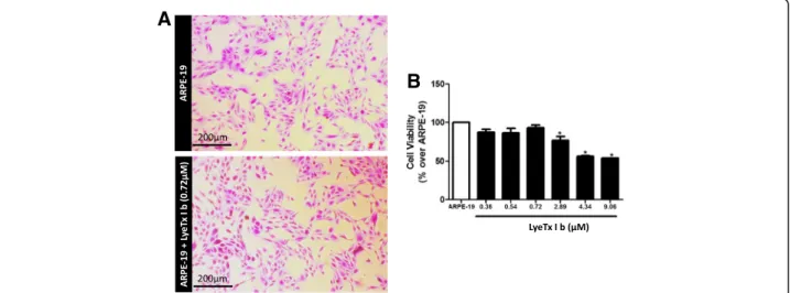
Fig. 1LyeTx I b does not alter the morphology of ARPE-19 cells and maintains cell viability above 50%.aFigure shows ARPE-19 cells in the absence or presence of LyeTx I b (2.89μM) indicating a healthy culture.bGraph shows cells not exposed (ARPE-19) or exposed to LyeTx I b (0.36, 0.54, 0.72,
2.89, 4.34 or 9.06μM). Data represent the means ± SEM of three independent experiments. * indicates significant difference as compared to ARPE-19
multiple comparison testing for ARPE-19 cells and CAM assay experiments.
Results
LyeTx Ib maintains viability of ARPE-19 culture above 50%
The ARPE-19 cells are involved in many ocular inflamma-tory diseases that may end in loss of vision and blindness
[18]. Based on a study of LyeTx I activity [11], different
concentrations of LyeTx I b were tested on ARPE-19 cells:
0.36; 0.54; 0.72; 2.89; 4.34 and 9.06μM. Our findings show
that in the presence of LyeTx I b, cells morphology was
not affected (Fig.1a), indicating that the cell culture was
healthy. In addition, LyeTx I b at concentrations of 2.89,
4.34 and 9.06 μM, despite promoting reductions in the
number of cells, maintained respective cell viabilities of
76.89, 56.16 and 53.94% (Fig. 1b). It can be inferred that,
within the range of concentrations tested, LyeTx I b does not present significant cytotoxic effects that would be able to drastically reduce cell viability, suggesting the safety of this peptide for ocular use.
LyeTx I b intravitreal penetrates the retina of rabbits in a short time period
Verifying the absence of in vitro toxicity of LyeTx I b, we initiated the investigation of safety of intravitreal in-jection of this peptide and its affinity for the retina of rabbits. We injected LyeTx I b conjugated with FITC to certify of the presence of this peptide on retinal layers. Fluorescence promoted by FITC indicates that after intravitreal injection, LyeTx I b progressively increased
its penetration with time, such that 2 h (Fig. 2b) < 4 h
(Fig. 2c) < 6 h (Fig. 2d) < 8 h (Fig. 2e). The arrows
Fig. 2LyeTx I b–FITC intravitreal penetrates the retina.aThe retina layers without peptide.bRetina layer 2 h after intravitreal injection of LyeTx I b -FITC (2.89μM).c4 h after intravitreal injection.d6 h after intravitreal injection.e8 h after intravitreal injection. RPE- Retinal Pigment Epithelium,
ONL-Outer nuclear layer, INL- Inner nuclear layer, GCL-Ganglion cell layer. Digital images were obtained using a microscope (Apotome.2, ZEISS, Germany) equipped for epifluorescence and a standard fluorescein filter with a 20× objective. FITC was excited at 490 nm and presented emission at 526 nm
0.54 0.72 2.17 2.89
0 10 20 30
Before injection 28 days after injection Before
injection
er
u
s
s
er
p
r
al
u
c
o
ar
t
nI
)
g
H/
m
m(
A
B
Vehicle 0.54 0.72 2.17 2.89
Vehicle 0.54 0.72 2.17 2.89
Vehicle 0.54 0.72 2.17 2.89
0 10 20 30
7 days after injection 14 days after injection 28 days after injection
er
u
s
s
er
p
r
al
u
c
o
ar
t
nI
)
g
H/
m
m(
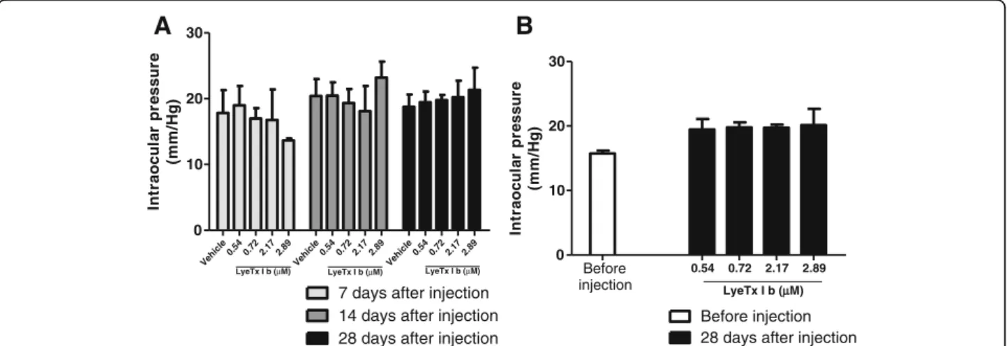
Fig. 3LyeTx I b intravitreal injection does not interfere with intraocular pressure:aGraph shows the safety of intravitreal injection of LyeTx I b (0.36, 0.54, 0.72 or 2.89μM) 28 days after injection indicating no alterations compared to control group (Before injection).bGraph shows the safety of intravitreal
injection of LyeTx I b (0.36, 0.54, 0.72 or 2.89μM) 7, 14 and 28 days after injection indicating no alterations compared to vehicle. Data represent the means
0 50 100 150 -50 0 50 100 150 Time (ms) ) V µ( e d uti l p m A
0 50 100 150
-50 0 50 100 150 200 Time (ms) A m plitud e (µ V )
0 50 100 150
-100 0 100 200 Time (ms) Am plitud e (µ V )
0 50 100 150
-50 0 50 100 150 Time (ms) ) V µ( e d uti l p m A
0 50 100 150
-100 -50 0 50 100 150 200 Time (ms) A m plitud e (µ V )
0 50 100 150
-200 -100 0 100 200 Time (ms) A m plitud e (µ V )
0 50 100 150
-50 0 50 100 150 200 Time (ms) ) V µ( e d uti l p m A
0 50 100 150
-100 0 100 200 Time (ms) Am p li tu d e ( µ V )
0 50 100 150
-200 -100 0 100 200 Time (ms) Am p li tu d e ( µ V ) Day 7 Day 14 Day 28 ROD
(0,01 cd.s/m2)
Combined Response
(3,0 cd.s/m2)
High Intensity
(10,0 cd.s/m2)
Vehicle
LyeTx I b(0.54 µM)
LyeTx I b(0.72 µM)
LyeTx I b (2.17 µM)
LyeTx I b (2.89 µM)
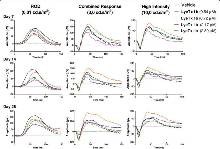
Fig. 4Representative ERG recordings of rabbit’s eye injected with different doses of LyeTx I b at different time points darkness-adapted (0.01, 3.0 and 10 cd.s/m2)
7 days
0 50 100 150 -50 0 50 100 150 Time (ms) ) V µ( e d uti l p m A 14 days
0 50 100 150 -50 0 50 100 150 Time (ms) Am plit ude ( µ V) 28 days
0 50 100 150
-50 0 50 100 150 Time (ms) Am pl it ude ( µ V) 7 days
0 50 100 150 -50 0 50 100 Time (ms) ) V µ( e d uti l p m A 14 days
0 50 100 150 -50 0 50 100 Time (ms) Am plit ude ( µ V ) 28 days
0 50 100 150 -50 0 50 100 Time (ms) Am plit ude ( µ V )
A
Light-adapted state (stimulus of 3 cd.s/m
2)
B
Light-adapted flicker (stimulus of 3 cd.s/m
2at 30 Hz)
Vehicle
LyeTx I b(0.72 µM)
LyeTx I b(2.17 µM)
LyeTx I b(2.89 µM)
LyeTx I b(0.54 µM)
7 days 14 days 28 days 0
50 100 150 200 250
)
V
µ(
e
d
uti
l
p
m
A
7 days 14 days 28 days
40 50 60 70 80 90
Implicit time (ms)
Vehicle
LyeTx I b(0.54 µM)
LyeTx I b(0.72 µM)
LyeTx I b (2.17 µM)
LyeTx I b(2.89 µM)
A
B
7 days 14 days 28 days 0
100 200 300 400
)
V
µ(
e
d
uti
l
p
m
A
7 days 14 days 28 days 0
20 40 60 80
Implicit time (ms)
7 days 14 days 28 days -150
-100 -50 0
)
V
µ(
e
d
uti
l
p
m
A
7 days 14 days 28 days 10
12 14 16 18
Implicit time (ms)
C
D
E
F
7 days 14 days 28 days 0
100 200 300 400
)
V
µ(
e
d
uti
l
p
m
A
7 days 14 days 28 days
0 20 40 60 80
Implicit time (ms)
7 days 14 days 28 days
-200 -150 -100 -50 0
)
V
µ(
e
d
uti
l
p
m
A
7 days 14 days 28 days
10 12 14 16 18
Implicit time (ms)
G
H
I
J
indicates the increasing of the fluorescence mainly in the Retinal Pigment Epithelium (RPE).
LyeTx I b is safe for intravitreal administration
The safety of retinal application of LyeTx I b could be ob-served by tonometer evaluation. We obob-served that when LyeTx I b was injected for 7, 14 or 28 days at the
concen-trations 0.54; 0.72; 2.17 or 2.89μM, the intravitreal
injec-tion did not affect intraocular pressure of the rabbits
(Fig.3a). Furthermore, we observed that LyeTx I b did not
alter the intraocular pressure after the procedure (Fig.3b).
LyeTx I b does not compromise visual acuity
Darkness and light-adapted representative ERG records obtained at 7, 14, and 28 days after the injection of
intra-vitreal LyeTx I b at doses of 0.54, 0.72, 2.17 and 2.89μM
are shown in Figs. 4 and 5, respectively. Amplitude and
implicit time are displayed in Fig 6. Our findings
indi-cate that the group injected with LyeTx I b 0.72 μM
showed a lower b-wave amplitude in the darkness-adapted rod mediated response 28 days after intravitreal
injection (Fig.5a) compared with the controls. No
statis-tically significant differences were found between vehicle values and post-injection values on days 7, 14, and 28 at
other LyeTx I b doses tested for the amplitude and the implicit wave time (which represents photoreceptor function) or b wave implicit time in the ERG response to single flash white light. We observed in the group
injected with LyeTx I b at 0.54 μM an increase in their
light adapted b-wave amplitude response to the
single-flash white light and to the 30 Hz flickering white light compared with the vehicle 28 days after intravitreal injection. At all other injected concentrations, no differ-ences were observed between the ERG responses of the experimental and control eyes in the light-adapted
con-dition. LyeTx I b 0.54 μM treatment caused an
increas-ing of b-wave amplitude in the darkness-adapted combined responses from photoreceptors and bipolar
cells (Fig.6e) and in the darkness-adapted high intensity
response (Fig. 6i) compared to the vehicle-injected
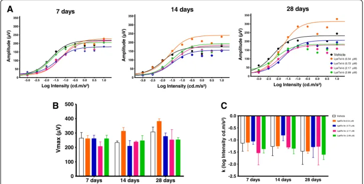
rab-bits 28 days after intravitreal injection. Naka–Rushton
parameters (Vmax: maximal b-wave amplitude and k:
semi-saturation constant) for each dose of LyeTx I b and time point were obtained from b-wave amplitude versus flash intensity curves in the darkness-adapted state
(Fig.7). We did not observe differences in b-wave
ampli-tude versus flash intensity curves in the
darkness-adapted state,Vmaxork.
(See figure on previous page.)
Fig. 6ERG darkness-adapted b-wave amplitude variation (a) and implicit time (b) in the experimental eyes with a stimulus of 0.01 cd.s/m2. ERG darkness-adapted a-wave amplitude variation (c), a-wave implicit time (d), a-wave amplitude variation (e) and b-wave implicit time (f) in the experimental eyes with a stimulus of 3 cd.s/m2. ERG darkness-adapted a-wave amplitude variation (g), a-wave implicit time (h), a-wave amplitude variation (i) and b-wave implicit time (j) in the experimental eyes with a stimulus of 10 cd.s/m2
7 days
-3.0 -2.5 -2.0 -1.5 -1.0 -0.5 0.0 0.5 1.0 0
50 100 150 200 250 300 350
Log Intensity (cd.m/s²)
)
V
µ(
e
d
uti
l
p
m
A
14 days
-3.0 -2.5 -2.0 -1.5 -1.0 -0.5 0.0 0.5 1.0 0
50 100 150 200 250 300 350
Log Intensity (cd.m/s²)
Amplitude (µ
V
)
28 days
-3.0 -2.5 -2.0 -1.5 -1.0 -0.5 0.0 0.5 1.0 0
50 100 150 200 250 300 350
Vehicle
LyeTxI-b (0.54 µM)
LyeTxI-b (0.72 µM)
LyeTxI-b (2.17 µM)
LyeTxI-b (2.89 µM)
Log Intensity (cd.m/s²)
Am
p
lit
u
d
e (
µ
V
)
7 days 14 days 28 days
0 100 200 300 400 500
Vm
ax (
µ
V)
7 days 14 days 28 days -2.5
-2.0 -1.5 -1.0 -0.5
0.0 Vehicle
LyeTx I b(0.54 µM)
LyeTx I b(0.72 µM)
LyeTx I b (2.17 µM)
LyeTx I b(2.89 µM)
k (l
og
I
n
ten
s
it
y cd.
m
/s
²)
A
B
C
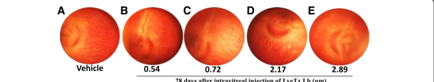
Retinal vasculature is not altered after intravitreal LyeTx I b
Eye fundus was performed after intravitreal injections of LyeTx I b at the following concentrations: 0.54; 0.72;
2.17 and 2.89 μM at 7, 14 and 28 days. We found that
LyeTx I b did not alter retinal vasculature at 7 or 14 days (data not shown) and for a long period (28 days) was safe at all concentrations studied compared to the
con-trol (Fig.8).
LyeTx I b does not alter retinal morphology integrity
Histological evaluation (Fig. 9) shows no alterations of
retinal layers, indicating that LyeTx I b is nontoxic to the retina.
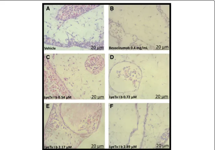
LyeTx I b prevent neovascularization on CAM model
Despite the absence of published studies on the role of the peptide in eye vascularization, it is important to in-vestigate the behavior of LyeTx I b through models com-patible with retinal vasculature. In this sense, CAM assays have been considered an appropriate model. Thus, we investigated primarily, in this traditional model of vascularization, whether CAM could present altered vascularization when exposed to LyeTx I b. Starting from the result that LyeTx I b did not affect viability of ARPE-19 cells, we tested its potential to reduce neovas-cularization. At the same concentrations used in animals the peptide was nontoxic in CAM at 0.54, 0.72, 2.17 and
2.89μM (Fig.10a, b, c, d, e and f, respectively).
Interest-ingly, LyeTx I b at 0.54μM promoted neovascularization
at the same levels as the vehicle (Fig. 10g); however, the
opposite effect was produced by the other
concentra-tions, where LyeTx I b at 0.72 μM, 2.17 μM and
2.89 μM was able to prevent neovascularization
(Fig. 10g). Importantly, LyeTx I b 2.89μM did not alter
the stromal layer of CAM at 0.54μM, 0.72μM, 2.17μM
or 2.89μM (Fig. 11c, d, e, and f respectively) compared
to vehicle (Fig.11a), indicating the peptide was not toxic.
Otherwise, the peptide was as effective at reducing
neo-vascularization as Bevacizumab (0.4 mg/mL) (Fig. 11b)
at a concentration one thousand times lower (Fig. 11f )
preventing 50% of neovascularization without promoting
toxicity to the embryo, thus indicating a safe LyeTx I b concentration for this purpose.
Discussion
Retinal pigment epithelial (RPE) cells are central to retinal health and homoeostasis. RPE damage occurs as part of the pathogenesis of age-related macular degeneration and
neo-vascular retinopathies [19]. In this study, we investigated
the safety of different doses of LyeTx I b, a synthetic peptide
derived from a toxin isolated from the venom of L.
ery-thrognatha, in the rabbit vitreous, until 28 days after injec-tion. Due to the absence of studies investigating the effects of this synthetic peptide on the eye, especially in retinal cells, we aimed to analyze the impact of LyeTx I b on the viability of ARPE-19 cells. LyeTx I b was active against
dif-ferent types of bacteria such asE. coli, whose minimum
in-hibitory concentration (MIC) was 0.71 μM, and against
different types of fungal such as Candida lusitanea
11.52μM of MIC [14] . To the best of our knowledge, the
safety of intraocular injection of LyeTx I b has never been reported. Our study was designed to investigate possible toxic effects of LyeTx I b on the retina, taking into account that ARPE-19 cells did not exhibit apparent modifications on morphology when exposed to all concentrations of this
peptide (Fig.1a), demonstrating the preservation of healthy
culture. Furthermore, ARPE-19 cells treated with LyeTx I b presented the same viability levels as untreated ARPE-19.
Importantly, the cell viability starts to decrease at 2.89μM
of the peptide, but those concentrations were able to keep
cell viability above 50% (Fig.1b).
We evaluated the penetration efficiency of the
FITC-labeled with LyeTx I b after intravitreal injection. It has been demonstrated that FITC is able to perfuse the
ret-ina [20]. In our study, retinal permeability, measured by the
fluorescence of LyeTx I b–FITC, at 2 h, 4 h, 6 h and 8 h
after injection (Fig.2), demonstrated that intravitreal
injec-tion was successful and LyeTx I b penetrates progressively
the retinal layers in this time range. (Fig.2, b, c, d and e).
These data are very important despite the absence of stud-ies assessing the capacity of LyeTx I b - FITC to penetrate eye structures. In addition, we observed that even after 8 h of treatment this peptide did not spread to other layers of
A
B
C
D
E
the retina, suggesting that LyeTx I b shows higher specifi-city for receptors in the RPE region.
Besides evaluating the efficiency of intravitreal injec-tion, we examined the effects of LyeTx I b on ocular pressure at 7 days, 14 days and 28 days after intravitreal injection. Our results demonstrated no alterations on ocular pressure at 7, 14 or 28 days after treatment with
the peptide, compared to vehicle (Fig.3a). In addition to
that, we verified the safety of intravitreal injection of LyeTx I b 28 days after, comparing it to eyes before
in-jection (Fig.3b). We did not observe alterations in
intra-ocular pressure.
Furthermore, we decided to evaluate visual impair-ment of intravitreal through eletroretinography for 7, 14 or 28 days. At 28 days after the LyeTx I b intravitreal in-jection, no alterations were detected. The analysis of the
b-wave amplitude variation according to the luminous stimulus intensity is a widely used method for the
func-tional evaluation of the retina [21,22].
Over the observation period the different doses of LyeTx I b did not affect overall retinal function. Nowadays, VEGF-inhibitors like bevacizumab, ranibizumab, pegapta-nib, are the first choice in therapies for the treatment of neovascular ocular diseases. Some studies demonstrated transient changes in the electroretinograms after intravitreal injection of VEGF-inhibitors, although clinical adverse
ef-fects in the adult human eye are not common [23–26].
In different animal models, a combination of electro-physiological and histological examinations has been employed to evaluate drug safety. Rabbits present rod-dominated retina due to differences in retinal anat-omy, which can explain why the ERG effects in rabbits
Vehicle 0.54 0.72 2.17 2.89 0.0
200000.0 400000.0 600000.0 800000.0
Cones and Rods External nuclear layer Internal nuclear layer Ganglionar layer
LyeTx I b (µM)
s
s
e
n
k
ci
t
r
e
y
a
L
)
sl
e
xi
p
f
o
r
e
b
m
u
n(
Fig. 9LyeTx I b does not alter the morphological integrity of the retina: Graph shows the measure of layer thickness of cones and rods, external, internal and ganglion layers 28 days after intravitreal injection of Vehicle, LyeTx I b 0.54μM, LyeTx I b 0.72μM, LyeTx I b 2.17μM or LyeTx I b
2.89μM. Data represent the means ± SEM of three independent experiments. No significant difference was observed as compared to vehicle
group (p > 0.05). Abbreviations: SEM, standard error of the mean
A
B
G
C
D
E
F
Fig. 10LyeTx I b prevents neovascularization on the CAM: Sequence of photographs illustrating the effect of (a) Vehicle, (b) Bevacizumab 0.5 mg/mL, (c) LyeTx I b 0.54μM, (d) LyeTx I b 0.72μM, (e) LyeTx I b 2.17μM and (f) LyeTx I b 2.89μM on the CAM over a 72-h period.gGraph shows the measure of
vascularization after exposure to untreated eggs (Vehicle) or treatment with LyeTx I b (0.54μM, 0.72μM, b 2.17μM or 2.89μM). Data represent the ± SEM of
number of pixels of twelve independent experiments. * indicates significant difference as compared to vehicle (p< 0.05).#indicates significant difference as
are predominantly on the rod-mediated response [27]. Furthermore, we found evidence that LyeTx I b does not alter retinal function for long periods of treatment,
ex-cept at 0.54μM as indicated by (Fig. 4) where LyeTx I b
promoted an increase of the wave times 14 and 28 days after, but interestingly, this finding is followed by the fact that cones and rods or ganglionic layers expression shows no alteration in the presence of LyeTx I b
com-pared to vehicle (Fig.9).
At the end of ERG responses of 7 days, 14 days and
28 days we evaluated the effects of LyeTx I b at 0.54μM,
0.72μM, 2.17μM and 2.89μM (Fig.8 b, c, d, and e,
re-spectively) in the eye fundus through Clear View®
assem-bly to vehicle (Fig.8a). Our findings indicate that LyeTx
I b did not alter retinal vascularization at 7 or 14 days after (data not shown) whereas, importantly, 28 days after, LyeTx I b remains nontoxic to the retina assembly to vehicle.
It has been proposed that spider venoms are able to
ameliorate retinal injury [28]. Despite the absence of
previous reports about the effect of LyeTx I b on the
eye, the results presented herein demonstrate that this peptide is biocompatible with the ocular system. Never-theless, our data indicate that LyeTx I b is likely to be implicated in the decreased vascularization observed in
CAM above 0.72μM (Fig.10g).
However, our data demonstrate for the first time that LyeTx I b is effective by itself at reducing neovasculariza-tion, at concentrations a thousand times lower than Bev-acizumab, the treatment of reference, In addition, LyeTx I b did not promote inflammatory reaction in CAM
(Fig. 10c, d, e and f ). It is important to note that we
ex-amined the interaction between LyeTx I b and neovascu-larization in a native organism, without an installed disease. Besides that, LyeTx I b was administered in a
single I.V. application, whereas Freitas et al (2013) [29]
performed a clinical study using multiple intravitreal in-jections, during which all eyes developed cataract and one patient developed vitritis. One eye had mild persist-ent submacular fluid without active choroidal neovascu-larization, whereas the other eye had a persistent amount of intraretinal fluid due to active choroidal
A
B
C
D
E
F
Fig. 11LyeTx I b prevents neovascularization keeping the morphology integrity of CAM:A:Histological images illustrating CAM mesoderm after exposure to (a) Vehicle, (b) Bevacizumab 0.5 mg/mL, (c) LyeTx I b 0.54μM, (d) LyeTx I b 0.72μM, (e) LyeTx I b 2.17μM and (f) LyeTx I b 2.89μM over a 72-h period. (aandc) Vasculogenic reaction: simultaneous and co-localized vasculogenesis and hematopoiesis is observed.b,d,eandf
neovascularization. Our study of the synthetic peptide did not show the abovementioned alterations.
The treatment for retinopathies with bevacizumab
ap-pear to be good [30–33], but there are no studies
dem-onstrating their safety when injected into the eye. Moreover, we demonstrate that LyeTx I b is a safe pro-cedure useful for development of new studies focused on treatment of eye diseases that require I.V.I. for reduc-tion of retinal vascularizareduc-tion.
In conclusion, the findings of this study strongly indicate that LyeTx I b can facilitate reduction of neovasculariza-tion with a single intravitreal injecneovasculariza-tion and that, even 28 days post injection, no toxicity or morphological alter-ations of the retina were observed, up to a concentration
of 2.89μM. This finding suggests that the peptide is safe
for intraocular injection. Therefore, additional studies need to be performed to verify the long-term safety of high doses of LyeTx I b in the retina. If this peptide proves to be safe, intraocular LyeTx I b might be considered as a possible new agent for the treatment of neovascularization in ocular diseases, such as macular edema, diabetic macu-lar edema, and age-related macumacu-lar degeneration.
Abbreviations
AMD:Macular Age-Related Degeneration; CAM: Corio-allantoic membrane; ERG: Electroretinogram; FITC: Fluorescein Isothiocyanate; I.V.: Intravitreal; I.V.I.: Intravitreal injection; LyeTx I b: Synthetic peptide obtained fromLycosa erithrognathaspider venom; RPE: Retinal Pigment Epithelium; VEGF: Vascular Endothelial Growth Factor
Acknowledgements
INCT-NANOFARMA, Conselho Nacional de Desenvolvimento Científico e Tecnológico”(CNPq, Brazil), Coordenação de Aperfeiçoamento de Pessoal de Nível Superior (CAPES).
Funding
This study is part of the National Institute of Science and Technology in Pharmaceutical Nanotechnology: a transdisciplinary approach INCT-NANOFARMA, which is supported by São Paulo Research Foundation (FAPESP, Brazil) Grant #2014/50928–2, and by“Conselho Nacional de Desen-volvimento Científico e Tecnológico”(CNPq, Brazil) Grant # 465687/2014–8. Conselho Nacional de Pesquisa (CNPq), and by Coordenação de Aperfeiçoa-mento de Pessoal de Nível Superior (CAPES) Grant# 23038.000776/2017O54. Moreover, this publication was supported by the Coordination for the Im-provement of Higher Education Personnel (CAPES) through Programa Editor-ação CAPES–Edital No. 13/2016, No. do Auxílio 0722/2017, No. do Processo 88881.142062/2017–01 and from the National Council for Scientific and Technological Development (CNPq) Programa Editorial CNPq/CAPES process No. 26/2017, Proc. No. 440954/2017–7.
Availability of data and materials
The datasets used and/or analyzed during the current study are available from the corresponding author on reasonable request.
Authors’contributions
FRS designed the study and conducted CAM (Chorio-allantoic Membrane), Tonometer, Electroretinogram recordings and Histological analysis experiments; LFND and ROS performed Tonometer, Eye Fundus, Electroretinogram recordings and Histological slices; LFND contributed analysis and interpretation of data; CSN made substantial contributions to acquisition, analysis and interpretation of FITC experiment and critical revision of the manuscript for scientific and revised the manuscript critically for important intellectual content; Intravitreal injections were performed by BLC and contributed with Electroretinogram, eye fundus and critical review;
Cell culture experiment was conducted by CRT; MELP contributed peptide synthesis and made substantial and critical revision of the manuscript for scientific content and revised the manuscript critically for important intellectual content; ASCJ coordinated the study, provided the necessary infrastructure for the experiments and made substantial and critical revision of the manuscript for scientific content and revised the manuscript critically for important intellectual content. Results were analyzed by FRS, MBP, CRT and CSN. The article was written by FRS and all other authors revised the data and discussed the manuscript. All authors read and approved the final manuscript.
Ethics approval
The study was approved by the Committee for Ethics in the Use of Animals (CEUA, Belo Horizonte, Brazil, Protocol n° 298/2017). All the experiments were conducted in accordance with the Association for Research in Vision and Ophthalmology (ARVO).
Consent for publication
Not applicable.
Competing interests
The authors declare that they have no competing interests.
Publisher’s Note
Springer Nature remains neutral with regard to jurisdictional claims in published maps and institutional affiliations.
Author details
1Faculdade de Farmácia, Universidade Federal de Minas Gerais, Av Antônio Carlos, 6627, 2nd Floor, Room 2031, Pampulha, Belo Horizonte, Minas Gerais 31270-901, Brazil.2Departamento de Bioquímica e Imunologia, Instituto de Ciências Biológicas, Universidade Federal de Minas Gerais, Belo Horizonte 31270-901, Brazil.
Received: 7 May 2018 Accepted: 31 October 2018
References
1. Bharadwaj AS, Appukuttan B, Wilmarth PA, Pan Y, Stempel AJ, Chipps TJ, et al. Role of the retinal vascular endothelial cell in ocular disease. Prog Retin Eye Res. 2013;32:102–80.
2. Kempen JH, O'Colmain BJ, Leske MC, Haffner SM, Klein R, Moss SE, et al. The prevalence of diabetic retinopathy among adults in the United States. Arch Ophthalmol. 2004;122(4):552–63.
3. Kunkel JM, Hawksley CA. Cysticercosis presenting as a solitary dominant breast mass. Hum Pathol. 1987;18(11):1190–1.
4. Cheung N, Mitchell P, Wong TY. Diabetic retinopathy. Lancet. 2010; 376(9735):124–36.
5. Lim LS, Mitchell P, Seddon JM, Holz FG, Wong TY. Age-related macular degeneration. Lancet. 2012;379(9827):1728–38.
6. Laude A, Cackett PD, Vithana EN, Yeo IY, Wong D, Koh AH, et al. Polypoidal choroidal vasculopathy and neovascular age-related macular degeneration: same or different disease? Prog Retin Eye Res. 2010;29(1):19–29. 7. Sasaki M, Kato Y, Fujinami K, Hirakata T, Tsunoda K, Watanabe K, et al.
Advanced quantitative analysis of the sub-retinal pigment epithelial space in recurrent neovascular age-related macular degeneration. PLoS One. 2017; 12(11):e0186955.
8. Campbell JP, Bressler SB, Bressler NM. Impact of availability of anti-vascular endothelial growth factor therapy on visual impairment and blindness due to neovascular age-related macular degeneration. Arch Ophthalmol. 2012; 130(6):794–5.
9. Monge-Fuentes V, Gomes FMM, Campos GAA, Silva JC, Biolchi AM, dos Anjos LC, et al. Neuroactive compounds obtained from arthropod venoms as new therapeutic platforms for the treatment of neurological disorders. J Venom Anim Toxins incl Trop Dis. 2015;21:31.https://doi.org/10.1186/ s40409-015-0031-x.
11. Santos DM, Verly RM, Piló-Veloso D, de Maria M, de Carvalho MA, Cisalpino PS, et al. LyeTx I, a potent antimicrobial peptide from the venom of the spiderLycosa erythrognatha. Amino Acids. 2010;39(1):135–44.
12. Consuegra J, de Lima ME, Santos D, Sinisterra RD, Cortés ME. Peptides: beta-cyclodextrin inclusion compounds as highly effective antimicrobial and anti-epithelial proliferation agents. J Periodontol. 2013;84(12):1858–68. 13. Cruz Olivo EA, Santos D, de Lima ME, Dos Santos VL, Sinisterra RD, Cortes
ME. Antibacterial effect of synthetic peptide LyeTxI and LyeTxI/beta-Cyclodextrin association compound against planktonic and multispecies biofilms of periodontal pathogens. J Periodontol. 2017;88(6):e88–96. 14. Reis PVM, Boff D, Verly RM, Melo-Braga MN, Cortes ME, Santos DM, et al.
LyeTxI-b, a synthetic peptide derived fromLycosa erythrognathaspider venom, shows potent antibiotic activityin vitroandin vivo. Front Microbiol. 2018;9:667.
15. Agostini RM, do Nascimento Pinheiro AC, Binda NS, Romano Silva MA, do Nascimento Cordeiro M, Richardson M, et al. Phoneutria spider toxins block ischemia-induced glutamate release and neuronal death of cell layers of the retina. Retina. 2011;31(7):1392–9.
16. McCulloch DL, Marmor MF, Brigell MG, Hamilton R, Holder GE, Tzekov R, et al. ISCEV standard for full-field clinical electroretinography (2015 update). Doc Ophthalmol. 2015;130(1):1–12.
17. Kalweit S, Besoke R, Gerner I, Spielmann H. A national validation project of alternative methods to the Draize rabbit eye test. Toxicol in Vitro. 1990;4(4–5):702–6.
18. Mateos MV, Kamerbeek CB, Giusto NM, Salvador GA. The phospholipase D pathway mediates the inflammatory response of the retinal pigment epithelium. Int J Biochem Cell Biol. 2014;55:119–28.
19. Gong X, Draper CS, Allison GS, Marisiddaiah R, Rubin LP. Effects of the macular carotenoid lutein in human retinal pigment epithelial cells. Antioxidants (Basel). 2017;6(4):1–13.
20. Van Bergen T, Hu TT, Etienne I, Reyns GE, Moons L, Feyen JHM. Neutralization of placental growth factor as a novel treatment option in diabetic retinopathy. Exp Eye Res. 2017;165:136–50.
21. Damico FM, Scolari MR, Ioshimoto GL, Takahashi BS, Cunha AS Jr, Fialho SL, et al. Vitreous pharmacokinetics and electroretinographic findings after intravitreal injection of acyclovir in rabbits. Clinics (São Paulo). 2012;67(8):931–7.
22. Gasparin F, Aguiar RG, Ioshimoto GL, Silva-Cunha A, Fialho SL, Liber AM, et al. Pharmacokinetics, electrophysiological, and morphological effects of the intravitreal injection of mycophenolic acid in rabbits. J Ocul Pharmacol Ther. 2014;30(6):502–11.
23. Lipski A, Bornfeld N, Jurklies B. Multifocal electroretinography in patients with exudative amd and intravitreal treatment with pegaptanib sodium. Retina. 2007;27(7):864–72.
24. Maier M, Feucht N, Lanzl I, Kook P, Lohmann CP. Retinochoroidopathy after intravitreal anti-VEGF treatment. Ophthalmologe. 2009;106(8):729–34 [Article in German].
25. Stahl A, Feltgen N, Fuchs A, Bach M. Electrophysiological evaluation of retinal photoreceptor function after repeated bevacizumab injections. Doc Ophthalmol. 2009;118(2):81–8.
26. Pedersen KB, Moller F, Sjolie AK, Andréasson S. Electrophysiological assessment of retinal function during 6 months of bevacizumab treatment in neovascular age-related macular degeneration. Retina. 2010;30(7):1025–33.
27. Myers AC, Lövestam Adrian M, Bruun A, Ghosh F, Andreasson S, Ponjavic V. Retinal function and morphology in rabbit after intravitreal injection of VEGF inhibitors. Curr Eye Res. 2012;37(5):399–407.
28. Binda NS, Carayon CP, Agostini RM, Pinheiro AC, Cordeiro MN, Silva MA, et al. PhTx3–4, a spider toxin calcium channel blocker, reduces NMDA-induced injury of the retina. Toxins (Basel). 2016;8(3):1–14.
29. Freitas LG, Isaac DL, Tannure WT, Gabriel LA, Reis RG, Rassi AR, et al. Intravitreal bevacizumab combined with infliximab in the treatment of choroidal neovascularization secondary to age-related macular degeneration: case report series. Arq Bras Oftalmol. 2013;76(3):180–4. 30. Costa RA, Jorge R, Calucci D, Cardillo JA, Melo LA Jr, Scott IU. Intravitreal
bevacizumab for choroidal neovascularization caused by AMD (IBeNA study): results of a phase 1 dose-escalation study. Invest Ophthalmol Vis Sci. 2006;47(10):4569–78.
31. Rich RM, Rosenfeld PJ, Puliafito CA, Dubovy SR, Davis JL, Flynn HW Jr, et al. Short-term safety and efficacy of intravitreal bevacizumab (Avastin) for neovascular age-related macular degeneration. Retina. 2006;26(5):495–511.
32. Spaide RF, Laud K, Fine HF, Klancnik JM Jr, Meyerle CB, Yannuzzi LA, et al. Intravitreal bevacizumab treatment of choroidal neovascularization secondary to age-related macular degeneration. Retina. 2006;26(4):383–90. 33. Lazic R, Gabric N, Dekaris I, Gavric M, Bosnar D. Photodynamic therapy