*Corresponding author: Esmaeil Safinezhad.
Cornea Research Center, Khatam Al-Anbia Eye Hospital, Mashhad University of Medical Sciences, Mashhad, Iran.
E-mail: [email protected] Tel: 09173750423
This is an Open Access article distributed under the terms of the Creative Commons Attribution License (http://creativecommons. org/licenses/by/3.0), which permits unrestricted use, distribution, and reproduction in any medium, provided the original work is properly cited.
The effect of intravitreal bevacizumab injection on the corneal
endothelial cells : systematic review
Akbar Derakhshan (MD), Alireza Eslampour (MD), Esmaeil Safinezhad (MD)*, Samira Hasanzadeh (MD)
1Cornea Research Center, Department of Ophthalmology, Khatam Al-Anbia Eye Hospital, Mashhad University of Medical Sciences, Mashhad, Iran.
ARTICLE INFO ABSTRACT
Article type
Systematic review article
Article history
Received: 11 Mar 2015 Revised: 10 Apr 2015 Accepted: 15 Apr 2015
Keywords
Avastin Endothelial Cells Eye Diseases
Introduction:Bevacizumab (Avastin), as an effectiveness treatment modality, is
currently used in patients with various ocular disease. However the results have been promising, the use of bevacizumab in the treatment of ocular disease is an off-label application. Hence, the aim of this study was to systematically review the effectiveness of intravitreal injection of bevacizumab on various ocular tissues, especially corneal endothelial cells.
Methods: The articles related to the effect of application of Avastin in the treatment
of ophthalmic diseases and especially its effect on corneal endothelial cells were collected and reviewed. We searched PubMed, Google scholar, and Scopus databases and used Avastin, ocular diseases and corneal endothelial cells as search keywords.
Result: Of all 55 articles found in all databases, only 10 were relevant to the purpose
of this study, and 45 articles were excluded in several step by step process of article selection according to the inclusion/exclusion criteria. The results revealed that intracameral bevacizumab injection caused no changes in specular microscopy and corneal pachymetry. Moreover, it had no significant toxicity on corneal endothelial cells.
Discussion: Effectiveness of bevacizumab as a new modality in the treatment of
different ophthalmic diseases have been suggested. Recent data on both human and animal models showed that intravitreal injection of bevacizumab resulted in no significant toxicity on various ocular cells, and it could be considered as a suitable therapeutic approach in clinical use.
Conclusion: According to the results of included documents, bevacizumab was not
toxic to corneal endothelial cells at various clinically relevant doses.
Please cite this paper as:
Derakhshan A, Eslampour A, Safinezhad E, Hasanzadeh S. The effect of intravitreal bevacizumab injection on the corneal endothelial cells. Rev Clin Med. 2016;3(2):78-83.
Introduction
Bevacizumab with the trade name of Avastin is a humanized monoclonal antibody with various ther
-apeutic applications. Bevacizumab is mostly known
as an anti-angiogenesis agent, which inhibits the
growth and the development of new blood vessels. Its anti-angiogenesis property is thought to be medi
-ated through inhibiting vascular endothelial growth factor A (VEGF-A) (1). Hence, the most acceptable
mechanistic pathway of bevacizumab antitumor
ac-tivity is the binding to VEGF and inhibiting its signal pathway (2). The relative efficiency of bevacizum
-ab in the treatment of several tumor and non-tu
-mor diseases has been confirmed. Food and Drug Administration (FDA) of the United States has ap
included document was screened to include other additional suitable and related documents.
Study selection and inclusion/exclusion criteria No time limitation was defined for the includ
-ed articles, but only articles in English language were selected and used for further data extraction to avoid any possible errors and misinterpreta
-tion during the whole process of data extrac-tion. Different types of the articles with various study designs including case-controls, case reports, clin
-ical trials, cross-sectional, and prospective cohort studies were included in this review study. Howev
-er, conference papers, letters, review articles, and meta-analysis were excluded from further assess
-ment. Inclusion criteria were all the documents in which the efficacy of intravitreal bevacizumab in
-jection on corneal endothelium had been evaluat
-ed. Duplicated documents and papers with subject or language irrelevancy were excluded by review
-ing the title, keywords, and abstract in the first step of article selection. Studies on animals or in vitro studies were excluded from further assessment.
Data extraction
Data including publication date, study design, country of origin, name of the first author, method of assessment, and critical findings were extract
-ed and systematically sort-ed bas-ed on the main purpose of this study. All existing data including total number of patients and demographic data of studied individuals were collected in regard to the previously defined inclusion and exclusion criteria. Data were systematized based on the re
-sults of selected studies reporting the effects and
therapeutic value of intravitreal bevacizumab
in-jection on corneal endothelium. We extracted the data reporting the qualitative, quantitative, and morphological changes of ocular endothelium after administration of different doses of beva
-cizumab. All processes including data extraction and selection of the articles were performed ac
-cording to the recommendation of PRISMA 2009 checklist (11).
Results
Study search results
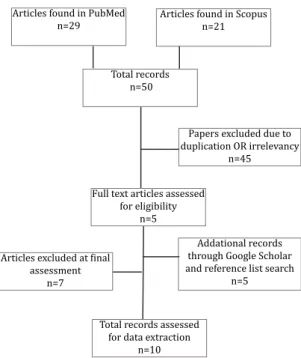
From total of 29 articles found in PubMed, and 21 records in Scopus search, we omitted 25 articles in the first step by reviewing the abstract of articles due to irrelevancy. Moreover, 20 duplicated docu -ments or articles with language irrelevancy were
excluded in subsequent process of article selec
-tion, and only 5 unique records seemed related to the main subject of this study. Two additional doc
-uments were also included by manual reference different types of cancers and eye diseases. In 2004,
FDA approved Avastin as an effective medication
for the treatment of metastatic colorectal cancer in combination with some other conventional
chemo-therapeutic agents (3). Soon after, and during 2006 to 2010, it was the first-line clinical management for
the treatment of other types of malignancies such as
lung, breast, brain, and renal cancers (4).
Bevacizumab is now widely used for treating several ophthalmic complications including retinal neovascularization, macular edema, neovascular glaucoma, and other ocular problems (5-7). More
-over, it is demonstrated that intravitreous injection
of bevacizumab is an effective therapy in patients
with intraocular neovascular complications and macular edema (8). However the results reported
for the clinical applications are promising, the use
of Avastin for therapeutic purpose in ocular diseas
-es is an off-label application. Therefore, it is of great importance to reevaluate its efficacy and safety in ocular tissues (2). Several studies have shown that bevacizumab at therapeutic quantities that is typ
-ically used for treating retinal disorders, had no significant toxicity to the retinal pigment epithelial (RPE) cells, retinal ganglion cells, or corneal endo
-thelial cells (2,7,9). Since VEGF and its receptors (Flt-1 and Flk-1) are highly expressed in inflamed and vascularized corneal tissues, and therefore they may have crucial role in corneal neovascularization. Bevacizumab is suggested as possible therapeutic
agent for the treatment of corneal
neovasculariza-tion, thus it is recommended to evaluate the effica
-cy and potentially -cytotoxicity of bevacizumab on corneal endothelial cells (2,9,10). According to the results in databases, we did not find any meta-anal -ysis or systematic review article about the effect of
Avastin injection on corneal endothelial cells. In this study, we aim to systematically review various clin
-ical studies to evaluate the effect of intravitreal bev
-acizumab injection on the corneal endothelial cells.
Methods
Search methodsWe studied the articles related to the application of Avastin in the treatment of ophthalmic diseases and specially its effect on corneal endothelial cells. We searched PubMed and Scopus databases using the terms: Avastin (bevacizumab), ocular diseases and corneal endothelial cells. Related articles with
the following search term “(Intravitreal
screening of the previously included articles. Three unique records were also included by searching Google Scholar. Finally, according to the previously defined inclusion/exclusion criteria, and after com
-prehensive reviewing of the included articles, only 10 relevant papers, which fully met the inclusion criteria, were selected and the desired data were carefully extracted based on the results reporting
the effects of bevacizumab injection on corneal
en-dothelium. Figure 1 shows the step by step process of literature search and study selection.
Figure 1. Flowchart for selection of studies
Articles found in Scopus n=21
Total records n=50
Full text articles assessed
for eligibility
n=5
Articles excluded at final
assessment n=7
Total records assessed for data extraction
n=10 Articles found in PubMed
n=29
Addational records through Google Scholar and reference list search
n=5 Papers excluded due to duplication OR irrelevancy
n=45
General characteristics of the included articles The total number of patients were 1375, partic
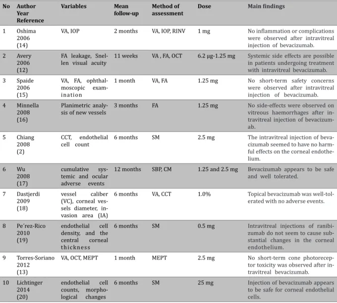
-ipated in the selected studies in which the effects of intravitreal bevacizumab injection had been evaluated on corneal endothelium. Mean age of the studied patients was 56.8 years, but it should be noted that the age of studied patients had not been mentioned in two studies (12,13). Minimum and maximum ages of all participants were 21 and 89 years, respectively. Of these patients, 565 were males and 778 were females. In one study with 32 studied patients, the sex ratio was unknown (12). The duration of the studies varied from 1 month to 1 year. Different doses of Avastin ranging from 0.5 mg to 25 mg were used for the treatment of different ocular diseases. In the selected articles, different methods including specular microscopy images, central corneal thickness (CCT) measuring,
multifocal electrophysiologic testing, intraocular
pressure (IOP) changes, the mean diameter of the corneal vessels, and endothelial cell counting were used to assess the effects of intravitreal injection
of bevacizumab on corneal endothelium. In Table 1, general characteristics of included studies are summarized.
Study results
Data showed complete safety for intravitreal bevacizumab injection in the corneal endothelium. In addition, according to the results of included studies, injection of different doses of bevacizumab seemed to have no destructive effects on the corneal endothelium. Up to 12-month follow-up in patients with various ocular diseases such as diabetic reti
-nopathy and macular degeneration who had been treated with intravitreal injection of bevacizumab showed no significant changes in the corneal en
-dothelium after bevacizumab injection (17). Table 2 shows the main clinical outcomes of treatment with different doses of Avastin. According to the results of included studies, bevacizumab injection does not cause ocular toxicity, especially on corneal endothelial cells. But, the results of a study that had been conducted on 45 eyes of 32 patients showed
that treatment with intravitreal bevacizumab may
possibly cause systemic side effects (12).
Discussion
So far, several clinical trials have been conducted
on the effect of Avastin injection in the treatment
of ophthalmic diseases (21,22). The results of some studies showed that bevacizumab did not cause considerable cytotoxicity on human retinal pigment epithelium, rat retinal ganglions, and pig choroidal endothelial cells (7,23). In addition, majority of experimental findings supported the efficacy and safety of intravitreal bevacizumab injection at different doses. However, some studies demonstrated that bevacizumab might have adverse effects on the proliferation of primary retinal epithelial cells and cell viability at higher concentration of around 2,500 microg/ ml (24). Moreover, findings were suggestive of clear anti-angiogenic activity and anti-fibrotic effects of bevacizumab after corneal burn (25). Pharmacological and toxicological assessment of the topical bevacizumab showed that topical administration of bevacizumab has no adverse effects on epithelium and endothelium if used in the treatment of corneal neovascularization (17).
Therapeutic potency of bevacizumab has also been
evaluated in iris rubeosis (12,26). The results of these studies demonstrated that no inflammation or relapse occurred within the short-term follow-up of follow-up to four weeks.
endothelial cells, and human corneal cells (23,24,31). Furthermore, studies suggested that bevacizumab could be considered as a safe and
alternative therapeutic strategy for iris rubeosis
in neovascular glaucoma. These findings were supported with several studies conducted on both human and animals. Because there are some limited documents reporting the adverse effects
of intravitreal bevacizumab injection on corneal
endothelial cells, the efficacy and therapeutic value of Avastin is suggested to be evaluated in a well-designed clinical trials.
Conclusion
In sum, the results of this systematic literature
review showed that the use of bevacizumab is
Table 1. General characteristics of the included articles.
No Author Year Reference
Country Number of patients
Sex ratio Age (year) (Mean) Study design *
1 Oshima
2006 (14)
Japan 5 Male: 3
Female: 2 55.8 (48.6-72) NCICS
2 Avery
2006 (12)
USA 32 NR © NR RCS
3 Spaide 2006 (15)
USA 2 Male: 1
Female: 1 68 and 38 CR
4 Minnella
2008 (16)
Italy 10 Male: 6
Female: 4 49.6 (33-65) PCS
5 Chiang 2008 (2)
Taiwan 50 Male: 37
Female: 13 68 (42-80) PCS
6 Wu 2008 (17)
Costa Rica ® 1173 Male: 470
Female: 703 54 (41-89) RUICS
7 Dastjerdi
2009 (18)
USA 10 Male: 4
Female: 6 46.7 (23-71) PNC
8 Pe´rez-Rico
2010 (19)
Spain 52 Male: 29
Female: 23 70.5 (61–80) PCS
9 Torres-Soriano
2012 (13)
Mexico 31 Male: 12
Female: 19
NR PIUCS
10 Lichtinger
2014 (20)
Canada 10 Male: 3
Female: 7 46 (21–85) CR
* NCICS: Non-comparative, interventional case series, RCS: Retrospective case series, CR: Case Report, PCS: Prospective case series, RUICS: Retrospective, uncontrolled interventional case series, PNC: Prospective, non-comparative, PIUCS: Prospective, interventional,
uncontrolled, clinical study. ® This study is conducted as a joint research in 7 countries including Mexico, Venezuela, Puerto Rico, Bra
-zil, Peru, Colombia and Costa Rica. © NR: Not reported.
concentrations (27). Similarly, no significant effect on cell proliferation was reported in cells treated with bevacizumab in combination with 50 ng/ml of VEGF. Morphological studies of cells showed that there was no significant changes in coroneal endothelial cells after the treatment with bevacizumab (27,28). Several studies conducted in vitro and on animal models demonstrated that bevacizumab was a safe and nontoxic agent to human fibroblast and corneal epithelial at pharmacological concentrations (22,29,30).
In vitro studies has confirmed the safety of bevacizumab in various ocular cells, including photoreceptor cells, human optic nerve head
Table 2. Variables and methods of assessment in the selected studies.
No Author Year Reference
Variables Mean follow-up
Method of assessment
Dose Main findings
1 Oshima
2006 (14)
VA, IOP 2 months VA, IOP, RINV 1 mg No inflammation or complications were observed after intravitreal injection of bevacizumab.
2 Avery
2006 (12)
FA leakage, Snel -len visual acuity
11 weeks VA , FA, OCT 6.2 µg-1.25 mg Systemic side effects are possible in patients undergoing treatment with intravitreal bevacizumab. 3 Spaide
2006 (15)
VA, FA, ophthal -moscopic exam -ination
1 month VA, FA 1.25 mg No short-term safety concerns were observed after intravitreal injection of bevacizumab.
4 Minnella
2008 (16)
Planimetric analy -sis of new vessels
3 months FA 1.25 mg No side-effects were observed on vitreous haemorrhages after in-travitreal injection of bevacizum-ab.
5 Chiang 2008 (2)
CCT, endothelial cell count
6 months SM 2.5 mg The intravitreal injection of
beva-cizumab seemed to have no harm
-ful effects on the corneal endothe
-lium. 6 Wu
2008 (17)
cumulative sys-temic and ocular adverse events
12 months SBP, CM 1.25 and 2.5 mg Bevacizumab appears to be safe and well tolerated.
7 Dastjerdi 2009 (18)
vessel caliber (VC), corneal ves -sels diameter, in -vasion area (IA)
6 months VA, CCT 1.0% Topical bevacizumab was well-tol-erated with no adverse events.
8 Pe´rez-Rico 2010 (19)
endothelial cell density, and the central corneal t h i c k n e s s
6 months SM 0.5 mg Intravitreal injections of
ranibi-zumab do not seem to cause sub
-stantial changes in the corneal endothelium.
9 Torres-Soriano
2012 (13)
VA, OCT, MEPT 1 month MEPT 2.5 mg No short-term cone
photorecep-tor toxicity was observed after in
-travitreal bevacizumab. 10 Lichtinger
2014 (20)
endothelial cell counts, morpho-logical changes
6 months SM 25 mg Injection of bevacizumab appears to be safe for corneal endothelial cells.
SM: Specular microscopy, VA: Visual acuity, FA: Fluorescein angiography, OCT: Optical coherence tomography, MEPT: Multifocal electro
-physiological testing, IOP: Intraocular pressure, RINV: Regression of iris neovascularization, SBP: Systemic blood pressure, CM: Cardiac measurement, CCT: Central corneal thickness.
a safe treatment method with no considerable toxicity to corneal endothelial cells at therapeutic doses.
Acknowledgement
We would like to thank Clinical Research Development Unit of Ghaem Hospital for their assistant in this manuscript.
Conflict of Interest
The authors declare no conflict of interest.
References
1. Los M, Roodhart JM, Voest EE. Target practice: lessons from phase III trials with bevacizumab and vatalanib in the treatment of advanced colorectal cancer. Oncologist. 2007;12:443-450.
2. hiang CC, Chen WL, Lin JM, et al. Effect of bevacizumab on human corneal endothelial cells: a six-month follow-up
study. Am J Ophthalmol. 2008;146:688-691. 3. Ellis LM. Bevacizumab. Nat Rev Drug Discov. 2005;8-9.
4. Shih T, Lindley C. Bevacizumab: an angiogenesis inhib
-itor for the treatment of solid malignancies. Clin Ther. 2006;28:1779-1802.
5. Gunther JB, Altaweel MM. Bevacizumab (Avastin) for the treatment of ocular disease. Surv Ophthalmol. 2009-;54:372-400
6. Spitzer MS, Yoeruek E, Sierra A, et al. Comparative anti
-proliferative and cytotoxic profile of bevacizumab (Avas
-tin), pegaptanib (Macugen) and ranibizumab (Lucentis)
on different ocular cells. Graefes Arch Clin Exp Ophthal
-mol. 2007;245:1837-1842.
7. Spitzer MS, Wallenfels-Thilo B, Sierra A, et al. Antiprolif
-erative and cytotoxic properties of bevacizumab on dif
-ferent ocular cells. Br J Ophthalmol. 2006;90:1316-1321.
8. Adamis AP, Shima DT. The role of vascular endotheli
-al growth factor in ocular he-alth and disease. Retina. 2005;25:111-118.
10. Zaki AA, Farid SF. Subconjunctival bevacizumab for corneal neovascularization. Acta Ophthalmol. 2010;88:868-871.
11. Liberati A, Altman DG, Tetzlaff J, et al. The PRISMA state
-ment for reporting systematic reviews and meta-analyses of studies that evaluate health care interventions: explanation and elaboration. Annals Intern Med. 2009;151:65-94.
12. Avery RL, Pearlman J, Pieramici DJ, et al. Intravitreal bev
-acizumab (Avastin) in the treatment of proliferative dia
-betic retinopathy. Ophthalmology. 2006;113:1695.1-15. 13. Torres-Soriano ME, Cubas-Lorenzo V, Garcí�a-Aguirre G, et al.
Multifocal electrophysiologic findings after intravitreal bev -acizumab (avastin) treatment. Retina. 2012;32:972-976. 14. Oshima Y, Sakaguchi H, Gomi F, et al. Regression of iris
neovascularization after intravitreal injection of bevaci-zumab in patients with proliferative diabetic retinopathy. Am J Ophthalmol. 2006;142:155-158.
15. Spaide RF, Fisher YL. Intravitreal bevacizumab (Avastin)
treatment of proliferative diabetic retinopathy complicat
-ed by vitreous hemorrhage. Retina. 2006;26:275-278. 16. Minnella AM, Savastano CM, Ziccardi L, et al. Intravitreal
bevacizumab (Avastin) treatment of proliferative diabetic retinopathy complicated by vitreous hemorrhage. Retina. 2006;26:275-278.
17. Wu L, Martí�nez-Castellanos MA, Quiroz-Mercado H, et al. Twelve-month safety of intravitreal injections of
bevaci-zumab (Avastin): results of the Pan-American Collabora
-tive Retina Study Group (PACORES). Graefes Arch Clin Exp Ophthalmol. 2008;246:81-87.
18. Dastjerdi MH, Al-Arfaj KM, Nallasamy N, et al. Topical bevacizumab in the treatment of corneal neovasculariza-tion: results of a prospective, open-label, noncomparative study. Arch Ophthalmol. 2009;127:381-389.
19. Pérez-Rico C, Bení�tez-Herreros J, Castro-Rebollo M, et al. Effect of intravitreal ranibizumab on corneal endo -thelium in age-related macular degeneration. Cornea. 2010;29:849-852.
20. Lichtinger A, Yeung SN, Kim P, et al. Corneal endothelial safety following subconjunctival and intrastromal injec -tion of bevacizumab for corneal neovasculariza-tion. Int
Ophthalmol.2014;34:597-601.
21. de Moraes CG, Facio AC, Costa JH, et al. Intracameral bev
-acizumab and mitomycin C Trabeculectomy for eyes with neovascular glaucoma: a case series. J Ocul Biol Dis Infor. 2009 31;2:40-46.
22. Shin JP, Lee JW, Sohn BJ, et al. In vivo corneal endothelial
safety of intracameral bevacizumab and effect in neovas
-cular glaucoma combined with Ahmed valve implanta
-tion. J Glaucoma. 2009;18:589-594.
23. Luthra S, Narayanan R, Marques LE, et al. Evaluation of in vitro effects of bevacizumab (avastin) on retinal pigment
epithelial, neurosensory retinal, and microvascular endo
-thelial cells. Retina. 2006;26:512-518.
24. Kernt M, Welge-Lussen U, Yu A, et al. Bevacizumab is not toxic to human anterior- and posterior-segment cultured cells. Ophthalmologe. 2007;104:965-971.
25. Yoeruek E, Ziemssen F, Henke-Fahle S, et al. Safety, pen
-etration and efficacy of topically applied bevacizumab: evaluation of eyedrops in corneal neovascularization after chemical burn. Acta Ophthalmol. 2008;86:322-328.
26. Grisanti S, Biester S, Peters S, et al. Intracameral bevaci
-zumab for iris rubeosis. Am J Ophthalmol. 2006;142:158-160.
27. Rusovici R, Sakhalkar M, Chalam KV. Evaluation of cyto
-toxicity of bevacizumab on VEGF-enriched corneal endo
-thelial cells. Mol Vis. 2011;17:3339-3346.
28. Park HY, Kim SJ, Lee HB, et al. Effect of intracameral beva
-cizumab injection on corneal endothelium in rabbits. Cor
-nea. 2008;27:1151-1155.
29. Shalam KV, Agarwal S, Brar VS, et al. Evaluation of Cytotox
-ic Effects of Bevacizumab on Human Corneal Cells. Cor
-nea. 2009;28:328-333.
30. Bayar SA, Altinors DD, Kucukerdonmez C, et al. Severe corneal changes following intravitreal injection of bevaci-zumab. Ocul Immunol Inflamm. 2010;18:268-274.
31. Maturi RK, Bleau LA, Wilson DL. Electrophysiologic find