Article
Printed in Brazil - ©2017 Sociedade Brasileira de Química0103 - 5053 $6.00+0.00
*e-mail: [email protected]
Analysis of Xanthine Oxidase Inhibitors from
Puerariae flos
Using Centrifugal
Ultrafiltration Coupled with HPLC-MS
Liangliang Liu,a Aiping Xiao,a Lei Mab and Defang Li*,a
aInstitute of Bast Fiber Crops, Chinese Academy of Agricultural Sciences, 410205 Changsha, China
bState Key Laboratory of Cotton Biology, Institute of Cotton Research of CAAS, 455000 Anyang, China
In this study, centrifugal ultrafiltration coupled with high performance liquid chromatography-mass spectrometry was utilized to screen and identify xanthine oxidase inhibitors from Puerariae flos extract. The experimental conditions of centrifugal ultrafiltration including xanthine oxidase concentration, incubation time, pH and temperature were optimized. At the optimum condition (xanthine oxidase concentration: 30.0 µg mL-1, incubation time: 20 min, pH 7.0 and temperature:
25 °C), four compounds were successfully screened from P. flos extract and identified as tectoridin, daidzin, ononin and biochanin A. The yields of tectoridin, daidzin, ononin and biochanin A were 0.231, 0.117, 0.303 and 0.089 g from 50.0 g crude P. flos samples. The inhibitory activities of these compounds were verified by xanthine oxidase inhibition assays. The experimental half maximal inhibitory concentration (IC50) values of tectoridin, daidzin, ononin and biochanin A were 88.5,
85.1, 88.8 and 87.0 µmol L-1, and the binding degree of them were 5.70, 8.28, 6.31 and 37.83%
at the optimum condition, respectively. The proposed method provided a rapid and effective way to screen and analyze active compounds from natural products.
Keywords: HPLC-MS, inhibitor, Puerariae flos, ultrafiltration, xanthine oxidase
Introduction
Natural products have been used as the most consistently successful resources of new drug discovery for a long time because of their great diversity of the chemical structures and better drug-like properties compared to the synthetic
compounds.1 However, natural products resources like
plant extracts were very complex and usually contained various kinds of components. Separation and purification of natural compounds were time-consuming and laborious processes.2 Hence, simple and effective methods aiming at
directly screening natural product extracts would be greatly helpful for drug discovery.3
Centrifugal ultrafiltration (CU) utilized centrifugal force and a semi-permeable membrane to retain suspended solids and high molecular weight solutes, while liquid and low molecular weight solutes were allowed to pass through depending on the nominal molecular weight cut-off of the membrane.4 Based on these features, active
compounds could be retained by membrane together with enzyme due to the binding with enzyme. Thus, CU became a useful technique for screening active compounds bound
to biomacromolecules such as bovine serum albumin,5
α-glucosidase,6 quinone reductase-2,7 deoxyribonucleic
acid (DNA) and liposomes.8,9 High performance liquid
chromatography-mass spectrometry (HPLC-MS or LC-MS) has been widely applied for the simultaneous separation and identification of active compounds in complex mixtures.10
The combination of CU and LC-MS (CU-LC-MS) became a powerful tool in analyzing active compounds due to its simple operation, high speed and low sample consumption. Compared with immobilized enzyme screening assay, complex synthesis procedures could be avoided in
CU-LC-MS as well.11 Moreover, the efficient screening and
identification of active constituents in natural product extracts could be accomplished by CU-LC-MS because of the high throughput screening ability, high sensitivity and selectivity for characterization of compounds at low concentrations without purification procedures.12
one of the therapeutic approaches to treat hyperuricemia by reducing or blocking the formation of uric acid. As one of the XO inhibitors used in clinical treating, Allopurinol showed many side effects such as hepatitis, nephropathy and allergic reactions.15,16 Therefore, new potential XO
inhibitors with better therapeutic activity and fewer side effects were still needed.
As a well known traditional Chinese medicine and food supplement for human health care, Puerariae flos was prepared from the dried flower of Pueraria lobata (Willd.) Ohwi. In China, P. flos was traditionally used to treat diabetes mellitus and alcoholic intoxication because of its activities including antioxidant,17 detoxification of alcohol,18
hepatoprotective19 and anticancer.20 The aqueous extract of
P. flos contained various isoflavonoids and triterpenoid
saponins possessing pharmacological activity.21 Some
studies concerning XO inhibitory effects of isoflavonoids from P.radix were reported.22 Nevertheless, systematic XO
inhibitory property researches on constituents from P. flos
were still in demand.
In this study, XO inhibitors from P. flos were screened and analyzed by CU-LC-MS. The experiment conditions including XO concentration, incubation time, temperature and pH were optimized and four compounds were identified as XO inhibitors. The results indicated this method permit rapid screening and analysis of XO inhibitors from natural products.
Experimental
Materials
Xanthine oxidase (XO; E.C. 1.17.3.2, from cow milk) was purchased from F. Hoffmann-La Roche Ltd.
(Basel, Switzerland). P. flos was purchased from Wan
Hua Cao Healthcare Products Co., Ltd. (Anhui, China). Xanthine was acquired from Sigma-Aldrich Chemicals (St. Louis, MO, USA). The ultrafiltration filter used was Nanosep MF Centrifugal filter (Pall, Ann Arbor, MI, USA), and the molecular weight cutoff was 10 kDa. The HPLC grade acetonitrile was bought from Tedia Company Inc. (Fairfield, Ohio, USA). Ultrapure water
(18.2 MΩ cm resistivity) was obtained from a Milli-Q
water purification system (Millipore, Bedford, MA, USA). All other chemicals were of analytical grade and purchased from Sinopharm Chemical Reagent Co., Ltd. (Shanghai, China). Tectoridin, daidzin, ononin and biochanin A were
isolated and characterized from Puerariae genus in our
laboratory (Figure 1). Their structures were identified by UV, MS/MS, 1D and 2D nuclear magnetic resonance (NMR) experiments. The purity of each compound was
determined to be ≥ 97% by HPLC analysis.
Preparation of crude extract
P. flos (50.0 g) was extracted three times (each for 3 h) with 90% ethanol under reflux. The combined extracts were filtrated and concentrated under reduced pressure and re-dissolved in 50 mL water (crude extract concentration
60 mg mL-1) and filtered through a 0.45 µm membrane
(Acrodisc® Syringe Filter, Pall, Ann Arbor, MI, USA). The filtrate was stored at 4 °C for further experiments.
Enzyme activity assay
The enzyme activity was measured spectro photo-metrically by continuously monitoring uric acid formation
at 295 nm with xanthine as the substrate.23 10 µL XO
solution (30 µg mL-1) and 1000 µL sample solution were
mixed. After 5 min incubation, 1000 µL xanthine solution (0.1 mg mL-1) was added and the mixtures were incubated
for an additional 3 min at 25 °C. The absorbance of mixture was measured at 295 nm using an UV-2450 UV-Vis
Spectrophotometer (Shimadzu, Kyoto, Japan). The same
mixture, with phosphate buffer instead of sample solution, was used as a control. The inhibition of XO activity was calculated according to equation 1.
Inhibition = (∆A0 – ∆A) / ∆A0 × 100% (1)
where ∆A0 is the absorbance increase of control solution
and ∆A is the absorbance increase of sample solution. The
extent of inhibition by sample solution was expressed as the concentration of sample needed to inhibit 50% of the enzymatic activity (IC50). All the assays were operated with
three replicates.
XO inhibitors screening assay
For fully interaction between the compounds and XO, 500 µL XO solution (30 µg mL-1) and 500 µL P. flos extract
(100 µg mL-1) were mixed and incubated at 25 °C for
20 min. Then, the mixture was ultrafiltrated using an Allegra 64R Centrifuge (Beckman Coulter, Brea, California, USA) with a Nanosep MF Centrifugal filter at 10,000 rpm for 10 min at room temperature. The filter was washed through centrifugation with 500 µL phosphate buffer (pH 7.0) three times to separate the unbound compounds. After washing, the bound active compounds were eluted from XO by adding 500 µL methanol-water solution (80:20, v/v), followed by centrifugation at 10,000 rpm for 15 min at room temperature. The final eluent was stored at 4 °C for analysis. The control experiment was carried out with denatured enzyme (incubated in 100 °C for 30 min).
The binding strength of compound to enzyme was defined as the binding degree, which can be calculated by equation 2.
Binding degree = (Ab – Ac) / Aa × 100% (2)
where Aa is the peak area of a compound in chromatogram of
P. flos, Ab is the peak area of a compound in chromatogram
of P. flos performing CU with XO and Ac is the peak area
of a compound in chromatogram of P. flos performing CU
with denatured XO.
HPLC-MS analysis
The HPLC analysis was carried out on an Agilent 1260 HPLC system (Agilent Technologies, Santa Clara, California, USA) which consisted of a G1311C quaternary pump equipped with on-line vacuum degasser, a G1329B auto-sampler, a G1316A column oven and a G1315D diode array detector. Chromatographic separations were
performed on a reversed phase XBrigeTM C18 column
(250 × 4.6 mm, internal diameter 5 µm, Waters, Milford, MA, USA). The mobile phase consisted of water containing 0.4% (v/v) acetic acid (A) and acetonitrile containing 0.4% acetic acid (B). A gradient elution program was used as follows: 0-5 min, 15% B; 5-15 min, 15-25% B; 15-40 min, 25-40% B. The column temperature was 25 °C and the
flow rate was maintained at 0.8 mL min-1. Spectra were
recorded from 200 to 400 nm and the chromatogram was recorded at 254 nm.
For HPLC-MS experiments, HPLC was performed on
AcquityTM UPLC system (Waters Corp., Milford, MA, USA)
with autosampler and column oven. The analysis parameters were the same as those in the above HPLC analysis. Triple quadrupole tandem mass spectrometric detection was
carried out on a Micromass® Quattro microTM API mass
spectrometer (Waters Corp., Milford, MA, USA) with an electrospray ionization (ESI) interface. The ESI source was set in negative ionization mode. The following settings
were applied to the instrument: capillary voltage, 3.00 kV; cone voltage, 40.0 V; extractor voltage, 3.00 V; source temperature, 120 °C; desolvation temperature, 400 °C; desolvation gas flow, 750 L h-1; cone gas flow, 50 L h-1, dwell
time, 0.05 s. Nitrogen was used as the desolvation and cone gas. Mass detection was performed in full scan mode for
m/z in the range 160-800. All data collected were acquired
and processes using MassLynxTM NT 4.1 software with
QuanLynxTM program (Waters Corp., Milford, MA, USA).
Results and Discussion
Optimization of HPLC analysis
To obtain the optimum HPLC analytical conditions, various mobile phase compositions with different concentrations of acetic acid, various flow rates and detection wavelengths were tested. The mixture of water containing 0.4% acetic acid (A) and acetonitrile containing 0.4% acetic acid (B) was chosen as the gradient eluting solvent system because of the acceptable separation achieved within the run time of 40 min. The column temperature was also tested between 20 and 35 °C because it would affect the chromatographic behavior, and most components achieved separation at the column temperature of 25 °C. The programmed was operated as follows, 0-5 min, 15% B; 5-15 min, 15-25% B; 15-40 min, 25-40% B. The flow rate was 0.8 mL min-1. Spectra were recorded
from 190 to 400 nm and detection wavelength was set at
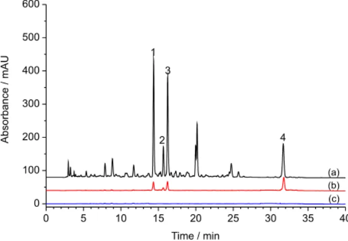
254 nm. Representative chromatogram of P. flos extract
was shown in Figure 2a.
XO inhibitors screening assay
According to XO activity and inhibition tests, the
P. flos extract showed XO inhibition with an IC50 value of
81.3 µg mL-1. The result suggested that there were compounds
with XO inhibition in the P. flos extract. Screening with denatured enzyme could exclude the nonspecific adsorption between compounds and enzyme and ensure the accuracy and authenticity of tests. Therefore, XO inhibitors screening assays from P. flos extract with active and denatured XO were
conducted. Figure 2 showed the chromatograms of P. flos
after performing CU with XO (Figure 2b) and denatured XO (Figure 2c). Compared with the chromatogram of P. flos
Optimization of screening conditions
Effect of XO concentration
Different concentrations of XO (10, 20, 30 and
40 µg mL-1) were incubated with the same amount of
P. flos extract to investigate the effect of XO concentration on binding degree. Figure 3a showed the binding degrees of four compounds incubated with different XO concentrations. The binding degrees of four compounds enhanced with the increase of XO concentration. When the XO concentration was higher than 30 µg mL-1, the binding
degrees remained unchanged and even decreased slightly. In consideration of the increase of experiment costs and the waste of XO solution, XO concentration was set as 30 µg mL-1.
Effect of incubation time
Screening experiments with different incubation times ranging from 5 to 60 min were conducted to investigate the effect on binding degree. The binding degrees of four compounds at different incubation times were calculated and shown in Figure 3b. When incubation time reached 20 min, the binding degrees of four compounds reached the highest levels. The results manifested that 20 min of incubation was sufficient for this screening experiment.
Figure 2. The chromatograms of (a) P. flos before and (b) after performing CU with XO, and (c) with denatured XO. 1: Tectoridin, 2: daidzin, 3: ononin, 4: biochanin A.
Effect of temperature
Enzyme was thermal sensitive in general. Its activity would decrease during both low and high temperature environment.24 The effect of temperature on binding degree
was investigated and the results were shown in Figure 3c. It was found that the highest binding degrees of four compounds were achieved at 25 °C. Thus, the temperature was set at 25 °C in order to ensure the activity of XO during experiments.
Effect of pH
The pH value of solution would affect the status of XO and its activity. The effect of pH on the binding degree was studied at different pH values ranging from 5.0 to 9.0. As shown in Figure 3d, the maximum binding degrees of four compounds were obtained at pH 7.0. As reported in the literatures, XO also showed optimum activity at pH 7.0 and the pH value was set at 7.0 in these experiments, which was in accordance with experiment results.25,26 Therefore,
the pH value was set at 7.0.
Structural identification
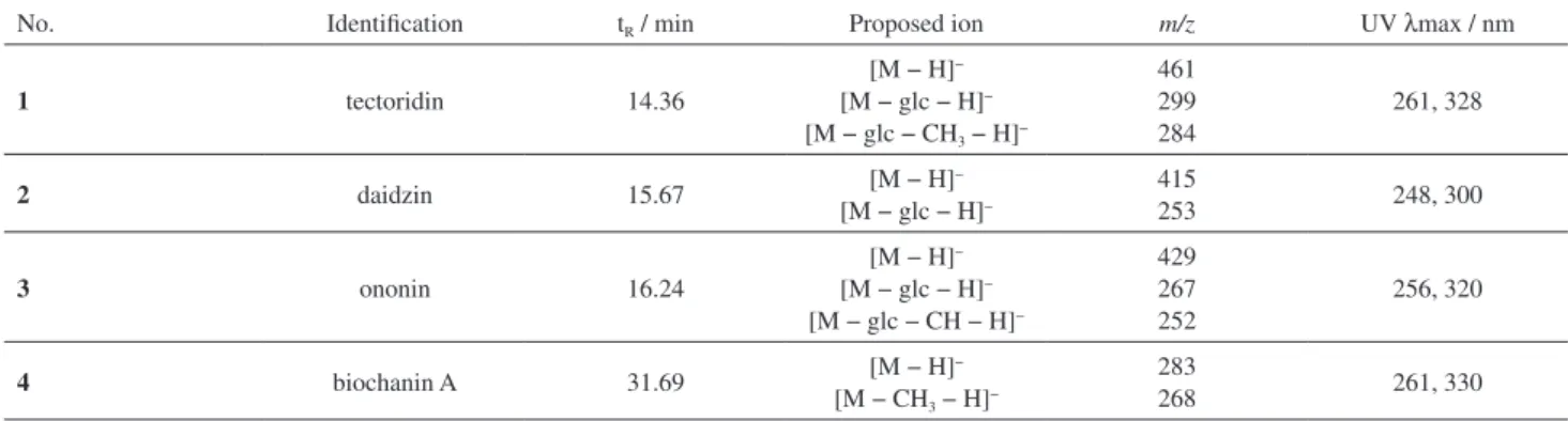
The chemical structures of these compounds were identified by HPLC-MS experiment and the analysis of their retention times, UV data and MS data is shown in
Table 1. By analysis of the UV spectra, all of the compounds typically had a maximum absorbance near 260 nm with a second maximum between 300 and 330 nm, which were the typical spectra of isoflavone derivatives. In the negative mode, all the isoflavones revealed deprotonated molecular ion [M − H]− in the MS spectrum. According to the studies
on the isoflavone glycosides in Puerariae lobata,27,28
the [M − 162 − H]− ion in MS spectra corresponded to
the presence of hexose sugar. The [M − 15 − H]− and
[M − 162 − 15 − H]− ion were observed in the fragments,
which were attributed to the neutral loss of CH3 caused
by the cleavage of methoxyl from the [M − H]− and
[M − 162 − H]−. The fragmentation pathways of these
four compounds were shown in Figure 4. Based on the differences that existed in the deprotonated molecular ion
[M − H]− in MS spectra and the maximum absorbance in
UV spectra, all of these four compounds showed typical molecular weights and were characterized as tectoridin (1), daidzin (2), ononin (3) and biochanin A (4).29-32
XO inhibition analysis of screened compounds
XO inhibitory activities assays of four screened compounds were carried out to evaluate the inhibition of each compound and verify the effectiveness of CU-LC-MS method. The binding degrees of four screened compounds
Table 1. The identification, retention time, UV and MS characteristics of compounds in P. flos
No. Identification tR / min Proposed ion m/z UV λmax / nm
1 tectoridin 14.36
[M − H]−
[M − glc − H]−
[M − glc − CH3 − H]−
461 299 284
261, 328
2 daidzin 15.67 [M − H]
−
[M − glc − H]−
415
253 248, 300
3 ononin 16.24
[M − H]−
[M − glc − H]−
[M − glc − CH − H]−
429 267 252
256, 320
4 biochanin A 31.69 [M − H]−
[M − CH3 − H]−
283
268 261, 330
were also calculated. As a result, tectoridin (1), daidzin (2), ononin (3) and biochanin A (4) authentically exhibited inhibitory activities on XO, and the IC50 values of them were
88.5, 85.1, 88.8 and 87.0 µmol L-1, respectively. Moreover,
the binding degrees of tectoridin (1), daidzin (2), ononin (3)
and biochanin A (4) at the optimum condition were
5.70, 8.28, 6.31 and 37.83%, respectively. These results demonstrated these four compounds possessed inhibition activities on XO. The groups on isoflavones and the derivatizations including glycosidation and methoxylation
would affect the inhibitory activities of compounds.33
According to reported literatures, the hydroxylation on isoflavones and the methylation or methoxylation of the hydroxyl group of flavonoids might affected the inhibitory activities.34-36 Based on current literatures, it has been
reported the inhibitory activity of tectoridin, daidzin and
biochanin A on XO.37-39 It demonstrated the screening
utilized by CU-LC-MS was effective and conclusive. This method exhibited acceptable screening efficiency and identification capability. It possessed advantages like high efficiency, simple procedures and low sample requirements. Therefore, the CU-LC-MS method was useful for systematical screening and analysis of active compounds from P. flos and other crude extracts.
Conclusions
In this study, a facile screening method based on CU-LC-MS was established for analyzing XO inhibitors
from P. flos. Four XO inhibitors including tectoridin,
daidzin, ononin and biochanin A were successfully screened and identified. All of these compounds exhibited inhibitory activities on XO. Results demonstrated that the proposed method is rapid and effective to screen and identify active compounds from natural products.
Acknowledgments
This work was supported by the risk assessment of agricultural products quality and safety project (GJFP2016010).
References
1. Li, F.; Zhang, Y.; Qiu, D.; Kang, J.; J. Chromatogr. A2015,
1400, 117.
2. Yang, Z.; Wu, Y.; Wu, S.; J. Chromatogr. A2016, 1431, 184. 3. Zhang, Y.; Li, F.; Li, M.; Kang, J.; J. Chromatogr. A2015, 1388,
267.
4. Luque-Garcia, J. L.; Neubert, T. A.; J. Chromatogr. A2007,
1153, 259.
5. Zhang, Y.; Peng, M. J.; Liu, L.; Shi, S.; Peng, S.; J. Agric. Food Chem.2012, 60, 3119.
6. Zhou, X.; Liang, J.; Zhang, Y.; Zhao, H.; Guo, Y.; Shi, S.; J. Chromatogr. B: Anal. Technol. Biomed. Life Sci.2015, 985, 149.
7. Choi, Y.; Jermihov, K.; Nam, S. J.; Sturdy, M.; Maloney, K.; Qiu, X.; Chadwick, L. R.; Main, M.; Chen, S. N.; Mesecar, A. D.; Farnsworth, N. R.; Pauli, G. F.; Fenical, W.; Pezzuto, J. M.; van Breemen, R. B.; Anal. Chem.2011, 83, 1048.
8. Zhou, J. L.; Qian, Z. M.; Luo, Y. D.; Tang, D.; Chen, H.; Yi, L.; Li, P.; Biomed. Chromatogr.2008, 22, 1164.
9. Chen, X.; Xia, Y.; Lu, Y.; Liang, J.; J. Pharm. Biomed. Anal.
2011, 54, 406.
10. Xiao, S.; Yu, R.; Ai, N.; Fan, X.; J. Pharm. Biomed. Anal. 2015,
104, 67.
11. Zeng, H. L.; Liu, Q.; Yu, J. G.; Wang, M.; Chen, M.; Wang, R.; He, X.; Gao, M.; Chen, X. Q.; J. Sep. Sci.2015, 38,3897. 12. Wang, J.; Liu, S.; Li, S.; Song, F.; Zhang, Y.; Liu, Z.; Liu, C.
M.; Anal. Methods2014, 6, 5918.
13. el Harrad, L.; Amine, A.; Enzyme Microb. Technol.2016, 85, 57.
14. Beedkar, S. D.; Khobragade, C. N.; Chobe, S. S.; Dawane, B. S.; Yemul, O. S.; Int. J. Biol. Macromol.2012, 50, 947. 15. Pacher, P.; Nivorozhkin, A.; Szabó, C.; Pharmacol. Rev.2006,
58, 87.
16. Li, S.; Tang, Y.; Liu, C.; Li, J.; Guo, L.; Zhang, Y.; Talanta2015,
134, 665.
17. Han, T.; Cheng, G.; Liu, Y.; Yang, H.; Hu, Y. T.; Huang, W.;
Food Chem. Toxicol.2012, 50, 409.
18. Shin, J. E.; Bae, E. A.; Lee, Y. C.; Ma, J. Y.; Kim, D. H.; Biol. Pharm. Bull.2006, 29, 1202.
19. Lee, H. U.; Bae, E. A.; Kim, D. H.; J. Pharmacol. Sci.2005,
97, 541.
20. Lee, K. T.; Sohn, I. C.; Kim, Y. K.; Choi, J. H.; Choi, J. W.; Park, H. J.; Itoh, Y.; Miyamoto, K. I.; Biol. Pharm. Bull.2001,
24, 1117.
21. Keung, W. M.; Vallee, B. L.; Phytochemistry1998, 47, 499. 22. Chang, W. S.; Lee, Y. J.; Lu, F. J.; Chiang, H. C.; Anticancer
Res.1993, 13, 2165.
23. Liu, H. X.; He, M. T.; Tan, H. B.; Gu, W.; Yang, S. X.; Wang, Y. H.; Li, L.; Long, C. L.; Phytochem. Lett.2015, 12, 133. 24. Martelli, G.; Folli, C.; Visai, L.; Daglia, M.; Ferrari, D.; Process
Biochem. 2014, 49, 154.
25. Dervisevic, M.; Dervisevic, E.; Azak, H.; Çevik, E.; Şenel, M.; Yildiz, H. B.; Sens. Actuators, B2016, 225, 181.
26. Ferrari, F. C.; Lima, R. C. L.; Ferraz Filha, Z. S.; Barros, C. H.; Araújo, M. C. P. M.; Saúde-Guimarães, D. A.;
J. Ethnopharmacol.2016, 180, 37.
27. Hirakura, K.; Morita, M.; Nakajima, K.; Sugama, K.; Takagi, K.; Niitsu, K.; Ikeya, Y.; Maruno, M.; Okada, M.; Phytochemistry
28. Ohshima, Y.; Okuyama, T.; Takahashi, K.; Takizawa, T.; Shibata, S.; Planta Med.1988, 54, 250.
29. Zhang, Y. Y.; Wang, Q.; Qi, L. W.; Qin, X. Y.; Qin, M. J.;
J. Pharm. Biomed. Anal. 2011, 56, 304.
30. Peng, J. B.; Jia, H. M.; Liu, Y. T.; Zhang, H. W.; Dong, S.; Zou, Z. M.; J. Pharm. Biomed. Anal. 2011, 55, 984.
31. Goto, H.; Terao, Y.; Akai, S.; Chem. Pharm. Bull.2009, 57, 346.
32. Farag, M. A.; Huhman, D. V.; Dixon, R. A.; Sumner, L. W.;
Plant Physiol.2008, 146, 387.
33. Promden, W.; Monthakantirat, O.; Umehara, K.; Noguchi, H.; de-Eknamkul, W.; Molecules2014, 19, 2226.
34. Xiao, J. B.; Ni, X. L.; Kai, G. Y.; Chen, X. Q.; Crit. Rev. Food Sci. Nutr.2015, 55, 16.
35. Hummelova, J.; Rondevaldova, J.; Balastikova, A.; Lapcik, O.; Kokoska, L.; Lett. Appl. Microbiol.2015, 60, 242.
36. Xiao, J. B.; Mao, F. F.; Yang, F.; Zhao, Y. L.; Zhang, C.; Yamamoto, K.; Mol. Nutr. Food Res.2011, 55, 1637. 37. Lee, K. T.; Sohn, I. C.; Kim, D. H.; Choi, J. W.; Kwon, S. H.;
Park, H. J.; Arch. Pharmacal Res.2000, 23, 461. 38. Toda, S.; Shirataki, Y.; Phytother. Res.1999, 13, 163. 39. Mo, S. F.; Zhou, F.; Lv, Y. Z.; Hu, Q. H.; Zhang, D. M.; Kong,
L. D.; Biol. Pharm. Bull.2007, 30, 1551.