2017
UNIVERSIDADE DE LISBOA
FACULDADE DE CIÊNCIAS
DEPARTAMENTO DE BIOLOGIA VEGETAL
Assessment of hiPSC derived cardiomyocytes maturation in
extended culture by analysis of alternative splicing expression
patterns
Rodrigo Paiva Gonçalves
Mestrado em Biologia Molecular e Genética
Dissertação orientada por:
Doutor Jorge Marques da Silva
i
Acknowledgements
The work I have developed for this thesis could not have been possible without so many of the people who have long been in my life and those who crossed my path during this journey, I wish to thank them all.
First of all, I want to thank my family, and especially my parents, not only for supporting me during this step of my life, but always. Without you I would never be where I am today, and no matter where I am, or how far apart we may have to be, I will always be grateful, and I will always hold you dear in my heart.
I must also thank my good old friends. They are also family, the family we choose, and who chooses you. Thank you all for standing by me and for being there
I thank Maria Fonseca for giving me the opportunity to work in her group and for the help and guidance.
A special thanks Teresa Carvalho and Sandra Martins, for all the guidance and knowledge they passed me, and for all the comprehension, motivation and joyfulness they showed. It was a pleasure having you as my advisors.
And last but not least a big thanks to Diogo Nogueira for teaching me all he knows about cardiac differentiation, to Marta Ribeiro for the huge help around the lab, and to all at Carmo’s group for being so welcoming and always willing to help.
Abstract
Heart failure is a major public health issue, being one of the main causes of death globally. Yet, the cellular and molecular mechanisms that trigger cardiovascular diseases are largely unknown. To date, basic research work has been hampered by lack of appropriate models. The emergence of patient-derived induced pluripotent stem cells (hiPSCs) that can be differentiated into functional cardiomyocytes holds great promise as an exciting new approach for cardiac disease modelling.
Determining whether or not cardiomyocytes differentiated in vitro from hiPSCs (hiPSC-CMs) recapitulate cardiac-specific characteristics is currently an area of intense research. In addition, recent evidence is revealing a critical role of alternative splicing in cardiac development and heart disease. Thus, understanding how alternative splicing contributes to regulation of cardiac gene expression will help elucidating the mechanisms underlying human heart failure.
Here, it was investigated the alternative splicing of core sarcomeric genes throughout differentiation of hiPSC-CMs. We analysed cardiac troponin 2 (TNNT2), myomesin 1 (MYOM1) and titin (TTN). We observed that these genes start to be expressed at day 8 of differentiation, with isoforms characteristic of early heart development representing the majority of the transcripts. By day 30, a shift in TNNT2 and TTN splicing isoforms was detected, with increased expression of adult isoforms. A further increase was observed at day 60 for TNNT2, but not for TTN. In the case of MYOM1, the ratio of immature isoform remains unchanged from day 13 to day 60, indicating this gene may not be adequate for maturation quantification.
In conclusion, the results indicate that an extended cell culture period is sufficient to induce a shift in the splicing pattern of TNNT2 and TTN mRNAs. Although hiPSC-CMs did not fully recapitulate the splicing patterns found in the human adult heart, this study highlights the relevance of long-term cultures for disease modeling and cardiac research.
ii
Key
words:
Heart disease; Cardiomyocytes; Development; Cardiacdifferentiation; Alternative splicing.
Resumo
A patologia cardiovascular é um dos maiores problemas de saúde pública e uma das principais causas de morte a nível mundial. Apesar de o impacto socioeconómico destas patologias ser tão demarcado, e ser previsto que a incidência das mesmas venha a aumentar drasticamente nos próximos anos, os mecanismos celulares e moleculares que desencadeiam as doenças cardiovasculares são amplamente desconhecidos. Uma forma de melhor compreender estas doenças é o uso de modelos de doença. Contudo, até à data, a investigação na área foi dificultada pela falta de modelos adequados.
As células pluripotentes induzidas são células com alta capacidade de proliferação e que podem ser diferenciadas em qualquer tipo celular de um organismo adulto. Estas células trazem enormes vantagens e carregam menos implicações éticas que alternativas derivadas de embriões.
O recente desenvolvimento de protocolos que permitem de forma eficiente diferenciar estas células em cardiomiócitos funcionais é uma nova e promissora forma de obter grandes quantidades de células cardíacas, que proporciona grandes vantagens na modelagem da doença cardíaca. No entanto, a necessidade de determinar se cardiomiócitos diferenciados in vitro de células pluripotentes induzidas recapitulam características cardíacas específicas, criou recentemente uma área de pesquisa intensa e os cardiomiócitos derivados de células pluripotentes têm sido caracterizados na literatura como sendo imaturos, tanto a níveis morfológicos como fisiológicos.
O “splicing” é um fenómeno que afeta a maioria dos genes humanos e permite a produção de proteínas diferentes a partir do mesmo gene. Estas diferentes isoformas proteicas permitem às células adaptarem-se as constantes alterações fisiológicas que enfrentam. Entender como o “splicing” alternativo contribui para a regulação da expressão dos genes cardíacos pode ajudar a elucidar os mecanismos subjacentes a inúmeras patologias cardíacas.
Trabalhos recentes revelam o papel crucial do “splicing” alternativo no desenvolvimento cardíaco e também doença cardíaca. Outras publicações mostram que o “splicing” em cardiomiócitos derivados de células pluripotentes induzidas é idêntico ao de cardiomiócitos fetais, mas que este “splicing” pode sofrer alterações e assemelhar-se mais ao “splicing” no coração adulto aumentando o tempo de cultura das células.
Aqui, foi investigado o “splicing” alternativo de genes sarcoméricos ao longo da diferenciação de cardiomiócitos derivados de células pluripotentes induzidas e foi avaliada a sua progressão ao longo de 60 dias em cultura. Foram também realizadas analises de imunofluorescência, para permitir aferir o grau de diferenciação dos cardiomiócitos obtidos por outros parâmetros.
Foi analisada a troponina cardíaca 2 (TNNT2), a miomesina 1 (MYOM1) e a titina (TTN). Observamos que esses genes começam a ser expressos no dia 8 da diferenciação, com isoformas características de baixa maturação a representar a maioria dos transcritos. No dia 30, foi detetada uma mudança nas isoformas de TNNT2 e TTN, com expressão aumentada de isoformas adultas. Observou-se um aumento adicional no dia 60 para o gene da TNNT2, mas não para o gene da titina o que pode implicar que culturas superiores a um mês não são vantajosas para a alteração de “splicing” deste gene. No caso do gene da miomesina o rácio de isoforma precoce permanece inalterado desde o dia 13 até ao dia 60. Isto pode significar que este gene não é adequado para a quantificação da maturação ou que períodos de cultura mais duradouros não são suficientes para induzir maturação ao nível deste gene.
iii Os resultados expressão génica que mostram maturação foram corroborados por analises de imunofluorescência realizadas. Estes mostram um aumento da dimensão dos cardiomiócitos e da organização das fibras do sarcómero quando comparando imagens de células no dia 13 de diferenciação e células no dia 30.
Em conclusão, os resultados indicam que um período prolongado até 30 dias de cultura de células é suficiente para induzir mudanças no “splicing” de TNNT2 e TTN, sendo que no caso do
TNNT2 culturas mais prolongadas podem ser vantajosas, o que não parece ser verdade no caso
da TTN.
Embora os cardimiócitos obtidos após longos períodos de cultura sejam mais semelhantes aos do coração humano adulto, estes não recapitulam completamente os seus padrões de “splicing”. Apesar disto, este estudo realça a relevância das culturas de longo prazo na investigação cardiovascular e no desenvolvimento de modelos de doença fidedignos.
No futuro seria interessante estudar o efeito de outros estímulos nos padrões de “splicing” destas células, como fatores químicos, estimulação elétrica, cultura em substratos mais rígidos e modelos em 3 dimensões, que têm sido mostrados como promotores de maturação, para características como a morfologia e a força contrátil.
Palavras
chave:
Patologia cardíaca; Cardiomiócitos; Desenvolvimento;iv
List of Contents
Acknowledgements ... i
Abstract ... i
Resumo ... ii
Tables and figures ... v
Abbreviations ... vi
Introduction ... 1
Cardiovascular diseases and disease modelling ... 1
Stem cells ... 1
Pluripotent Stem Cells ... 2
Pluripotent stem cell culture ... 2
Cardiac differentiation of hiPSCs ... 3
Differentiation by temporal modulation of the canonical WNT signalling pathway ... 3
hiPSC-CM maturation ... 3
Splicing in the developing human heart ... 4
Splicing in iPSC-CM ... 4
Objective ... 5
Materials and Methods ... 5
Cell line ... 5 Adhesion substrate ... 5 Culture media ... 5 Cell thawing ... 6 Cell passaging ... 6 Cell cryopreservation ... 6 Cell counting ... 6 Cardiac differentiation... 6
RNA extraction and isolation ... 7
DNase treatment ... 7
cDNA Synthesis ... 8
Qualitative polymerase chain reaction (PCR) and agarose gel electrophoreses ... 8
Quantitative real-time polymerase chain reaction (qPCR) ... 8
Immunofluorescence assays after cellular staining ... 9
Results and Discussion ... 9
Conclusions ... 16
v
List of figures
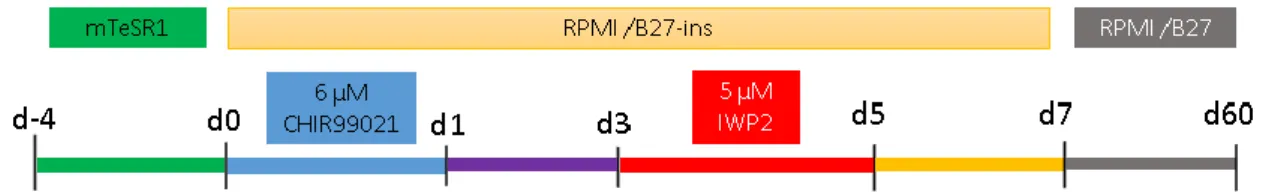
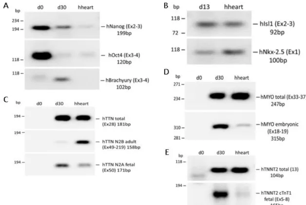
Figure 1: Scheme of the mediums used at the different stages of the differentiation ... 6 Figure 2: Schematic representation of the sites of annealing for the primers chosen to analyse alternative splicing. The purple arrows represent the primer pair intended for amplification of all isoforms, whereas in orange are represented the exons that show alternative splicing and the primer pairs specific to that isoform. ... 10 Figure 3: Agarose gel electrophoresis of the semi-quantitative PCRs. (A) Pluripotency markers Nanog and Oct4, and mesendodermal marker Brachyury at days 0 and 30 of differentiation. (B) Cardiac progenitor markers Isl1 and Nkx-2.5 at day 13. (C) Primer pair that amplifies both isoform classes, and primer pairs for each of the isoform classes. (D) Total and embryonic exclusive primer pairs for Myomesin at days 0 and 30. (E) Total and embryonic exclusive primer pairs for Tropomiosin T at days 0 and 30. Total human heart RNA was used as a control for all primer pairs. ... 10 Figure 4: Expression of the stemness markers Nanog and Oct 4 throughout cardiac differentiation and in the adult human heart. Expression values were normalized to the GAPDH house keeping gene. ... 11 Figure 5: Expression of the mesoendoderm markers Brachyury and cardiac precursors Isl1 and Nkx-2.5 throughout cardiac differentiation and in the adult human heart. Expression values were normalized to the GAPDH house keeping gene. ... 11 Figure 6: Expression of all the TTN transcripts and of the fetal N2BA isoforms and of the adult N2B isoforms throughout cardiac differentiation and in the adult human heart. Expression values were normalized to the GAPDH house keeping gene. ... 12 Figure 7 - Expression of all the MYOM1 transcripts and of the embryonic specific EH-MYOM1 isoform throughout cardiac differentiation and in the adult human heart. Expression values were normalized to the GAPDH house keeping gene. ... 13 Figure 8 - Expression of all the TNNT2 transcripts and of the fetal specific cTnT1 isoform throughout cardiac differentiation and in the adult human heart. Expression values were normalized to the GAPDH house keeping gene. ... 13 Figure 9: A - Schematic of the primer pair that allows semi-quantification of the adult TNNT2 isoform. The alternatively spliced exon is shown in red. B - Agarose gel electrophoresis using the primer pair shown in A with the house keeping gene GAPDH as a loading control. C - Western blot of the TNNT2 protein with Tubulin as a loading control. ... 14 Figure 10: Immunofluorescent of iPSC-CM at day 13 and 30 of differentiation stained using DAPI and a TNNT2 specific antibody. ... 15
vi
Abbreviations
CM – Cardiomyocytes
CVD – Cardiovascular disease cTnI – Cardiac troponin I cTnT – Cardiac troponin T
DMEM - Dulbecco's modified Eagle's medium DAPI - 4’,6-diamidino-2-phenylindole
EDTA - ethylenediaminetetraacetic acid ESC – Embryonic stem cell
GAPDH - glyceraldehyde-3-phosphate dehydrogenase hiPSC – Human induced pluripotent stem cell
iPSC – Induced plutipotent stem cell iPSC-CM – iPSC derived cardiomyocytes ISL - LIM-homeobox transcription factor islet KLF - Krüppel-like factor
MYOM - Myomesin
mRNA - messenger ribonucleic acid OCT - octamer-binding transcription factor PBS -Phosphate sodium buffer
PSC – Pluripotent stem cell
qPCR - quantitative real-time polymerase chain reaction SOX - sex determining region Y-box
RNA - ribonucleic acid
ROCK - rho-associated coiled-coil protein kinase RPMI - Roswell Park Memorial Institute (medium) TFC -T-cell factor
TNNT - troponin T
TTN - Titin
1
Introduction
Cardiovascular diseases and disease modelling
Cardiovascular diseases (CVDs) are diseases that affect the heart and the circulatory system. These include numerous pathologies, such as ischemia, cerebrovascular disease and hypertrophy1.
CVDs are the leading cause of death in the world, taking more than 17,3 million lives in 20132,
31 % of the years deaths3. These deaths are increasing and are expected to reach 23,6 million
by 20304.
The high incidence of these diseases correlates to a great and increasing socioeconomic burden, with costs amounting to 393,5 billion dollars in 20051, ramping up to 863 billion in 2010 and
estimated to go up to 1044 billion dollars by 20305.
In the more severe cases of CVD there can be a great amount of heart muscle cell death, and even though the heart has some regenerative capabilities, these are often not sufficient to repair the lost tissue6. Although there are many types of treatment, they commonly present low
efficiency, meaning a full cardiac transplant is necessary7, but even this may not present a
solution, because of rejection risks even though these can be minimised by immuno-suppression8. Adding to that, the high prevalence of CVDs, means cardiac transplant waiting
lists are long, and most of the patients can’t have access to a compatible heart1.
All of these facts elucidate the urgency to find better treatments for cardiac disease, meaning it is crucial to further understand them. For this, disease modelling tools present themselves as great assets, since they can offer a more practical and convenient way of studying these diseases.
These disease models can have a great impact in studying diseases like cardiac hypertrophy, a hereditary disease where an early diagnosis is crucial but approximately 50% of individuals with a clinical diagnosis have no specific mutation identified9. With a suitable disease model, a
large number of genetic variants can be tested, to assess whether or not they can be pathological, an important and unmet medical need.
The most convenient way of generating cardiac disease models is to obtain cardiomyocytes in
vitro, since cardiomyocytes have low proliferative capabilities and are difficult to collect1.
Stem cells present a convenient way to generate cardiomyocytes, as they can be expanded into large numbers and then differentiated into the desired cell type10.
Stem cells
Stem cells are self-renewing cells, that can be differentiated in culture through the use of several types of stimuli, having the potential to differentiate into different cell types11.
The stem cells potential to differentiate can be classified as unipotent, if the cells can only differentiate into one cell type, multipotent if they are able to differentiate into discrete cell types, pluripotent if they have the ability to differentiate into cells of any of the 3 germ layers (endoderm, mesoderm and ectoderm), and totipotent if a cell is able to differentiate into a whole organism1.
An example of unipotent cells are the spermatogonial stem cells, as they can only originate one cell type, sperm cells12 10. On the other hand, haematopoietic stem cells are multipotent, since
2
Pluripotent Stem Cells
When it comes to pluripotent stem cells these are usually either embryonic stem cells (ESCs), or induced pluripotent stem cells (iPSC). These cells are in theory immortal because of their capacity to regenerate telomeres, although continuous amplification may cause karyotypic alterations13. As they are able to differentiate into all adult cell types, and can be obtained in
great quantities, they hold a great potential for biomedical applications and for disease modelling1.
Embryonic stem cells are obtained from the inner cell mass of the blastocyst. ESCs were first derived from the inner cellular mass of mice blastocysts by Gail Martin14, and Evans and
Kaufman15 in 1981 by culturing them in a feeder layer of mouse embryonic fibroblasts.
The first human ESCs were produced in 1998 by in vitro fertilisation and culture with mouse embryonic fibroblasts by Thomson and colleagues16.
Human and mouse ESC are able to form teratomas when injected into immunodeficient mice. This ability to form tumours with the three germ layers comes from their embryo-like nature16.
ESCs pluripotency is what makes them attractive for biomedical applications, but at the same time makes them also dangerous, since their implantation always carries a tumourgenic danger. When it comes to disease models though, this does not present itself so much as a concern, despite ethical concerns being always present.
ESCs pluripotent alternative, iPSCs, are obtained by reprogramming terminally differentiated cells. This was first done by Takashi and Yamanaka in 2006, when they used a retroviral vector to transform embryonic and adult mouse fibroblasts with octamer-binding transcription factor (OCT) 3/4, sex determining region Y-box (SOX) 2, Krüppel-like factor (KLF) 4 and c-MYC, commonly known as the Yamanaka factors17.
The first human iPSCs (hiPSC) where generated in 2007 by Takashi, Yamanaka and colleagues by transfection oh the Yamanaka factors into adult human fibroblasts18.
That same year, Thomson and colleagues achieved similar results using a different combination of factors, OCT 3/4, SOX2, NANOG and LIN2819.
Although ESCs and iPSCs have really different ways of being obtained, they present similar characteristics, like self-renewal, morphology, surface markers and pluripotency1820.
Despite that, iPSCs carry less ethical concerns than ESCs, and on top of that, they can be patient-specific. This means they avoid rejection in case of biomedical applications, and make it possible to develop patient specific disease models21.
Pluripotent stem cell culture
While culturing PSCs, it is vital to maintain their pluripotency and avoid spontaneous differentiation.
One of the mediums developed to this end is mTeSR1. It is composed of DMEM/F12 basal medium, supplemented with bovine serum albumin, vitamins, antioxidants, trace minerals, lipids and growth factors. mTeSR1 maintains the cells pluripotency since it contains fibroblast growth factor, transforming growth factor β, lithium chloride, pipecolic acid and γ-aminobutyric acid.
PSCs need to be passaged as colonies, using ethylenediaminetetraacetic acid (EDTA) instead of enzymes, to avoid single-cell passaging, which causes the cells to suffer apoptosis due to lack of cell to cell contact, known as anoikis. This process is mainly mediated by the rho-associated coiled-coil protein kinase (ROCK) signalling pathway22. If a single-cell suspension is required
3 for protocols like fluorescence activated cell sorting, an inhibitor of this pathway, such as Y-27362 , can be used to stop apoptosis until the cells are no longer at a single cell state23.
Cardiac differentiation of hiPSCs
As stated before, PSCs can be differentiated into the three germ layers, meaning that under the right stimuli, they can be differentiated into mesoderm progenitors, and then into cardiomyocytes.
Cardiac differentiation protocols attempt to emulate cues from the embryonic heart development to commit cells to a cardiac lineage.
To this end, several different protocols have been developed, all with distinct differentiation efficiencies. Many of them, however, do not present elevated differentiation efficiencies (>90% cTnT+ cells) without any added step of purification after the differentiation.
Differentiation by temporal modulation o f the canonical WNT signalling
pathway
One of the protocols that shows higher differentiation efficiencies was developed in 2012 by Lian and colleagues2425.
This protocol aims at regulating exclusively elements of the canonical WNT signalling pathway. This signalling pathway is highly conserved between species and controls numerous cellular processes, including cell polarity and embryonic induction.
WNT proteins are able to induce gene expression through β-catenin, which is degraded in the absence of these proteins. In the presence of WNT proteins, β-catenin accumulates in the cytoplasm and translocates to the nucleus, interacting with the T-cell factor (TCF)/ lymphoid enhancer of WNT target genes26.
In human PCS, WNT signalling is maintained at a basal level, leading to self-renewal together with other proliferation factors. But when the culture conditions do not favour pluripotency, up-regulation of WNT signalling induces proliferation and differentiation27.
In cardiac differentiation in particular, the role of the WNT signalling pathway is related to one of its downstream targets, Brachyury, an important mesendodermal maker28. WNT signalling
induces the formation of the primitive streak crucial to the formation of mesoderm29.
Although important in mesendoderm induction, WNT signalling prevents cardiomyogenesis in mesoderm-committed cells. This means that a protocol for cardiac differentiation that only manipulates the WNT pathway needs to first activate it, and then inhibit it25. This is exactly
what Lian and co-workers did to develop a protocol that is highly efficient and factor-free.
hiPSC-CM maturation
A crucial downside of cardiac differentiation protocols is the fact that the produced cardiomyocytes are largely immature.
Lian and colleagues 25 quantified the expression of myosin light chain (MLC) and found that
after 20 days of differentiation, cardiomyocytes were expressing the MLC2a isoform, and not the MLC2v isoform, showing their immaturity. At day 60, more than 50% of the cardiomyocytes expressed MLC2v; however 80% of these cells still expressed MLC2a, meaning these cells suffered some maturation, but still differ from adult cardiomyocytes. To overcome this issue, many strategies have been successfully applied to improve the maturation of cardiomyocytes obtained in vitro. These include the use of specific mediums30,
4 Maturation protocols can originate cardiomyocytes that better resemble adult cardiac cells in characteristics like sarcomeric organisation, electrophysiological properties, higher contractile force and higher intracellular calcium levels.
Splicing in the developing human heart
More than 90 % of the human genes express several mRNAs, a consequence of alternative splicing, which is a post-transcriptional mechanism that allows the synthesis of multiple proteins from a single gene33.
Alternative splicing is not a rigid mechanism, as many proteins may switch to meet the cells changing needs. During cardiac development, changes in alternative splicing allow the cell to adapt to the changing physiological conditions34. There are many genes that suffer these
changes, but here I will focus in three in particular, as they were the emphasis of the work presented in this thesis.
The first of these genes is TTN, whose expression gradually shifts to stiffer isoforms to increase passive stiffness in developing mammalian hearts35. Titin is the largest protein in the human
body with a weight of ~3000 kDa and over 1 µm in length, and most of its 363 exons are alternatively spliced. The splicing of the gene occurs mainly in the elastic regions, which gives rise to two main classes of titin: the stiff and short N2B isoforms and the longer and more elastic N2BA isoforms36. Both types of isoforms can be coexpressed allowing a full spectrum of
stiffness. Fetal hearts express only the N2BA isoforms, which is gradually replaced by the N2B isoforms as it becomes about 70% of total titin in the developed heart35.
TNNT2 and MYOM1, suffer similar changes in splicing, as each their mRNAs loses one exon as
the heart develops.
Myomesin 1 is described to have an embryonic heart isoform (EH-myomesin), which has an exon known as the EH exon. The embryonic heart expresses high levels of EH-myomesin, whereas the adult heart expresses low levels37.
In the case of TNNT2, the characteristic fetal isoform is the cTnT1 isoform, which though the loss of one exon gives rise to the cTnT3 isoform, the only one expressed in the adult heart38.
Splicing in iPSC-CM
Cardiomyocytes derived from iPSCs are known to have splicing patterns that resemble those of fetal hearts, and changes in alternative splicing of these cardiomyocytes have already been shown.
One of these cases is the case of MLC already mentioned before. At the end of the differentiation, cells express only the MLC2a immature isoform, but after 60 days of culture 50% of the cells start to express the MLC2v mature isoform and about 20% of those cells stop expressing MLC2a25.
Another example of isoform switching in long culture cardiomyocytes is show in the work of Bedada and colleagues, where they show that iPSC-derived cardiomyocytes express only the immature Troponin I 1 isoform. These cardiomyocytes start to express the mature isoform Troponin I 3 after 2 months in culture although these levels are low and do not change even if the cardiomyocytes are kept in culture for 9,5 months39.
5
Objective
In this project, I sought out study the splicing patterns of three genes, Titin (TTN), Troponin T2 (TNNT2) and Myomesin 1 (MYOM1) to assess if these can be used as markers of hiPSC-CM maturation.
Materials and Methods
Cell line
The DF6-9-9T.B hiPSC line (WiCell®) was used in every experiment described. This is a
vector-free cell line with a 46, XY karyotype. This line was reprogrammed from healthy donor’s foreskin fibroblasts, using defined factors in the laboratory of Dr James Thomson, at the University of Wisconsin.
Adhesion substrate
Matrigel (Corning®) was used as the adhesion substrate in all experiments. This coating was
stored as aliquots at –20 °C and was thawed in ice and diluted 1:100 in cold DMEM/F12 medium (Gibco®) to avoid any jellification. The tissue culture plates (TPP®) were coated with
diluted Matrigel and left at room temperature for a minimum of 2 h prior to use, or stored at 4 °C for later use, for a maximum of 2 weeks.
Culture media
mTeSR1 (STEMCELL TechnologiesTM) was the medium used to maintain the hiPSCs. To
prepare the complete medium, mTeSR1 5× supplement was thawed overnight at 4 °C and diluted in 5 times the volume of mTeSR1 basal medium. The complete medium was stored in aliquots at –20 °C, being thawed overnight and stored at 4 °C, as well as being supplemented with 0.5% penicillin/streptomycin (Gibco®) before use. The medium was warmed at room
temperature before each medium change.
To maintain cells when these were processed in suspension, and to stop enzymatic digestions, what is commonly called as washing medium was used. Its basal formulation consists of DMEM/F12 containing L-glutamine (Gibco®), buffered with 2.44 g/L of sodium bicarbonate,
and supplemented with KO-SR (Gibco®), MEM non-essential amino acids (Gibco®) and
penicillin/streptomycin. This medium was stored at 4 °C and protected from light, while being warmed at room temperature before use.
RPMI 1640 medium (Gibco®) was the basal medium used for the cardiac differentiation of
hiPSCs. This was supplemented with either B-27 minus insulin (Gibco®) or B-27 (Gibco®)
supplements. B-27 minus insulin and B-27 were thawed at room temperature in the dark and added at 2% to RPMI 1640 medium along with 0.5% penicillin/streptomycin. The complete medium was stored at 4 °C and protected from light for a maximum of 30 days and was warmed at room temperature before use.
6
Cell thawing
Cryovials (with approximately 1 million cells) where removed from liquid nitrogen (–196 °C), and partially thawed in a 37 °C bath. These where then fully thawed with 1 mL of washing medium by slowly mixing.
This cell suspension was added to tube a containing 3 mL of washing medium, and was centrifuged for 3 minutes at 1500 g. The supernatant was discarded and the cells where resuspended in 1 mL of mTeSR1 medium and seeded onto a 9,6 cm2 tissue culture plate coated
with Matrigel containing 0,5 mL of mTeSR1, added previously, after removing the coating solution, to stop the substrate from drying.
Cell passaging
For passaging, cells were washed twice with 1 mL/well of EDTA dissociation solution (0,5 mM EDTA and 1,8 g/L sodium chloride in phosphate-buffered saline (PBS) solution), and left for 5 minutes in 1 mL/well of EDTA.
The EDTA was removed and the cells were flushed twice with 1 mL/well of culture medium and collected to a tube to which culture medium was added in order to get 1 mL/well for plating. In the new plate 0,5 mL/well of culture medium was added, as the Matrigel solution was removed, to avoid the Matrigel drying. Afterwards, 1 mL/well of cell suspension was added. Usually cells were passed in 1:3 or 1:4 dilutions.
Cell cryopreservation
To cryopreserve the hiPSCs these were washed twice with 1 mL/well of EDTA and then incubated for 5 minutes in 1 mL/well of EDTA. After removing the EDTA, cells were flushed twice with 1 mL/well of washing medium, collected to a Falcon tube, and centrifuged for 3 min at 1,500 g.
The resulting supernatant was discarded, and cells corresponding to 2 wells of a 6 well plate were resuspended in 250 μL of KO-SR containing 10% dimethylsulfoxide (DMSO). This volume was then transferred to a cryovial, and kept at –80 °C for 48 hours, before being moved to liquid nitrogen (–196 °C).
Cell counting
To count the hiPSCs for seeding, a volume of 10-20 μL of sample was mixed with 10 μL of accutase, if the cells were not subjected to enzymatic digestion beforehand. Trypan blue was added in the necessary volume to achieve the desired dilution and 10 μL of the resulting mixture were then loaded into a haemocytometer for counting.
Cardiac differentiation
7 The protocol used for the cardiac differentiation of hiPSCs was adapted from the work of Lian and co-workers 25, and is schematically presented in Figure 1.
To start cells where plated in 3.8 cm2 tissue culture plates (12 well plates) at a density of
100,000 cells/cm2 (380,000 cells/well). For 4 days, mTeSR1 medium was changed daily (1
mL/well). After this period, cells should achieve 90 % to 95 % confluence, ideal for differentiation.
To induce the differentiation, on day 0 of differentiation, 1.5 mL/well of RPMI/B27-insulin medium supplemented with 6 μM of CHIR99021 was added to each well. After 24 h, the medium was changed to RPMI/B27-insulin. At day 3, 750 μL/well of medium is removed, and replaced with medium containing IWP4, to obtain a final concentration of 5 μM in each well. IWP4 is removed in the medium change at day 5. At day 7, the medium was changed to RPMI/B27 and changed every 3 days until the end of the experiment.
RNA extraction and isolation
For each condition cells in 1 well from a 12 well plate were collected.
For this, each well was washed with 1ml of PBS and incubated for 5 minutes with 0,5 mL of trypsin. The cells were then collected to a 1,5 mL tube and centrifuged at 1500 g for 5 minutes, with the supernatant being discarded. The resulting cell pellet was resuspended in 1ml of Nyzol (Nzytech®), by pipetting several times up and down until the solution was clear and them
incubated for 5 minutes at RT. After that samples were either frozen at -20 °C or treated right away.
RNA isolation was continued by adding 200 μL of Chloroform and mixing well by shaking. This was followed by an incubation for 3 minutes at RT and then centrifugation at 12000 g for 15 minutes at 4 °C.
After that two phases are separated, a lower, organic phase that contains hydrophobic molecules (mostly cell membrane lipids) which was discarded, and the upper, aqueous phase, containing hydrophilic molecules, such as nucleic acids, which was transferred to a new 1,5 mL tube, always being careful to avoid disturbing the interface between the two phases, which contains mostly proteins, that form a film due to their amphipathic properties.
To the new tube were added 500 μL of isopropanol, and it was mixed well by shaking and then incubated 10 minutes at RT.
Afterwards, a centrifugation at 12000 g for 10 minutes at 4 °C was performed, with the resulting supernatant being very carefully discarded. The resulting RNA pellet was washed with 1 mL of 75 % ethanol and vortexed before being centrifuged at 12000 g for 10 minutes at 4 °C.
The supernatant was discarded very carefully and the RNA pellet was air dried, with an open lid, for 10 minutes at 37 °C. This RNA pellet was resuspended very well in 23 μL of RNase – free water.
The RNA in that suspension was then quantified by Nanodrop®.
DNase treatment
The quantified RNA was then added to a new eppendorf tube in a volume correspondent to 1 to 5 μg of RNA, along with 5 μL of 10x Dnase I buffer (Roche®), DNase I (Roche®) (1 μg/per1
μgRNA) (1-5 μL) and RNase free water up to 50 μL. This was then incubated for 2 hours at 37 °C
8 To the end reaction 150 μL of 100% EtOH was added, 5 μL sodium acetate and 1 μL GlycoblueTM (Ambion®) (for easier visualisation of the pellet). This was then vortexed and
incubated over night at -20 °C.
Afterwards the tubes where centrifuged at full speed (13000 g) for 30 minutes at 4 °C and the supernatant was discarded very carefully.
The RNA pellet was washed with 1 mL of 75 % ethanol and vortexed. Another centrifugation at full speed was done for 15 minutes at 4 °C.
After discarding supernatant very carefully the RNA pellet was air dried, with open lid, for 10 minutes at 37 °C.
The RNA pellet was resuspended in 13 μL of RNase –free water very well and a Nanodrop® quantification was performed.
cDNA Synthesis
Reverse transcription of the isolated RNA was made using the Transcription High Fidelity cDNA Synthesis Kit (Roche®), using 600 ng of DNase treated RNA, per reaction.
The 600ng of RNA where added to a 0.2 µL tube and RNase free water was added up to 9,4 µL. Then 2 μL of random primers were added to each tube, with this mixture being vortexed and shortly centrifuged before being incubated for 10 minutes at 65 ºC on a thermocycler (BioRad®), to allow primer annealing.
Then, 8,6 μL of reverse trancription mixture (4 μL of RT buffer 5x ; 0,5 μL of RNAse Out (40 u/μL); 1 μL dNTP mix (10 mM each); 1 μL DTT (Ditiotreitol); 1,1 μL Reverse Transcriptase) was added to each tube and these were then vortexed and shortly centrifuged.
The thermocycler (BioRad®) was then used to incubate these with the following programme:
29 ºC for 10 minutes 48 ºC for 60 minutes 85 ºC for 5 minutes 14 ºC until stored at -20 ºC
Semi-quantitative polymerase chain reaction (PCR) and agarose gel
electrophoreses
Semi-quantitative PCR and subsequent analyses of the PCR products by electrophoreses were used to verify the specificity of the primers used in the qPCRs.
The annealing temperature chosen was 60 °C and the reaction program was set to 30 cycles. The agarose gel was made at a concentration of 2 % and ethidium bromide (EtBr) was used to visualize the DNA fragment bands, being added before casting the gel. The migrations were performed at 100 Volts.
After the electrophoresis, the gels were visualized using a AlphaImager® HP system (Alpha
Innotech).
Quantitative real-time polymerase chain reaction (qPCR)
The cDNA samples were placed in 384-well plates, along with iTaqTM Universal SYBR®
Supermix (Bio-Rad) and the respective pair of primer for each of the genes analyzed.
The ViiA 7 System (Life Technologies) was used to processes the samples, with the annealing temperature being set to 60 °C.
9 Results were analyzed with the QuantStudioTM Real-Time PCR Software v1.1 (Life
Technologies) and the housekeeping gene glyceraldehyde-3-phosphate dehydrogenase (GAPDH) was used as an endogenous control, with threshold cycles (CT) for each sample being compared to the respective endogenous control CT. This resulted in a ΔCT, that allows the comparison of levels of expression between samples.
Immunofluorescence assays after cellular staining
For the immunofluorescence, cells where grown on coverslips places inside wells from a 12 well plate.
The coverslips where transferred to wells of a 6 well plate previously filled with PBS.
These where then wash twice with 1 mL/well of PBS and fixed by incubation in 1,5 mL/well of 3,7 % PFA (Paraformaldeíde) in PBS for 10 min at RT.
Cells were then washed twice with 1 mL/well of PBS and used right away or stored at 4 ºC for up to 2 weeks.
To permeabilize the cells, they were incubated with 0,5% Tritonin PBS for 10 minutes (12 minutes is the samples where thicker) and then washed with 0,05% Tween in PBS 3 times for 10 minutes each.
This was followed by an incubation with 6 µL of the primary antibody at a 1:200 dilution for 1 hour at room temperature and then over night at 4 ºC.
After this three 10 minute washes were made with 1 mL/well of PBS-Tween each and an incubation with 6 µL of the secondary antibody at the dilution of 1:200 for 60 min at RT and in the dark.
Ten minutes before the end of this incubation 5 μL of 4’,6-diamidino-2-phenylindole (DAPI), diluted 1: 100,000 in sodium bicarbonate, was added to label DNA.
Finally, these were washed 3 times for 10 minutes in 1 mL/well of PBS Twin.
Results
and
Discussion
After RNA was extracted at critical time points of the cardiac differentiation protocol (d0, d1, d5, d8, d13, d30, and d60) expression of pluripotency and cardiac markers was analyzed. The products amplified by the primer pairs selected to analyze alternative splicing are schematically shown in Figure 2.
It was decided to not design primers specific for the TNNT2 and MYOM1 mature isoforms, since it is arguable that exon-junction primers would only amplify that isoform as some of the primer sequence would be present in both splicing isoforms. This problem does not exist in the case of titin because the spliced region if too large to be amplified by the primers flanking it (Fig. 2).
To test the primers for qPCR, semi-quantitative PCRs and agarose gel electrophoresis of the obtained product were analyzed as shown in Figure 3. All primer pairs were show to be specific as only one band was observed for each primer pair.
10
Figure 2: Schematic representation of the sites of annealing for the primers chosen to analyse alternative splicing. The purple arrows represent the primer pair intended for amplification of all isoforms, whereas in orange are represented the exons that show alternative splicing and the primer pairs specific to that isoform.
Figure 3: Electrophoresis in agarose gell of the semi-quantitative PCRs. (A) Pluripotency markers Nanog and Oct4, and mesodermal marker Brachyury at days 0 and 30 of differentiation. (B) Cardiac progenitor markers Isl1 and Nkx-2.5 at day 13. (C) Primer pair that amplifies both TTN isoforms, and primer pairs for each of the isoform. (D) Total and embryonic exclusive primer pairs for Myomesin at days 0 and 30. (E) Total and embryonic primer pairs for TNNT2 at days 0 and 30. Total human heart RNA was used as a control for all primer pairs.
11 Once primer specificity was confirmed, mRNA expression of hiPSCs along cardiac differentiation was assessed by qRT-PCR analysis as shown in Figures 4 through 8. A human heart RNA was used as a positive cardiac control.
Figure 4: Expression of the stemness markers Nanog and Oct 4 throughout cardiac differentiation and in the adult human heart. Expression values were normalized to the GAPDH house keeping gene.
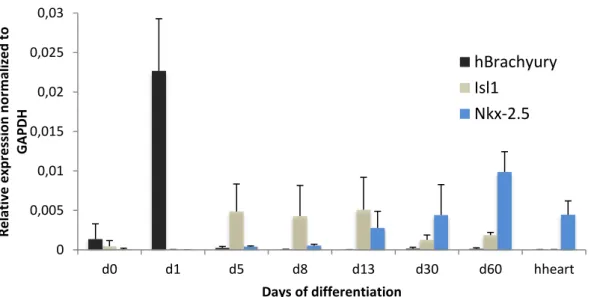
Figure 5: Expression of the mesoderm marker Brachyury and cardiac precursors Isl1 and Nkx-2.5 throughout cardiac differentiation and in the adult human heart. Expression values were normalized to the GAPDH house keeping gene.
Since the main goal of this work was to analyse the level of iPSC-CM maturation through analysis of splicing patterns. For that the efficiency of the differentiation is crucial to validate any results.
In Figure 4 it is possible to see the gradual decrease in expression of Nanog and Oct 4, correlating to a expected decrease of stemness and commitment to a specific lineage, signalling a successful differentiation12. This if further corroborated by the pick of brachyury at day one
0 0,04 0,08 0,12 0,16 0,2 d0 d1 d5 d8 d13 d30 d60 hheart R e lativ e e xp re ssi o n n o rm al ize d to G A PD H Days of differentiation
hNanog
hOct4
0 0,005 0,01 0,015 0,02 0,025 0,03 d0 d1 d5 d8 d13 d30 d60 hheart R e lativ e e xp re ssi o n n o rm al ize d to GAP D H Days of differentiationhBrachyury
Isl1
Nkx-2.5
12 showing the cells commitment to the mesendoderme28, and by the gradual increase of the
cardiac progenitor markers Isl1 and Nkx-2.5 (Fig. 5)40.
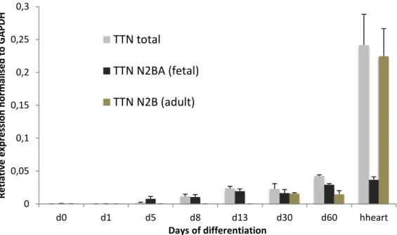
Figure 6: Expression of all the TTN transcripts and of the fetal N2BA isoforms and of the adult N2B isoforms throughout cardiac differentiation and in the adult human heart. Expression values were normalized to the GAPDH house keeping gene.
The titin gene starts to be expressed at day8, and until day 13 the adult N2B isoforms are not expressed, with the fetal N2BA isoforms being prevalent. At day 30 the N2B isoforms are already expressed, but the N2BA isoforms are still the majority of the transcripts. This is still true at day 60 when the splicing patter is still very different from the found in the adult human heart (Fig. 6). It is possible that keeping cardiomyocytes in culture for longer than a month is not advantageous if trying to achieve maturity on the splicing of this gene, much like the case of TNNI whose expression of the adult isoform stars after 2 months of culture, but remains low even after 9 months of culture39.
0 0,05 0,1 0,15 0,2 0,25 0,3 d0 d1 d5 d8 d13 d30 d60 hheart R e tlati ve e xp re ssi o n n o rm al ised to G A PD H Days of differentiation
TTN total
TTN N2BA (fetal)
TTN N2B (adult)
13
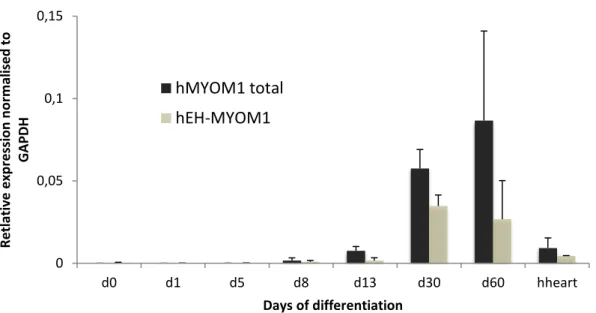
Figure 7 - Expression of all the MYOM1 transcripts and of the embryonic specific EH-MYOM1 isoform throughout cardiac differentiation and in the adult human heart. Expression values were normalized to the GAPDH house keeping gene.
In the case of myomesin the expression patter differs. At day 13 the embryonic EH isoform is only a small fraction of the transcripts, and the ratio of embryonic isoform does not seem to change (Fig. 7). This could mean that a short culture time is enough to achieve maturation when it comes to the splicing of this gene, or that this gene is not an adequate marker of maturation.
Figure 8 - Expression of all the TNNT2 transcripts and of the fetal specific cTnT1 isoform throughout cardiac differentiation and in the adult human heart. Expression values were normalized to the GAPDH house keeping gene.
When it comes to the expression of TNNT2, Figure 8 shows that much like titin, the gene starts to be expressed at day 8. At days 8 and 13 the cTnT1 fetal isoform represent the entirety of the cells transcripts. By day 30 there is a shift in splicing, and the ratio between the fetal isoform and the total number of transcripts greatly decreases, and this difference is accentuated at day 60 (Fig. 8). This implies an increase of the cTnT3 adult isoform. To confirm that a semi-quantitative PCR was done with a primer pair that amplifies both isoforms but with different fragment sizes, as shown in Figure 9(B). These results show that keeping the cells in culture for
0 0,05 0,1 0,15 d0 d1 d5 d8 d13 d30 d60 hheart R e tlati ve e xp re ssi o n n o rm al ised to GAP D H Days of differentiation
hMYOM1 total
hEH-MYOM1
-0,1 0,1 0,3 0,5 0,7 0,9 1,1 1,3 1,5 1,7 d0 d1 d5 d8 d13 d30 d60 hheart R e tlati ve e xp re ssi o n n o rm al ised to GAP D H Days of diferentiationhTNNT2 total
hTNNT2 cTnT1 (fetal)
14 up to 60 days allows the splicing of this gene to better resemble its splicing in the adult human heart, and further culture periods may decrease the slight gap that is still observed.
Figure 9: A - Schematic of the primer pair that allows semi-quantification of the adult TNNT2 isoform. The
alternatively spliced exon is shown in red. B - Agarose gel electrophoresis using the primer pair shown in A with the house keeping gene GAPDH as a loading control. C - Western blot of the TNNT2 protein with Tubulin as a loading control.
15
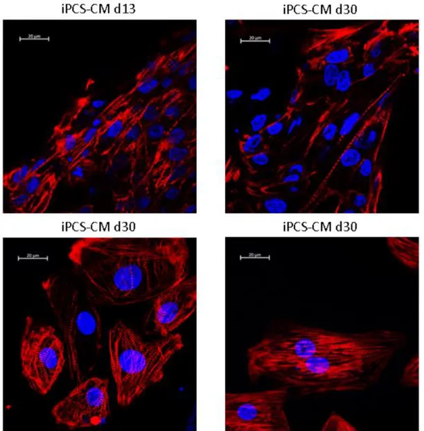
Figure 10: Immunofluorescent of iPSC-CM at day 13 and 30 of differentiation stained using DAPI and a TNNT2 specific antibody.
These results are partially supported in part by the immunofluorescence results, where between day 13 and day 30 cell show increased maturation in morphology, as their size increases and as the sarcomere becomes better organized (Fig. 10). The TNNT2 staining shows more distinct and cardiac fibres and with a higher level of striation, that tend, in general to present and increased degree of orientation. This shows a correlation in changes in splicing and changes in morphology.
16
Conclusions
In conclusion, these results indicate that extended the cells culture period is sufficient to induce a shift in the splicing pattern of TNNT2 and TTN mRNAs. Although hiPSC-CMs did not fully recapitulate the splicing patterns found in the human adult heart, this study highlights the relevance of long-term cultures for disease modeling and cardiac research.
This work only demonstrates the impact of long-term culture in splicing. As there are many other ways to improve iPSC-CM maturation, such as chemical, electrical and physical stimulation, it would be interesting to test some of these techniques and their impact in the splicing of these genes41. If these techniques are efficient in changing splicing, they may present
many advantages, the most striking one being a reduction in the amount of time needed to be able produce a model that better recapitulates the adult human heart.
References
1. Health, N. I. of. Regenerative Medicine 2006. Natl. Institutes Heal. 1–106 (2006). at <papers2://publication/uuid/05ECFE2C-F165-4F6C-8222-FCE8CDB6A13C> 2. Roth, G. A. et al. Demographic and Epidemiologic Drivers of Global Cardiovascular
Mortality. N. Engl. J. Med. 372, 1333–1341 (2015).
3. Naghavi, M. et al. Global, regional, and national age-sex specific all-cause and cause-specific mortality for 240 causes of death, 1990-2013: A systematic analysis for the Global Burden of Disease Study 2013. Lancet 385, 117–171 (2015).
4. World Health Organization. Global status report on noncommunicable diseases 2010.
World Health 176 (2010).
5. Forum, W. E. The global economic burden of non-communicable diseases. Lancet 1–20 (2011).
6. Bergmann, O. et al. Evidence for Cardiomyocyte Renewal in Humans. Science. 324, 98– 102 (2009).
7. Hunt, S. A. Current status of cardiac transplantation. J. Am. Med. Assoc. 280, 1692–1698 (1998).
8. Of, P. et al. Prevention of Rejection in Cardiac Transplantation By Blockade of the Interleukin-2 Receptor With a Monoclonal Antibody. N. Engl. J. Med. 342, 613–619 (2000).
9. National Heart, Lung, and Blood Institute.
10. Jaenisch, R. & Young, R. Stem cells, the molecular circuitry of pluripotency and nuclear reprogramming. 132, 567–582 (2014).
11. Young, H. E. et al. Adult reserve stem cells and their potential for tissue engineering.
Cell Biochem. Biophys. 40, 1–80 (2004).
12. De Los Angeles, A. et al. Hallmarks of pluripotency. Nature 525, 469–478 (2015). 13. Draper, J. S. et al. Recurrent gain of chromosomes 17q and 12 in cultured human
embryonic stem cells. 22, 2003–2004 (2004).
14. Martin, G. R. Isolation of a pluripotent cell line from early mouse embryos cultured in medium conditioned by teratocarcinoma stem cells Developmental Biology : Proc. Natl.
Acad. Sci. USA 78, 7634–7638 (1981).
15. Evans, M. J. & Kaufman, M. H. Establishment in culture of pluripotential cells from mouse embryos. Nature 292, 154–156 (1981).
16. Thomson, J. A. et al. Embryonic Stem Cell Lines Derived from Human Blastocysts.
Science (80-. ). 282, 1145–1148 (1998).
17. Takahashi, K. & Yamanaka, S. Induction of Pluripotent Stem Cells from Mouse Embryonic and Adult Fibroblast Cultures by Defined Factors. Cell 2, 663–676 (2006).
17 18. Takahashi, K. et al. Induction of Pluripotent Stem Cells from Adult Human Fibroblasts
by Defined Factors. Cell 131, 861–872 (2007).
19. Yu, J. et al. Induced pluripotent stem cell lines derived from human somatic cells.
Science (80-. ). 318, 301–302 (2007).
20. Yu, J. et al. Induced Pluripotent Stem Cell Lines Derived from Human Somatic Cells.
Science (80-. ). 318, 1917–1920 (2007).
21. Narsinh, K. H., Plews, J. & Wu, J. C. Comparison of Human Induced Pluripotent and Embryonic Stem Cells: Fraternal or Identical Twins? Mol. Ther. 19, 635–638 (2011). 22. Ohgushi, M. et al. Molecular pathway and cell state responsible for dissociation-induced
apoptosis in human pluripotent stem cells. Cell Stem Cell 7, 225–239 (2010).
23. Watanabe, K. et al. A ROCK inhibitor permits survival of dissociated human embryonic stem cells. Nat. Biotechnol. 25, 681–686 (2007).
24. Lian, X. et al. Cozzarelli Prize Winner: Robust cardiomyocyte differentiation from human pluripotent stem cells via temporal modulation of canonical Wnt signaling. Proc.
Natl. Acad. Sci. 109, E1848–E1857 (2012).
25. Lian, X. et al. Directed cardiomyocyte differentiation from human pluripotent stem cells by modulating Wnt/β-catenin signaling under fully defined conditions. Nat. Protoc. 8, 162–75 (2012).
26. Kwon, C. et al. Canonical Wnt signaling is a positive regulator of mammalian cardiac progenitors. Proc Natl Acad Sci U S A 104, 10894–10899 (2007).
27. Dravid, G. et al. Defining the Role of Wnt/β-Catenin Signaling in the Survival,
Proliferation, and Self-Renewal of Human Embryonic Stem Cells. Stem Cells 23, 1489– 1501 (2005).
28. Arnold, S. J. et al. Brachyury is a target gene of the Wnt/β-catenin signaling pathway.
Mech. Dev. 91, 249–258 (2000).
29. Lindsley, R. C. Canonical Wnt signaling is required for development of embryonic stem cell-derived mesoderm. Development 133, 3787–3796 (2006).
30. Ribeiro, M. C. et al. Functional maturation of human pluripotent stem cell derived cardiomyocytes invitro - Correlation between contraction force andelectrophysiology.
Biomaterials 51, 138–150 (2015).
31. Rodriguez, A. G., Han, S. J., Regnier, M. & Sniadecki, N. J. Substrate stiffness increases twitch power of neonatal cardiomyocytes in correlation with changes in myofibril structure and intracellular calcium. Biophys. J. 101, 2455–2464 (2011).
32. Nunes, S. S. et al. Biowire: a platform for maturation of human pluripotent stem cell-derived cardiomyocytes. Nat Methods 49, 781–787 (2013).
33. Wang, E. T. et al. Alternative isoform regulation in human tissue transcriptomes. Nature 456, 470–476 (2008).
34. Kalsotra, A. et al. A postnatal switch of CELF and MBNL proteins reprograms alternative splicing in the developing heart. Proc. Natl. Acad. Sci. 105, 20333–20338 (2008).
35. Lahmers, S., Wu, Y., Call, D. R., Labeit, S. & Granzier, H. Developmental Control of Titin Isoform Expression and Passive Stiffness in Fetal and Neonatal Myocardium. Circ.
Res. 94, 505–513 (2004).
36. Freiburg, A. et al. Series of exon-skipping events in the elastic spring region of titin as the structural basis for myofibrillar elastic diversity. Circ. Res. 86, 1114–21 (2000). 37. Schoenauer, R. et al. EH-myomesin splice isoform is a novel marker for dilated
cardiomyopathy. Basic Res. Cardiol. 106, 233–247 (2011).
38. Anderson, P. A. W., Malouf, N. N., Oakeley, A. E., Pagani, E. D. & Allen, P. D. Troponin T Isoform Expression in Humans. Circ. Res. 69, 1226–1233 (1991). 39. Bedada, F. B. et al. Acquisition of a quantitative, stoichiometrically conserved
ratiometric marker of maturation status in stem cell-derived cardiac myocytes. Stem Cell
Reports 3, 594–605 (2014).
18 human pluripotent stem cells. Nat. Biotechnol. 33, 970–979 (2015).
41. Denning, C. et al. Cardiomyocytes from human pluripotent stem cells: From laboratory curiosity to industrial biomedical platform. Biochim. Biophys. Acta - Mol. Cell Res. 1863, 1728–1748 (2016).