junho de 2014
Dalila Morais Teixeira
Are there structural differences in
Williams Syndrome social brains?
Universidade do Minho
Dissertação de Mestrado
Mestrado Integrado em Psicologia
Trabalho realizado sob orientação da
Professora Doutora Adriana Sampaio
e coorientação da
Professora Doutora Ana Mesquita
junho de 2014
Dalila Morais Teixeira
Are there structural differences in
Williams Syndrome social brains?
Universidade do Minho
iii Index Agradecimentos ... iv RESUMO ... vi ABSTRACT ... vii Introduction ... 8
Materials and Methods ... 14
Participants ... 14
Neurocognitive assessment ... 14
MRI Acquisition and Processing ... 14
Image Processing ... 14
Regions of Interest ... 15
posterior Superior Temporal Sulcus ... 15
Temporo-Parietal Junction ... 16
Subregions of the Cingulate Cortex ... 16
Data Analysis ... 17
Results ... 17
Overall Intracranial Volume ... 17
posterior Superior Temporal Sulcus ... 17
Temporo-Parietal Junction ... 18 Cingulate Cortex ... 19 Discussion ... 19 Future Directions ... 21 References ... 22 List of Tables ... 34 List of Figures ... 38
iv
Agradecimentos
Nesta dissertação estão inscritas diversas páginas que me ajudaram a construir enquanto ser e que me vão acompanhar para a vida. Apesar de individual não seria possível sem ajuda e apoio de muitas pessoas que contribuíram para que esta fosse alcançada com a melhor qualidade.
O meu grande agradecimento e carinho à professora Doutora Adriana Sampaio por me ter aceitado como orientanda, pelos incentivos, apoio, sugestões e correções pertinentes que fizeram com que fosse possível atingir esta grande meta, elevar os meus sonhos para outros patamares das Neurociências.
À professora Doutora Ana Mesquita, um grande obrigado, pelo apoio e orientação que me foi concedendo na minha etapa inicial no laboratório de Neuropsicofisiologia e ao longo deste percurso cheio de dúvidas e desejos em saber mais.
À professora Doutora Joana Coutinho pelo enorme tempo dispensado em que me ouviu as dúvidas, sonhos e frustrações, por me fazer acreditar nos meus sonhos e por me apaziguar nos momentos de maior stress, com a sua calma e serenidade sempre presente.
À sra. Engenheira Liliana Maia, um enorme e infinito obrigado por toda a ajuda, apoio, paciência e teres-me dado o gosto pela Neuroimagem.
À doutora Ana Pinheiro, uma enorme gratidão pelo apoio fundamental que me deu.
À Ana Carolina, Eduardo, Helga, Patrícia, Catarina, Alberto e Sónia pelas correcções e encorajamentos.
À equipa constituinte do Laboratório de Neuropsicofisiologia o meu muito obrigada por tudo. Pela companhia, boa disposição, integração e ajuda desde o início do meu percurso no Laboratório e por me fazerem acreditar cada vez mais que quero ser uma Neurocientista. É um obrigada que se manterá guardado para sempre no meu coração com muito carinho.
v
À Cátia Alexandra e ao Nuno Oliveira agradeço por os ter conhecido, por me terem ajudado nos bons e maus momentos e me terem acompanhado nesta última grande etapa sempre com um grande sorriso e um MMORPG.
Aos meus amigos de Viana do Castelo por me fazerem apreciar os bons momentos de amizade na vida e aos meus amigos da Banda Filarmónica Vila Nova de Anha pela boa companhia e disposição nos nossos momentos musicais.
Ao Nuno, por me teres acompanhado sempre em cada momento da minha vida ao longo destes três anos. A alegria, conforto, apoio incondicional e força que me dás tornam-me cada vez mais apaixonada pelo teu ser.
À minha família galega por toda a compreensão, carinho e acompanhamento que tiveram sempre comigo desde pequenina. À Amelia desejo o melhor futuro para ela, e que quando crescer lhe ensinarei o “bichinho” pelas Neurociências.
Ao meu maninho sr.Engenheiro Igor, o meu grande e emocionado agradecimento, pela ajuda, amor, paciência, troca de brincadeiras, risos que sempre trocamos e por seres o MELHOR irmão do universo.
Aos meus pais por serem SEMPRE o pilar da minha vida. Sem vocês eu não sou nada, simplesmente não vivo, são essenciais para a continuidade da minha existência. Agradeço-vos imensamente por me tornarem na mulher com ideias fortes e feministas que sou hoje.
vi
Mestrado Integrado em Psicologia da Universidade do Minho
Existem diferenças estruturais nos cérebros sociais em pacientes com Síndrome de Williams?
Dalila Morais Teixeira Adriana Sampaio
Ana Mesquita
RESUMO
A Teoria da Mente (ToM) é um processo crucial da cognição social e refere-se à capacidade de fazer predições acerca das ações das pessoas com base nos seus estados mentais, que representam a base da comunicação e interacção social humana. A característica mais relevante dos pacientes com Síndrome de Williams (WS) é a hipersociabilidade, que tem sido relatada em áreas como o pSTS(Sulco Temporal Superior posterior), TPJ(Junção Temporo-Parietal) e sub-regiões do CC(Córtex Cingulado).
O objetivo principal é avaliar volumes e assimetria do pSTS, TPJ e volumes das sub-regiões do CC no síndrome de Williams (WS) e no grupo controlo emparelhado por sexo e idade.
As imagens de ressonância magnética (N=34; 17 WS e 17 Controlos) foram exploradas, de forma a medir o volume da substância cinzenta total, substância branca total, e do líquido encefalorraquidiano nas áreas corticais mencionadas anteriormente.
Verificou-se um volume inferior intracraniano e nas áreas pSTS, TPJ esquerdo, Anterorostral, Anterodorsal e pCC (Córtex Cingulado Posterior) nos pacientes com WS, em comparação com os controlos.
Os resultados mostram que os WS apresentam uma simetria hemisférica, reflectindo-se na baixa capacidade de atribuir estados mentais e dificuldades em controlar a ansiedade emocional nas interações sociais.
vii
Mestrado Integrado em Psicologia da Universidade do Minho Are there structural differences in Williams Syndrome social brains?
Dalila Morais Teixeira Adriana Sampaio
Ana Mesquita
ABSTRACT
Theory of Mind (ToM), a crucial process of social cognition, refers to making predictions about people’s actions based on their mental states, representing the basis of human social communication and interaction. The most relevant feature of Williams Syndrome (WS) patients is the hypersociability, which has been implicated with specific neural signatures involving the posterior Superior Temporal Sulcus (pSTS), Temporo-Parietal Junction (TPJ) and subregions of Cingulate Cortex (CC).
The main goal is to evaluate volumes and asymmetry of pSTS, TPJ and volumes of subregions of CC in WS and in a typically developing age and sex matched group.
The acquired images from magnetic resonance imaging (N=34; 17 WS and 17 Controls) have been explored, in order to measure the whole gray matter, white matter, and cerebrospinal fluid volumes in the previous reported brain areas.
It has been verified a reduced intracranial volume and in the left pSTS, TPJ, Anterorostral, Anterodorsal and Posterior Cingulate Cortex in WS patients, compared with controls.
The results showed that WS present a high symmetry pattern, reflecting their poor ability to attribute mental states and difficulties in emotional control anxiety in social interactions.
8
Introduction
Brothers (1990) described “the social brain” as the higher cognitive and affective systems in the brain that evolved as a result of increasingly complex social selective pressures. These systems underlie our ability to function as highly social animals and provide the substrate for intact social cognition, social behavior and affective responsiveness.
Social cognition refers to the processes that subserves behavior in response to conspecifics (other individuals of the same species), and, in particular, to those higher cognitive processes subserving the extremely diverse and flexible social behaviors that are seen in primates (Adolphs, 1999). From infancy, humans display early signs of social cognition (Kovács et al., 2010; Striano and Reid, 2006). In fact, it is the social brain that allows us to interact with other people. As with all our interactions with the world, we can do much better if we can predict what is going to happen next in the social environment. The better we can predict what someone is going to do next, the more successful our interactions with that person will be (Frith, 2007a).
Perhaps the most important attribute of the social brain is that it allows us to make predictions about people's actions on the basis of their mental states. This assumption that behavior is caused by mental states has been called taking an ‘intentional stance’ (Dennett, 1987) or “having a theory of mind” (Premack & Woodruff, 1978a). One type of causality is the link between intentions and actions. Understanding that intentions cause actions and inferring intentions from actions might be a precursor to understanding the minds of others (Theory of Mind or ”mentalizing”; Leslie, 1994; Premack and Woodruff, 1978b).
Theory of Mind (ToM), the ability to attribute mental states to others is an important process in social cognition and thus forms the very basis of social interaction and communication (Burns, 2006). ToM involves activation of a widespread neural network comprising the medial prefrontal cortex (mPFC), the Posterior Cingulate Cortex (PCC)/ precuneus region (PC), as well as the middle temporal lobes (MT), superior temporal sulcus (STS), and the temporo-parietal junction (TPJ) (Saxe et al., 2004; Amodio and Frith, 2006; Saxe, 2006). In fact, several functional neuroimaging studies using a wide variety of mentalising tasks, evidenced that the process of mental state attribution has been associated with a network of brain regions that include the medial prefrontal cortex (mPFC), temporoparietal junction (TPJ), posterior Superior Temporal Sulcus (pSTS) and anterior temporal cortex (ATC) (Frith and Frith, 2003; Blakemore, 2012).
9
In fact, convergent evidence suggests therefore four specific brain regions, which are considered to have a role in social cognition: the posterior Superior Temporal Sulcus (pSTS) and the adjacent temporo-parietal junction (TPJ), the amygdala, the temporal poles, and the medial prefrontal cortex (mPFC) and the adjacent anterior cingulate cortex (ACC) (Frith, 2007b). In some studies, higher activity in more posterior regions, such as the pSTS/TPJ (Blakemore et al., 2007), and in the ATC (Burnett et al., 2009), was observed in adults as compared to adolescents. According to Gallagher and Frith (2003a) and Abu-Akel (2003), theory of mind can be connected to the anterior paracingulate cortex, the anterior cingulate gyrus, the ventral and medial prefrontal cortex, the inferolateral frontal cortex, the superior temporal sulcus, the temporal poles, and the amygdala too besides the orbitofrontal cortex.
Structural studies using voxel-based morphometry, have been reporting additional evidence that theory of mind was correlated with the volumes of the frontal and the temporal lobes (Lewis et al., 2011) have been reported.
Additionally, a correlation between social network size and mentalizing ability with gray matter density in the amygdala, ventromedial prefrontal cortex, anterior cingulate cortex (ACC), and in the temporal lobe, including the superior temporal sulcus (STS), in healthy subjects with larger social networks (Bickart et al., 2011; Kanai et al., 2012; Lewis et al., 2011). It is not just gray matter in STS, amygdala, and dorsal and anterior prefrontal cortex that is correlated with social network size but also its activity, measured with functional MRI (fMRI) (Sallet et al., 2011a; Bickart et al., 2012).
Curiously, spontaneous coupling of activity between STS and dorsal prefrontal cortex and ACC has been described as increased with social group size (Sallet et al., 2011b; Mars et al., 2012). Activity in these areas, and interactions between them, may be occurring more frequently when animals are in larger social groups because they have to make and adjust more predictions about what their cage mates, and groups of their cage mates, will do (Rushworth et al., 2013).
The STS has also consistently been described in ToM studies and this region therefore seems to be involved in the initial analysis of social cues and the detection of intentional activity. Specifically, the pSTS region plays an important role in the perception of social acts (Vander Wyk et al.,2009) and it was also found, that the right STS is significantly deeper on the right posterior segment as compared to the left (Barrick et al., 2005, Ochiai et al., 2004a, Van Essen, 2005 and Watkins et al., 2001) in human healthy brains. This difference is due to the deeper
10
development of the posterior sulcal root on the right (Ochiai et al., 2004b). A larger right volume in the depth of the STS might favor right hemispheric regions involved in theory of mind, voice perception, biological motion perception and visual processing (Glasel, 2011).
Brain regions near the temporo-parietal junction (TPJ), have been also implicated in a broad range of social cognition tasks (Allison et al., 2000; Gallagher and Frith, 2003b; Green and Haidt, 2003). Theory of mind tasks, which ask subjects to reason about the intentions and beliefs of others, activate medial prefrontal cortex and the TPJ (Adolphs, 2009). It is suggested that the TPJ is activated specifically in situations when one is inferring the mental states of others, rather than just information known about another (Saxe and Kanwisher, 2003a).
The TPJ, identified by responses to (false) belief stories, may play a broad role in social and even moral cognition (Moll 2002; Greene & Haidt, 2003a; Frith & Frith, 1999). The role of the TPJ in understanding other people appears to be specific to reasoning about the content of mental states (Saxe and Kanwisher, 2003).
Lesion (Samson et al., 2004; Apperly et al., 2007, Smith et al., 2013) and stimulation (transcranial magnetic stimulation – TMS and transcranial Direct Current Stimulation - tDCS) (Young et al, 2010; Santiesteban et al., 2012) studies provided further evidence suggesting that for social function is associated with the TPJ, but more generally, findings are highly dependent on spatial location. As described above, the best evidence for social function in the TPJ region comes from neuroimaging studies (Carrington and Bailey, 2009).
In accordance, neuroimaging research in social neuroscience suggests that the right TPJ (rTPJ) encodes imagined goals of others’ actions (Hamilton and Grafton, 2008) and contributes to social cognition by specifically representing others’ mental states such as thoughts and intentions (i.e., theory of mind, Saxe and Wexler, 2005) while left TPJ (lTPJ) seems to be associated with a broader range of tasks including mental states (false belief) as well as non-mental entities (false signs), as also left TPJ is necessary for the representation of non-mental states (Perner et al., 2006)
Finally, the cingulate cortex, including both anterior and posterior sectors (as well as the posteriorly adjacent retrosplenial cortex), is also described as playings a key role in emotion and in social behavior (Devinsky et al., 1995; Maddock, 1999)
The cingulate gyrus is a cortical area of mixed cytoarchitectonics that links to the limbic system and neocortex (Mesulam, 2000). The subcomponents of the cingulate gyrus serve a range
11
of functions, including emotional, cognitive and attentional, nociceptive, and motor processing (Drevets, 2000; Mesulam, 2001). Grossly, the anterior cingulate cortex (ACC) is differentiated from the posterior cingulate cortex (PCC) on the basis of cytoarchitecture, projection patterns, and functions (Vogt, 2008). For example, the anterior cingulate gyrus is activated by emotional stimuli, whereas the posterior cingulate gyrus is activated by both emotional and non-emotional stimuli and plays an important role in memory access and visuospatial orientation (Vogt et al., 1992a). Within the ACC, further parcellation can be made on the basis of functional and anatomical studies (Bush et al, 2000a). The rostral area of the ACC (the affective subregion) is connected to the nucleus accumbens, amygdala, insula, hippocampus, and orbitofrontal cortex and assesses the salience of emotional and motivational information regulating emotional responses (Bush et al, 2000b). The caudal (dorsal) area of the ACC (the cognitive subregion) has strong reciprocal interconnections with the lateral prefrontal cortex, parietal cortex, and premotor and supplementary motor areas (Devinsky et al., 1995) and modulates attention and executive functions, error detection, and working memory (Vogt et al., 1992b; Bush et al., 1999; Whalen et al., 1998). The portion of the ACC located inferior to the genu of the corpus callosum (subgenual subregion) has extensive connections to structures implicated in emotional behavior, mood, and autonomic responses to stressors and is the region used for deep-brain stimulation for treatment-resistant depression (Mayberg et al., 2003).
Many studies have shown that ACC responds to a variety of cognitive and affective ToM tasks (Chiu et al., 2008; Lombardo et al., 2009; Stuss et al., 2001; Völlm et al., 2006;Young et al., 2007). However, there is evidence suggesting that the ventral ACC (vACC) is involved in the emotional aspects of self-reflection (Moran et al., 2006; van der Meer et al., 2010), and that the dorsal ACC (dACC) is preferentially involved in processing cognitive ToM (Sommer et al., 2007).
Taking into account this three main areas role in social cognition, it is important to research these regions with key roles in social cognition, in populations with pro-social behavioral profiles, as patients with Williams Syndrome.
Williams syndrome(WS) is a neurodevelopmental disorder caused by a microdeletion of ~26 genes on chromosome 7q11.23 (Hillier et al., 2003; Osborne, 2012). This syndrome, which has a prevalence of 1/7,500 (Strømme et al., 2002), is associated with a recognizable pattern of physical characteristics, including a specific set of facial features, heart disease (most commonly
12
supravalvar aortic stenosis), connective tissue abnormalities, failure to thrive, and growth deficiency (Morris, 2006). The WS cognitive profile is characterized by deficits in visuospatial, motor and visuomotor abilities and relative strengths in face and object recognition, verbal short-term memory, expressive language and music processing skills (Morris, 2010; Dykens et al., 2005; Mobbs et al., 2007a; Tsai et al., 2008; Hocking et al., 2011).
WS profile is marked by overfriendliness, disinhibition and hypersociability, and are often impulsive (Bellugi et al., 1999; Klein-Tasman & Mervis 2003). Social behavior in WS may involve interaction of several factors including social cognition and social desire, regulation of emotions and disinhibition (Bellugi et al., 2007; Porter et al., 2007). The etiology of WS hypersociability is still unknown, however several studies have providing important informations regarding the neural and genetic mechanisms underlying WS social phenotype (Capitão et al., 2011).
The cognitive/behavioral phenotype has also been associated with several neuroanatomic changes, including volumetric and morphological changes, as well as abnormal asymmetry patterns (Sampaio et al., 2008a; Holinger et al., 2005). Importantly, brain alterations in individuals with WS, demonstrated a smaller overall brain volume (by 11–13%) and a smaller size of particular sub regions of the cerebral cortex, cerebellar cortex, and subcortical structures (Reiss et al., 2000; Eckert et al., 2005; Thompson et al., 2005). It was also observed differences in gray matter distribution (particularly in the left hemisphere) white matter abnormalities, as reduction in subcortical white matter (18%) (Hoeft et al., 2007; Marenco et al., 2007).
The anomalous brain structure in WS is largely consistent with the atypical social cognition and social behavior profile that characterizes the syndrome. Specifically, the cingulate, OFC, MPFC, insula, as well as the amygdala, are regions of the limbic system, which subserve functions related to emotion regulation, for example, heightened arousal, anxiety, enhanced emotional reactivity, and social impulsivity (Price, 1999a). Reiss et al. (2004b) observed in patients with WS, an increased volume and graymatter density in the anterior cingulate of participants with WS. However, volume increases were limited to the dorsal anterior (cognitive) segment, whereas increased density occurred within the ventral anterior (emotion-related) region (Bush et al., 2000c).
13
Williams syndrome patients show difficulties in higher order social-cognitive functions, manifested by atypical non-verbal communication, imagination, and problems in understanding the mental states of others (i.e., theory of mind) (Haas & Reiss, 2012).
This leads to a reduced ability to understand the mental states of others with emotional area preserved (Porter et al., 2007a).
While ToM and intellecttual abilities are often commensurate, dissociations occur within certain disorders. For example, people with autism or schizophrenia typically display deficits in their understanding of mental states such as intentions, thoughts and beliefs beyond their general level of intellectual functioning not surprisingly, their day-to-day social functioning is also impaired. ToM and intellectual abilities may also dissociate in people with Williams syndrome. Unlike people with autism or schizophrenia, people with Williams syndrome (WS) are sometimes said to show strengths in their day-to-day social functions, so one might expect that ToM abilities in WS are above their general intellectual abilities(Porter, 2007b).
The hypotheses for each ROI are:
1) Is rpSTS a region in WS that is involved in the analysis of the social cues and in the ability of inferring predictions in larger social groups?
2) Is the rTPJ in WS that encodes imagined goals of others’ actions? 3) Is the Anterrostral in WS capable of regulating emotional responses?
4) Is the Anterodorsal area in WS capable of establishing reciprocal interconnections between emotion and action?
5) Is the Subgenual implicated in regulation of emotional behavior?
6) Is the Posterior CC in WS capable of modulate attention to emotional stimulus?
The current study is the first to provide a detailed characterization of Cingulate Cortex (CC) using an automatic gray matter segmentation plus it´s posterior manual correction in 3D Slicer in WS.
Therefore, the main goal of this research study is to evaluate the volume and asymmetry of the pSTS, TPJ and subregions of CC in WS (N=17) and in a typically developing group (N=17) matched on age and sex.
14
Materials and Methods
Participants
Study participants included 17subjects (7 males and 10 females), diagnosed with WS (19,24 ± 6,037; age range: 11 to 32 years) and 17 healthy controls individually matched for sex, and age (7 males and 10 females) (20,53 ± 6,424; age range: 11 to 32 years). Mean full scale IQ was (50,12 ± 6,754) for WS and (110,13 ± 11,495) for controls.
Participants with WS were recruited at the Genetic Medical Institute (Portugal) and the Genomic Foundation in Galicia (Spain). WS diagnoses were made by fluorescent in situ hybridization for confirmation of elastin gene deletion (Ewart et al. 1993).
Controls were typically developing individuals without evidence of psychiatric, neurological disorder or cognitive impairment. Each participant gave written informed consent for their participation in the study via consent forms, after a complete description of the study. All participants were right-handed, determined through clinical interview.
Neurocognitive assessment
To assess general cognitive functioning, participants 8-16 years of age were administered the Wechsler Intelligence Scale for Children-Third Edition (WISC–III) (Wechsler 1991, 1997), while subjects over 16 years old were administered the Wechsler Adult Intelligence Scale- Third Edition (WAIS-III).
MRI Acquisition and Processing
MRI images were obtained on a 1.5-T General Electric system (GE Medical Systems). The scans acquisition protocol consisted of contiguous 1.5-mm coronal T1 (Spoiled gradient - SPGR) slices of the whole brain and an axial PD/T2 sequence (proton density and T2-weighted). The parameters used were: echo time: 5.0 msec, repetition time 35 msec, flip angle 45º, acquisition matrix 256x192, voxel dimensions: 9375x0.9374x1.5mm.
Image Processing
Cortical reconstruction was performed with the Freesurfer 5.3 image analysis suite along with APARC Atlas (Fischl et al., 2002), which is documented and freely available for download online (http://surfer.nmr.mgh.harvard.edu/). The utilization of this software for the analysis of
15
MRI structural images is an application well developed and largely wide used in the literature (Klauschen et al. 2009; Reuter et al. 2012).
In the automatic reconstruction process of structural images it can occur some mistakes, being at this way necessary the manual verification of these same errors. The steps for verification includes: verification of the co-sign in the Tailarach template (template based on the distance of the posterior and anterior commissure), verification of the skull remove (because it can occur errors at the remotion of the cerebral tissue or in the no- totally remove of the brain), errors in the boundaries definition of the cortical and sub-cortical surfaces. For correction, it´s necessary to remove or enhance voxels manually.
After this steps for the reconstruction verification, it was possible to obtain the images already processed (with the contrasts corrected, aligned by the Tailarach template and with the skull removed) for this work.
Then, each cortical structure (except for Subgenual subregion) reconstruction obtained was manually corrected by using 3D slicer version 4.3.1. The subgenual subregion of ACC was manually segmented (Koo et al., 2008) using the 3D Slicer Software in the realigned images. It was used the label zero (0) to erase and the label one (1) to paint effect to add labels. From 3D slicer it has been extracted the measurements of grey matter volume (mm3) for each region of interest plus the gray matter, white matter and CSF (mm3) of each brain for TIV calculation.
Regions of Interest
In this work it has been extracted from FreeSurfer six labels, each one correspondent to each ROI in my study involved in Theory of Mind. Then, as described these were all manually corrected by using 3D Slicer except the subgenual subregion that was manually segmented by using the same tool.
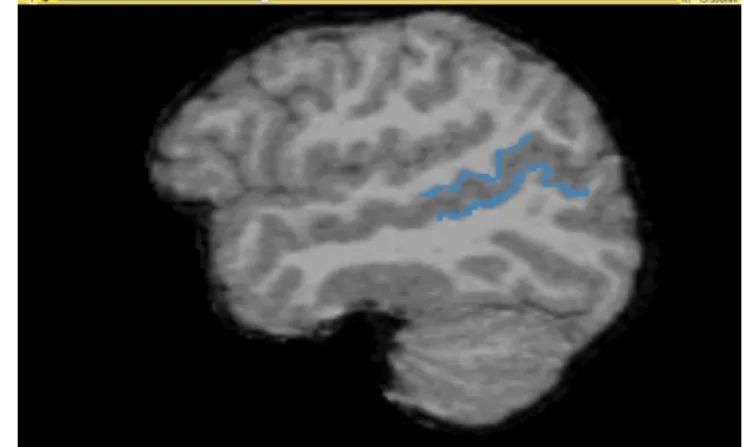
posterior Superior Temporal Sulcus
The posterior Superior Temporal Sulcus label was extracted from the files
rh.pSTS.ANNOT and lh.pSTS.Annot. Kathryn Mills et al.(2013), was created the pSTS region by
extending the Desikan-Killiany Atlas defined bank STS (Fischl et al., 2004; Desikan et al., 2006) to the border of the TPJ.
16
Temporo-Parietal Junction
The Temporo-Parietal junction label was extracted from the files rh.TPJp.ANNOT and
lh.TPJp.ANNOT. It was used the border coordinates of a functional subdivision generated in a
previous study (Mars et al., 2012). Subregions of the Cingulate Cortex
1) Anterrostral: the affective subregion (anterorostral) (Bush et al., 2000a) was bounded anteriorly by the cingulate sulcus and posteriorly above the corpus callosum by a line (Crespo-Facorro et al., 1999) passing through the most anterior point of the inner surface of the genu of the corpus callosum, and anterior to the subgenual division below the corpus callosum. This region was extracted from the label 2023.
2) Anterodorsal: the cognitive subregion (anterodorsal) (Bush et al., 2000b) is defined as the remaining ACC between the affective subregion and posterior cingulate gyrus. This region was extracted from the label 2002.
3) Subgenual: the subgenual subregion (Drevets et al., 1997) was segmented by using the following boundaries defined as the cingulate area under the corpus callosum bounded anteriorly by the line passing through the anterior margin of the genu of corpus callosum and posteriorly 1 section anterior to the internal capsule that divides the striatum.
4) Posterior CC: the posterior cingulate extends to the line passing through the most posterior end of the corpus callosum (Noga et al., 1995). This region was extracted from the label 2026.
A relative measure of pSTS, TPJ and all four subregions of CC were computed as the ratio between cortical volume and total gray matter volume. Asymmetry index of pSTS and TPJ were computed according to the following expression: (L−R)/0.5 (L+R), where L and R refer to left and right hemispheres.
17
Data Analysis
All volumetric data met the criteria for the use of parametric tests, including normality (Kolmogorov-Smirnov and Shapiro-Wilk tests) and variance homogeneity (Levene test). A repeated-measure analysis of variance (Mixed ANOVA) was used to determine pSTS and TPJ absolute, relative volumes differences between the WS and control subjects. Thus, diagnosis (WS and controls) was used as the between- subject factor and hemispheric side (left vs. right) as the within factor. If a main effect for group was found, then a Post Hoc t-test was used to test the mean difference between groups. A Post Hoc t-test it was also used to test the mean difference in relative volumes of CC subregions between groups as also the asymmetry in pSTS and TPJ. A p value less than 0.05 was assumed to denote a significant difference.
Results
Overall Intracranial Volume
WS participants showed absolute values of gray matter [t(32)= -2.582, p= 0.015], white matter [t(32)= -5,030, p=0,000], and CSF [t(32)= -3.477, p=0.001] volumes that were significantly reduced compared with controls. As a consequence, TIV was significantly reduced in the clinical group [t(32) = -4.998, p= 0.000] (Table 2).
When relative volume was estimated (ie, ratio between tissue volume and TIV), no significant differences were found for CSF volume [t(32)= -1.358, p=0.184]. However, gray matter volume [t(32)=2.358, p=0.025] was significantly increased and white matter volume [t(32)=-2.329, p=0.026] was significantly reduced (Table 2).
posterior Superior Temporal Sulcus
Repeated-measures analysis of variance of absolute volumes revealed a significant difference, showing main effect of side (left vs. right) [F(1,32) = 9.090, p=0.005)], and a marginal interaction between side and diagnosis [F(1,32) = 3.396, p= 0.075)]. No diagnosis effect was found [F(1,32) = 2.577 , p=0.118)].
18
Post Hoc t-test showed that absolute pSTS volumes (Table 3) were significantly reduced
in WS in the left hemisphere [t(32) = −2.425, p=0.021] when comparing with control group, than in the right hemisphere [t(32) = -0.270, p= 0.789].
However when relative volumes of pSTS were computed (ratio between pSTS volume and total gray matter volume) (Table 3), a main effect of side [F(1,32) = 9.650, p = 0.004)] was found. An interaction between side and diagnosis was not found [F(1,32) = 2.555, p = 0.120)]. No diagnosis effect was found [F (1,32) = 0.545, p = 0.466)]. Indeed, t tests yielded no statistical significant difference between the 2 groups, for either right hemisphere [t(32) = 1.630, p = 0.113] or left hemisphere [t(32) = −0.403, p = 0.690] (Table 3).
The cortical asymmetry between left-right pSTS (Table 4) was no significant [t(32) = −1.572, p = 0.126] between groups.
Temporo-Parietal Junction
Repeated-measures analysis of variance of absolute volumes revealed no significant difference in effect of side (left vs. right) [F(1,32) = 0.076, p = 0.785)], diagnosis [F(1,32) = 2.883, p = 0.099)], neither in interaction between side and diagnosis [F(1,32) = 1.357, p = 0.253)].
Post Hoc t-test showed that absolute TPJ volumes(Table 3) were significantly reduced in
WS in the left hemisphere [t(32) = −2.151, p= 0.039] but not in the right hemisphere [t(32)= -0.902, p= 0.374].
Relative volumes of TPJ were computed (ratio between TPJ volume and total gray matter volume). It was not found a side effect [F(1,32) = 0.071, p = 0.791)] neither an interaction between side and diagnosis [F(1,32) = 0.939, p = 0.340)]. No diagnosis effect was found [F (1,32) = 0.032, p = 0.859)]. Indeed, t tests yielded no statistical significant difference between the 2 groups, for either right hemisphere [t(32) = 0.433, p = 0.668] or left hemisphere [t(32) = −0.762, p = 0.451] (Table 3).
The cortical asymmetry between left-right TPJ (Table 4). was no significant [t(32) = −1.168, p = 0.252] (Table 4).
19
Cingulate Cortex
Anterorostral subregion
T-test showed that absolute Anterorostral volumes were significantly reduced in WS, when comparing with control group, [t(32) = -3,412, p=0,002]. Relative volume revealed group differences [t(32)= -2,041, p= 0,050] (Table 3) in WS participants.
Anterodorsal subregion
T-test showed that absolute Anterodorsal volumes were significantly reduced in WS, when comparing with control group, [t(32) = −2,595, p=0,014]. No group differences were observed for Anterodorsal relative volume [t(32)= -1,349, p= 0,187] (Table 3).
Subgenual subregion
T-test showed that were no statistical significant differences in absolute Subgenual volumes [t(32) = −0,296, p=0,769]. It was not observed any group differences in subgenual relative volume [t(32)= 0,426, p= 0,673] (Table 3).
Posterior Cingulate Cortex
T-test showed that absolute Posterior CC volumes were significantly reduced in WS, when comparing with control group, [t(32) = −3,115, p=0,004]. No group differences in Posterior Cingulate Cortex relative volumes were found [t(32)= -1,483, p= 0,148] (Table 3).
Discussion
This study provides an insight as to how altered or delayed brain cortical structures contributes from atypical social behavior in WS.
The main goal of this study was to assess structural alterations in three of the main brain regions involved in social cognition – pSTS, TPJ and CC in Williams syndrome.
20
The present study confirms an overall reduction in brain volumes in WS patients, including a reduction in overall gray matter, white matter, and CSF compared with typically developmental controls. Most importantly, this reduction was found to be disproportionate. That is, when relative volumes were computed, the WS patients showed a gray matter volume increase, in parallel with a decrease in white matter volumes although it wasn´t observed any difference in CSF volumes in WS patients. These facts are in accordance with previous studies related by Reiss et al. (2000; 2004a) and Sampaio et al.(2008b).
A volumetric reduction in the left pSTS in the WS group was evidenced, which suggest that the ability of perception the acts of other´s including the intentions that are intrinsic to them, is weak. However the preservation of rpSTS is consistent with evidence of the relationship between the observed (mental states) and the structure of the surrounding environment involved in the representation of observed intentional actions is affected (Saxe et al.,2004)
The relative preservation of right TPJ in WS participants, reported in the current study predicts the need to think about thoughts. This combines with the reduced ability to understand the mental states of others (Santos and Deruelle, 2009) in WS. However it was observed a volumetric decrease in left TPJ, which suggests that this area is poor when implicated in processing representations that refers to the presence of persons in relational contexts in general (Takahashi et al.,2008).
Patients with WS present reduced volumes in anterorostral and the anterodorsal subregions of the Anterior Cingulate Cortex volumes. These data suggest that WS might not have the capacity of regulating emotional responses. Specifically, during intense emotional states it might exist a reciprocal suppression of the cognitive subregion, which corresponds to previous data where refer enhanced emotional reactivity in this patients (Price, 1999b).The Posterior Cingulate Cortex is also reduced in WS, which is possibly related to difficulties in memory at accessing to emotional stimulus.
When compared with relative volumes of each Cingulate Cortex, only the anterorostral subregion has shown a marginal group effect, which suggests that this specific subregion in WS is associated with difficulties poor in regulating emotional responses. This subregion is activated during emotional processing in normal healthy volunteers and symptom provocation studies in a number of psychiatric disorders as anxiety (Drevets et al., 1997; Whalen et al., 1998; Bush et al.,
21
1998). This is consistent with data showing increased anxiety and social impulsivity levels in WS.
In conclusion, the present study reveals that WS show a high symmetry brain, where the rpSTS and rTPJ are relatively preserved, suggesting that these regions may be partially associated with the ability to attribute mental states to others. Reduced volumes in the lpSTS, lTPJ, anterrostral, anterodorsal and Posterior Cingulate Cortex are consistent with the hypothesis that WS present difficulties in control their emotions and anxiety which leads to the difficulty in maintain a social interaction without be “too” spontaneous. This provides evidence for deficits in inhibitory capacity. The link between intentions and actions is at this way maintained.
Future Directions
Future studies are needed to more closely evaluate the implications of structural brain anomalies in WS at this specifically area (social brain). It will be interesting to made a structural and functional MRI in the field of auditory processing, related to social interactions in WS. It will be also interesting to compare these results with other studies in other neurodevelopmental disorders group, such as ADHD, Down Syndrome and Fragile X syndrome.
22
References
Abu-Akel, A. (2003). A neurobiological mapping of theory of mind. Brain Res. Rev, 43, 29–40.
Adolphs, R. (1999). Social Cognition and the Human Brain. Trends in Cognitive Science, 3(12), 469-479.
Adolphs R (2009). The social brain: Neural basis of social knowledge. Annu Rev Psychol, 60, 693–716.
Amodio, D.M., Frith, C.D. (2006). Meeting of minds: the medial frontal cortex and social cognition. Nat. Rev. Neurosci, 7 (4), 268–277.
Allison, T., Puce, A., McCarthy, G. (2000). Social perception from visual cues; role of the STS region. Trends Cogn. Sci, 4, 267-278.
Apperly, I.A.. et al. (2007). Testing the domain-specificity of a theory of mind deficit in brain injured patients: evidence for consistent performance on non-verbal, “reality-unknown” false belief and false photograph tasks. Cognition, 103, 300-321.
Barrick, T.R., Mackay, C.E., Prima, S., Maes, F., Vandermeulen, D., Crow, T.J., Roberts, N. (2005). Automatic analysis of cerebral asymmetry: an exploratory study of the relationship between brain torque and planum temporale asymmetry. NeuroImage, 24, 678-691.
Bellugi, U., Adolphs, R., Cassady, C., Chiles, M. (1999). Towards the neural basis for hypersociability in a genetic syndrome. Neuro-Report,10(8),1653.
Bellugi, U., Jarvinen-Pasley, A., Doyle, T.F., Reilly, J., Reiss, A.L., Korenberg, J.R., (2007). Affect, social behavior and the brain in Williams syndrome. Current Directions in
23
Bickart, K.C., Wright, C.I., Dautoff, R.J., Dickerson, B.C., Barrett, L.F. (2011). Amygdala volume and social network size in humans. Nat Neurosci , 14, 163-164.
Bickart, K.C., Hollenbeck, M.C., Barrett, L.F., Dickerson, B.C. (2012). Intrinsic amygdala cortical functional connectivity predicts social network size in humans. J Neurosci, 32 , 14729 14741.
Brothers, L. (1990). The social brain: a project for integrating primate behaviour and neurophysiology in a new domain. Concepts in Neuroscience, 1, 27-51.
Burns, J. (2006). The social brain hypothesis of schizophrenia. World Psychiatry, 5(2), 77–81.
Burnett, S., Bird, G., Moll, J., Frith, C., Blakemore, S.J. (2009). Development during ado- lescence of the neural processing of social emotion. Journal of Cognitive Neuroscience, 21, 1736-50.
Blakemore, S.J., den Ouden, H., Choudhury, S., Frith, C. (2007). Adolescent development of the neural circuitry for thinking about intentions. Social Cognitive and Affective Neuroscience, 2, 130-9.
Blakemore, S.J. (2012). Development of the social brain in adolescence. Journal of the Royal
Society of Medicine, 105, 111-6.
Bush, G. et al. (1998). The counting Stroop: an interference task specialized for functional neuroimaging – validation study with functional MRI. Hum. Brain Mapp, 6, 270–282.
Bush, G. et al. (1999). Anterior cingulate cortex dysfunction in attention deficit/hyperactivity disorder revealed by fMRI and the counting Stroop. Biol. Psychiatry, 45, 1542–1552.
Bush G, Luu P, Posner MI. (2000a). Cognitive and emotional influences in anterior cingulate cortex. Trends Cogn Sci, 4(6), 215–222.
24
Bush G, Luu P, Posner MI. (2000b). Cognitive and emotional influences in anterior cingulate cortex. Trends Cogn Sci, 4(6), 215–222.
Bush G, Luu P, Posner MI. (2000c). Cognitive and emotional influences in anterior cingulate cortex. Trends Cogn Sci, 4(6), 215–222.
Capitão, L., Sampaio, A., Férnandez, M., Sousa, N., Pinheiro, A., & Gonçalves, O. (2011). Williams syndrome hypersociability: A neuropsychological study of the amygdala and prefrontal cortex hypotheses. Research in Developmental Disabilities, 32, 1169–1179.
Carrington, S.J. and Bailey, A.J. (2009). Are there theory of mind regions in the brain? A review of the neuroimaging literature. Hum. Brain Mapp, 30, 2313-2335.
Chiu., P.H, Kayali, M.A., Kishida, K.T., Tomlin, D., Klinger, L.G., Klinger, M. et al. (2008).
Self responses along cingulate cortex reveal quantitative neural phenotype for high functioning
autism. Neuron, 57 463–473.
Crespo-Facorro B, Kim JJ, Andreasen NC, O'Leary DS, Wiser AK, Bailey JM, Harris G,
Magnotta VA. (1999). Human frontal cortex: an MRI-based parcellation method. Neuroimage, 10(5):500–519.
Dennett, D.C. (1987). The intentional stance. Cambridge, MA: The MIT Press.
Devinsky, O., Morrell, M.J., Vogt, B.A. (1995). Contributions of anterior cingulate cortex to behavior. Brain, 118, 279–306.
Desikan, R.S., Ségonne, F., Fischl, B., et al. (2006). An automated labeling system for subdividing the human cerebral cortex on MRI scans into gyral based regions of interest. NeuroImage, 31, 968–80.
25
prefrontal cortex abnormalities in mood disorders. Nature 1997, 386(6627),824–827.
Drevets., WC. (2000). Neuroimaging studies of mood disorders. Biol Psychiatry, 48(8), 813-29.
Dunbar, R. I. M. (2009). Darwin and the ghost of Phineas Gage: Neuro-evolution and the social brain. Cortex, 45, 1119-1125.
Dykens, E.M., Rosner, B.A., Ly, T., Sagun, J. (2005). Music and anxiety in Williams syndrome: a harmonious or discordant relationship? Am J Ment Retard. 110, 346–358.
Eckert, M.A., Hu, D., Eliez, S., et al. (2005). Evidence for superior parietal impairment in Williams syndrome. Neurology, 64, 152–153.
Fischl B, Salat DH, Busa E, Albert M, Dieterich M, Haselgrove C, van der Kouwe A, Killiany R, Kennedy D, Klaveness S, Montillo A, Makris N, Rosen B, Dale AM. (2002). Whole brain segmentation: automated labeling of neuroanatomical structures in the human brain. Neuron, 33, 341–355.
Fischl, B., van der Kouwe, A., Destrieux, C., et al. (2004). Automatically parcellating the human cerebral cortex. Cerebral Cortex, 14, 11 –22.
Frith, C.D., & Frith, U. (1999). Interacting minds—a biological basis. Science, 286, 1692–1695.
Frith, U., Frith, C.D. (2003). Development and neurophysiology of mentalizing. Philosophical
Transactions: Biological Sciences, 358, 459-73.
Frith, C. (2007a). The social brain? Phil. Trans. R. Soc. B, 362, 671–678.
Frith, C. (2007b) The social brain? Phil. Trans. R. Soc. B, 362, 671–678.
26
Sci, 7, 77-83.
Gallagher, H.L., & Frith, C.D. (2003b). Functional imaging of “theory of mind”. Trends Cogn.
Sci, 7, 77-83.
Glasel, H., Leroy, F., Dubois, J.,Hertz-Pannier, L., Mangin, J.F., Dehaene-Lambertz, G. (2011). A robust cerebral asymmetry in the infant brain: The rightward superior temporal sulcus.
NeuroImage, 58, 716-723.
Greene, J., & Haidt, J. (2003a). How (and where) does moral judgement work? Trends Cogn.
Sci, 6, 517–523.
Greene, J., & Haidt, J. (2003b). How (and where) does moral judgement work? Trends Cogn.
Sci, 6, 517–523.
Hamilton, A.F., Grafton, S.T. (2008). Action outcomes are represented in human inferior frontoparietal cortex. Cereb Cortex, 18, 1160-1168.
Hillier, L.W., Fulton, R.S., Fulton L.A., et al. (2003). The DNA sequence of chromosome 7. Nature, 42, 157–164.
Hocking, D.R., Rinehart, N.J., McGinley, J.L., Moss, S.A., Bradshaw, J.L. (2011). A kinematic analysis of visually-guided movement in Williams syndrome. J Neurol Sci, 301, 51–58.
Hoeft, F., Barnea-Goraly, N., Haas, B.W., Golarai, G., Ng, D., Mills, D., Korenberg, J., Bellugi, U., Galaburda, A., Reiss, A.L. (2007). More is not always better: increased fractional
anisotropy of superior longitudinal fasciculus associated with poor visuospatial abilities in Williams syndrome. J Neurosci, 27(44), 11960–11965.
Holinger, D.P., Bellugi, U., Mills, D.L., Korenberg, J.R., Reiss, A.L., Sherman, G.F., Galaburda, A.M. (2005). Relative sparing of primary auditory cortex in Williams Syndrome. Brain Res,
27
1037(1–2), 35–42.
Kanai, R., Bahrami, B., Roylance, R., Rees, G. (2012). Online social network size is reflected in human brain structure. Proc Biol Sci, 279, 1327-1334.
Klein-Tasman, B., & Mervis, C. (2003). Distinctive personality characteristics of 8, 9, and 10 year-olds with Williams syndrome. Dev Neuropsychol, 23(1), 269–290.
Kovács, Á. M., Téglás, E., & Endress, A. D. (2010). The social sense: susceptibility to others' beliefs in human infants and adults. Science, 330, 1830-4.
Leslie, A.M. (1994). Pretending and believing: issues in the theory of tomm. Cognition, 50 (1 – 3), 211 – 238.
Lewis, P.A., Rezaie, R., Brown, R., Roberts, N., Dunbar, R.I. (2011). Ventromedial prefrontal volume predicts understanding of others and social network size. Neuroimage, 57, 1624-1629.
Lombardo, M.V., Chakrabarti, B., Bullmore, E.T., Sadek, S.A., Pasco, G., Wheelwright ,S.J. et al. (2009). Atypical neural self-representation in autism. Brain, 132, 306–320.
Maddock, R.J. (1999). The retrosplenial cortex and emotion: new insights from functional neuroimaging of the human brain . Trends Neurosci, 22, 310–316.
Marenco, S., Siuta, M.A., Kippenhan, J.S., Grodofsky, S., Chang, W.L., Kohn, P., Mervis, C.B., Morris, C.A., Weinberger, D.R., Meyer-Lindenberg, A., Pierpaoli, C., Berman, K.F. (2007). Genetic contributions to white matter architecture revealed by diffusion tensor imaging in Williams syndrome. Proc Natl Acad Sci USA, 104(38), 15117–15122.
Mars RB, Neubert FX, Noonan MP, Sallet J, Toni I, Rushworth MF. (2012). On the relationship between the ‘‘default mode network’’ and the ‘‘social brain’’. Front Hum Neurosci, 6,189. Mars, RB., Sallet, J., Schuffelgen, U., Jbabdi, S., Toni, I., Rushworth, MF. (2012).
28
“Connectivity- based subdivisions of the human right “temporoparietal junction area”: evidence for different areas participating in different cortical networks. Cereb Cortex, 22, 1894-1903.
Mayberg HS.(2003). Modulatingdysfunctionallimbic-cortical circuits in depression: towards development of brain-based algorithms for diagnosis and optimised treatment. Br Med Bull, 65, 193–207.
Mesulam, M.M. (2000). Principles of behavioral and cognitive neurology. Oxford, 1(2), pp 1 120.
Mesulam M.M. (2001). Primary progressive aphasia. Ann Neurol, 49, 425–32.
Mills KL, Lalonde F, Clasen LS, Giedd JN, Blakemore S-J. (2013). Developmental changes in the structure of the social brain in late childhood and adolescence. Soc. Cogn. Affect. Neurosci, 9(1), 123-131.
Mobbs, D., Eckert, M.A., Mills, D., Korenberg, J., Bellugi, U., et al. (2007). Frontostriatal dysfunction during response inhibition in Williams syndrome. Biol Psychiatry, 62, 256–261.
Moll, J., Oliveira-Souza, R., Eslinger, P.J., Bramati, I.E., Mourao-Miranda, J., Andreiuolo, P.A., Pessoa, L. (2002). The neural correlates of moral sensivity: a functional magnetic resonance imaging investigation of basic and moral emotions. J. Neurosci, 22, 2730-2736.
Moran, J.M., Macrae, C.N., Heatherton, T.F., Wyland, C.L, & Kelley, W.M. (2006).
Neuroanatomical evidence for distinct cognitive and affective components of self. Journal of
Cognitive Neurosciences, 18, 1586-1594.
Morris, C. A. (2006). The dysmorphology, genetics, and natural history of Williams–Beuren syndrome. In C. A. Morris, H. M. Lenhoff, & P. P. Wang (Eds.) Williams - Beuren syndrome:
29
Morris, C.A. (2010). The behavioral phenotype of Williams syndrome: A recognizable pattern of neurodevelopment. Am J Med Genet C Semin Med Genet, 154C, 427–431.
Noga, J.T., Aylward, E., Barta, P.E., Pearlson, G.D. (1995). Cingulate gyrus in schizophrenic patients and normal volunteers. Psychiatry Res, 61(4),201–208.
Ochiai, T., Grimault, S., Scavarda, D., Roch, G., Hori, T., Riviere, D., Mangin, J.F., Regis, J. (2004a). Sulcal pattern and morphology of the superior temporal sulcus. NeuroImage, 22, 706 719.
Ochiai, T., Grimault, S., Scavarda, D., Roch, G., Hori, T., Riviere, D., Mangin, J.F., Regis, J. (2004b). Sulcal pattern and morphology of the superior temporal sulcus. NeuroImage, 22, 706 719.
Osborne, LR. (2012). Genes: The gene expression approach. In: Farran, EK.; Karmiloff-Smith, A., editors. Neurodevelopmental disorders across the lifespan: A neuroconstructivist approach, 59-81.Oxford, UK: Oxford University Press.
Price JL. (1999a). Prefrontal cortical networks related to visceral function and mood. Annals of
the New York Academy of Sciences. 877, 383–396.
Price JL. (1999b). Prefrontal cortical networks related to visceral function and mood. Annals of
the New York Academy of Sciences. 877, 383–396.
Perner, J., Aichhorn, M., Kronbichler, M., Staffen, W., Ladurner, G. (2006): Thinking of mental and other representations: The role of left and right temporo-parietal junction. Soc Neurosci 1, 245– 258.
Premack, D., Woodruff, G. (1978). Does the chimpanzee have a theory of mind? Behav. Brain
Sci., 4, 515 – 526.
30
hypersociability in Williams and Down syndrome. Neuropsychologia . 45, 2839-49.
Porter, M.A., Coltheart, M., & Langdon, R. (2007b). The neuropsychological basis of hypersociability in Williams and Down syndrome. Neuropsychologia . 45, 2839-49.
Reiss, A.L., Eliez, S., Schmitt, J.E., et al. (2000). Neuroanatomy of Williams syndrome: a high resolution MRI study. J Cognit Neurosci,12(1), 65–73.
Reiss, A.L., Eckert, M.A., Rose, F.E., et al. (2004a) An experiment of nature: brain anatomy parallels cognition and behavior in Williams syndrome. J Neurosci, 24, 5009–5015.
Reiss, A.L., Eckert, M.A., Rose, F.E., et al. (2004b) An experiment of nature: brain anatomy parallels cognition and behavior in Williams syndrome. J Neurosci, 24, 5009–5015.
Rushworth, M.F., Mars, R.B., and Sallet, J. (2013). Are there specialized circuits for social cognition and are they unique to humans? Curr. Opin. Neurobiol, 23, 436–442.
Sallet, J., Mars, R.B., Noonan, M.P., Andersson, J.L., O´Reilly, J.X., Jbabdi, S., Croxson, P.L., Jenkinson, M., Miller, K.L., Rushworth, M.F. (2011a). Social network size affects neural circuits in macaques. Science, 334, 697–700.
Sallet, J., Mars, R.B., Noonan, M.P., Andersson, J.L., O´Reilly, J.X., Jbabdi, S., Croxson, P.L., Jenkinson, M., Miller, K.L., Rushworth, M.F. (2011b). Social network size affects neural circuits in macaques. Science, 334, 697–700.
Sampaio, A., Sousa, N., Fernandez, M., Vasconcelos, C., Shenton, M.E., Gonçalves, O.F. (2008a). MRI assessment of superior temporal gyrus in Williams syndrome. Cogn Behav
Neurol, 21(3), 150–156.
Sampaio, A., Sousa, N., Fernandez, M., Vasconcelos, C., Shenton, M.E., Gonçalves, O.F. (2008b). MRI assessment of superior temporal gyrus in Williams syndrome. Cogn Behav
31
Neurol, 21(3), 150–156.
Samson, D., Apperly, I., Humphreys, G. (2004). Left temporoparietal junction is necessary for representing someone else's belief. Nat Neurosci, 7, 499–500.
Santiesteban, I. et al. (2012) Enhancing social ability by stimulating right temporoparietal junction. Curr. Biol. 22, 2274-2277.
Saxe, R., & Kanwisher, N. (2003a). People thinking about thinking people: the role of the temporo-parietal junction in “theory of mind”. NeuroImage, 19, 1835-1842.
Saxe, R., & Kanwisher, N. (2003b). People thinking about thinking people: the role of the temporo-parietal junction in “theory of mind”. NeuroImage, 19, 1835-1842.
Saxe, R., & Kanwisher, N. (2003). People thinking about thinking people: the role of the temporo-parietal junction in “theory of mind”. NeuroImage, 19, 1835-1842.
Saxe, R., Xiao, D.-K., Kovacs, G., Perrett, D.I., and Kanwisher, N. (2004). A region of right posterior superior temporal sulcus responds to observed intentional actions. Neuropsychologia,
42, 1435–1446.
Saxe, R., Wexler, A .(2005). Making sense of another mind: the role of the right temporo-parietal junction. Neuropsychologia, 43(10), 1391-1399.
Saxe, R.R. (2006). Uniquely human social cognition. Curr. Opin. Neurobiol, 16, 235–239.
Smith, D.V. et al. (2013). Decoding the anatomical network of spatial attention. Proc. Natl. Acad.
Sci. U.S.A., 110(4), 1518–1523.
Sommer, M., Dohnel, K., Sodian, B., Meinhardt, J., Thoermer, C., & Hajak, G. (2007). Neural correlates of true and false reasoning. NeuroImage, 35, 1378-1384.
32
Striano, T., & Reid, V. M. (2006). Social cognition in the first year. Trends in Cognitive Science, 10, 471-6.
Strømme, P., Bjørnstad, P.G., & Ramstad, K. (2002). Prevalence estimation of Williams syndrome. Journal of Child Neurology. 17, 269–271.
Stuss, D.T., Gallup, G.G., Alexander, M.P. (2001). The frontal lobes are necessary for ‘Theory of Mind’. Brain, 124, 279–286.
Takahashi, H., Matsuura, M., Koeda, M., Yahata, N., Suhara, T., Kato, M.C. & Okubo, Y. (2008). Brain activations during judgments of positive self-conscious emotion and positive basic emotion: pride and joy. Cereb Cortex, 18(4), 898-903.
Tsai, S.W., Wu, S.K., Liou, Y.M., Shu, S.G. (2008). Early development in Williams syndrome. Pediatr Int., 50, 221–224.
Thompson, P.M., Lee, A.D., Dutton, R.A., et al. (2005). Abnormal cortical complexity and thickness profiles mapped in Williams syndrome. J Neurosci, 25, 4146–4158.
van der Meer, L., Costafreda, S., Aleman, A., & David, A.S. (2010). Self reflection and the brain: A theoretical review and meta- analysis of neuroimaging studies with implications for
schizophrenia. Neuroscience and Biobehavioral Reviews, 34, 935-946.
Van Essen, D.C. (2005). A Population-Average, Landmark-and Surface-based (PALS) atlas of human cerebral cortex. NeuroImage, 28, 635-662.
Vander Wyk, B.C., Hudac, C.M., Carter, E.J., Sobel, D.M., Pelphrey, K.A. (2009). Action understanding in the superior temporal sulcus region. Psychol. Sci., 20, 771–777.
Vogt, B.A. (2008). Architecture, cytology and comparative organization of primate cingulate
33
Vogt, B.A, Finch DM, Olson CR. (1992a). Functional heterogeneity in cingulate cortex: the anterior executive and posterior evaluative regions. Cereb Cortex, 2, 435–443.
Vogt, B.A, Finch DM, Olson CR. (1992b). Functional heterogeneity in cingulate cortex: the anterior executive and posterior evaluative regions. Cereb Cortex, 2, 435–443.
Völlm, B.A., Taylor, A.N.W., Richardson, P., Corcoran, R., Stirling, J., McKie, J. et al. (2006). Neuronal correlates Theory of Mind and empathy: A functional magnetic resonance imaging study in a nonverbal task. NeuroImage, 29, 90–98.
Whalen, P.J. et al. (1998). The emotional counting Stroop paradigm: a functional magnetic resonance imaging probe of the anterior cingulate affective division. Biol. Psychiatry, 44, 1219–1228.
Watkins, K.E., Paus, T., Lerch, J.P., Zijdenbos, A., Collins, D.I., Neelin, P., Taylor, J., Worsley, K.J., Evans, A.C. (2001). Structural asymmetries in the human brain: a voxel-based statistical analysis of 142 MRI scans. Cereb. Cortex, 11, 868-877.
Young, L., Cushman, F., Hauser, M., Saxe, R. (2007). The neural basis of the interaction between Theory of Mind and moral judgment. Proceedings of the National Academy of Sciences, 104, 8235–8240.
Young, L. et al. (2010). Disruption of the right temporoparietal junction with transcranial magnetic stimulation reduces the role of beliefs in moral judgments. Proc. Natl. Acad. Sci.
34
List of Tables
Table 1.
IQ indicates intelligence quotient; Mdn, median; WS, Williams syndrome; TD, Typically Developing WS (N=17) TD Group (N=17) M SD M SD Age 19,24 6,037 20,53 6,424 Full scale IQ 50,12 1,091 110,13 11,495 Mdn Mdn Level of Education 9 11 Sex Male 7 7 Female 10 10
35
Table 2.
CSF indicates cerebrospinal fluid; TIV, total intracranial volume; WS, Williams syndrome
Volume mm3 WS (N=17) TD Group (N=17) M SD M SD t(32) p TIV 1234,028 110,011 1451,010 141,183 -4,998 0,000 Gray Matter Absolute 744,309 90,679 823,412 87,905 -2,582 0,015 Relative 0,603 0,046 0,568 0,039 2,358 0,025 White Matter Absolute 371,486 52,791 475,277 66,725 -5,030 0,000 Relative 0,301 0,037 0,327 0,025 -2,329 0,026 CSF Absolute 118,233 25,187 152,321 31,612 -3,477 0,001 Relative 0,096 0,019 0,105 0,021 -1,358 0,184
36 Table 3. Volume mm3 WS (N=17) TD Group (N=17) M SD M SD t(32) p rh_pSTS Absolute 1,814 0,377 1,844 0,246 -0,270 0,789 Relative 0,002 0,000 0,002 0,000 1,630 0,113 lh_pSTS Absolute 1,885 0,253 2,138 0,348 -2,425 0,021* Relative 0,002 0,000 0,002 0,000 -0,403 0,690 rh_TPJ Absolute 1,601 0,323 1,721 0,439 -0,902 0,374 Relative 0,002 0,000 0,002 0,000 0,433 0,668 lh_TPJ Absolute 1,548 0,282 1,809 0,410 -2,151 0,039* Relative 0,002 0 ,000 0,002 0,000 -0,762 0,451 Anterorostral Absolute 1,466 0,354 1,898 0,384 -3,412 0,002* Relative 1,466 0,354 1,898 0,384 -2,041 0,050* Anterodorsal Absolute 1,892 ,433 2,330 0,545 -2,595 0,014* Relative 0,002 0,000 0,003 0,001 -1,349 0,187 Subgenual Absolute 0,375 0,156 0,394 0,217 -0,296 0,769 Relative 0,000 0,000 0,000 0,000 0,426 0,673 PosteriorCC Absolute 2,736 0,539 3,316 0,546 -3,115 0,004* Relative 0,004 0,001 0,004 0,001 -1,483 0,148 p<0,005*
37
Table 4.
Left-right asymmetry in pSTS and in TPJ
Assymetry WS (N=17) TD Group (N=17) M SD M SD t(32) p Left-right assymetry in pSTS 0,049 0,204 0,143 0,139 -1,572 0,126 Left-right asymmetry in TPJ -0,030 0,200 0,054 0,220 -1,168 0,252
38
List of Figures
Figure 1. In the left side is the lpSTS and in the right the rpSTS.
Figure 2. On the sagittal view of the cingulate gyrus, the subgenual subdivision is red; the
39