Ciências da Saúde
Serum Biomarkers in Elderly Asthma
João Pedro Cavaleiro Rufo
Dissertação para obtenção do Grau de Mestre em
Ciências Biomédicas
(2º ciclo de estudos)
Orientador: Prof. Doutora Olga Lourenço
iii “Victoria discentium, gloria docentium”
iv
Acknowledgements
À minha orientadora, a Professora Doutora Olga Lourenço, um profundo agradecimento por toda a orientação, dedicação, e paciência que para comigo demonstrou, nos bons e nos maus momentos. Um grande obrigado por todos os segredos de citometria que comigo partilhou, permitindo-me ver e compreender aquilo que realmente podemos encontrar na folha de aquisição, para além de pontos e eixos em fundo branco.
Ao Professor Doutor Luís Taborda Barata, por toda a ajuda que forneceu para a submissão do nosso artigo, e por ter estimulado o meu interesse nesta área da Alergologia.
À Professora Doutora Mafalda Fonseca pela fundamental transmissão de conhecimentos de imunologia e pelas pequenas mas valiosas sugestões sobre citometria de fluxo.
À minha família, em especial aos meus pais, por toda a preocupação e ajuda que disponibilizaram. Sem eles nunca teria conseguido chegar onde cheguei.
Ao David e ao Paulo, por terem partilhado comigo, durante cinco anos, os bons momentos da vida e do espírito académico, contribuindo de igual parte para a coleção de boas memórias que adornam o topo dos armários da nossa cozinha.
À Laura, pelo seu sempre presente espírito crítico, por rever este manuscrito e dar a sua opinião imparcial, como se da sua própria dissertação se tratasse, e pela ajuda que me deu nos momentos mais difíceis do último ano.
A todos os amigos e colegas com os quais partilhei a magia da Covilhã no decorrer desta importante fase da minha vida. Foram uma verdadeira família.
Ao Gonçalo Nogueira, pela ajuda que me deu com a chave dos questionários que contribuíram para a seleção dos voluntários deste estudo.
À Doutora Graça e Doutora Patrícia por me terem feito sentir “em casa” no Laboratório de Patologia Clínica, no Hospital da Guarda, e por todas as instruções que tornaram tão fácil operar o ImmunoCAP250™.
À Fundação para a Ciência e Tecnologia pelo financiamento deste estudo (PTDCU/ESA/100666/2008 – FCT).
vi
Resumo Alargado
A asma é uma doença crónica das vias aéreas que afeta todas as idades e é incorretamente tratada ou sub-diagnosticada na população idosa, resultando em complicações para o doente e numa diminuição da qualidade de vida. Diferentes fenótipos podem requerer diferentes tratamentos e, portanto, é necessário diagnosticar corretamente qual o tipo de asma de que o doente sofre. Apesar de grande parte do tratamento e pesquisa se concentrarem na asma alérgica, existem várias outras formas da doença que requerem atenção, tal como a asma neutrofílica ou asma não-alérgica, que afeta normalmente os doentes mais velhos. A doença pulmonar obstrutiva crónica (DPOC), também muito comum em idosos, pode prejudicar ainda mais o diagnóstico da asma, devido às semelhanças entre as duas doenças. Biomarcadores da asma estão a ser estudados clinicamente com vista a aumentar o poder discriminativo entre os diferentes fenótipos da doença, estando as citocinas dentro dos mais promissores.
Assim sendo, o nosso principal objetivo foi medir seis citocinas, nomeadamente a IL-1β, IL-6, IL-8, IL-10, IL-12p70 e TNF-α, no sangue periférico de uma população de idosos asmáticos. Pretendíamos também usar a citometria de fluxo para quantificar as citocinas, de modo a podermos comparar facilmente os resultados e estabelecer padrões citocínicos. Também tínhamos como objectivo avaliar alterações nos padrões citocínicos relacionadas com a idade, usando um grupo controlo de adultos jovens. Por fim, pretendíamos investigar possíveis alterações nos padrões citocínicos relacionados com cada fenótipo da asma (alérgica ou não-alérgica).
A população deste estudo foi constituída por 16 idosos com asma alérgica, 20 idosos com asma não-alérgica, 9 idosos não asmáticos para controlo, assim como 11 adultos jovens não asmáticos para controlo, recrutados através de uma base de dados. Os indivíduos foram inseridos nos respectivos grupos pelos resultados de testes cutâneos, medição da IgE total e específica, e pelas respostas dadas a um questionário. As citocinas previamente referidas foram quantificadas no soro dos indivíduos por citometria de fluxo, usando o Cytometric Bead Array (CBA) Human Inflammatory Cytokines Kit (BD Biosciences).
Os resultados obtidos mostraram que o TNF-α, a IL-10 e a IL-8 se encontravam elevadas no grupo dos asmáticos não alérgicos, apesar da diferença não ser significativa. A IL-6 encontrava-se significativamente elevada no grupo dos idosos com asma não-alérgica, quando comparada com ambos os grupos de controlo e quando comparada com o grupo dos adultos jovens individualmente, embora não tenham sido encontradas diferenças significativas quando comparada com o grupo de controlo dos idosos, separadamente. Os níveis de IL-1β encontravam-se elevados no grupo dos adultos jovens, porém sem significado estatístico. A IL-8 encontrava-se geralmente mais elevada nos idosos. A IL12p70 não foi detectada em nenhum indivíduo.
vii Concluindo, medimos as citocinas previamente referidas no sangue periférico de uma população idosa, cumprindo assim o nosso objectivo principal. Porém, não foi possível definir um padrão de altercações a nível das citocinas relacionado com diferenças entre os fenótipos ou relacionadas com o envelhecimento, embora tenha sido possível notar uma tendência para um aumento das citocinas ligadas à severidade (TNF-α, IL-6 e IL-8) no grupo dos idosos com asma não-alérgica, o que nos leva a concluir que este fenótipo da asma é, de facto, mais severo, tal como sugere a bibliografia.
O principal ponto forte é o facto deste ser o primeiro estudo a medir estas seis citocinas no soro de idosos com asma. Uma limitação deste estudo centra-se no facto dos voluntários terem sido classificados como asmáticos através das respostas a um questionário, não tendo, portanto, nenhuma confirmação clínica da existência da patologia. Isto pode ter levado à inclusão de voluntários nos grupos errados.
Para obtenção de melhores resultados os voluntários deveriam ser recrutados após o diagnóstico efetuado por um especialista e submetidos a testes complementares, como por exemplo a espirometria, de modo a garantir a correta inserção dos indivíduos nos grupos. Outros dos marcadores referenciados neste trabalho, como a proteína catiónica dos eosinófilos (ECP) e a YKL-40, também aparentam ter algum potencial para a monitorização e diagnóstico da asma, devendo ser investigados.
Palavras-chave
ix
Abstract
Asthma is a chronic disease of the airways that can affect all ages. It is usually undertreated in the elderly population, resulting in complications and increased severity for the patient. Different phenotypes may imply different treatments and therefore it becomes important to correctly determine which type of asthma the patient is suffering from. Biomarkers of asthma have been clinically studied in order to help discriminate among the different phenotypes of the disease, cytokines being among the most promising. Our main goal was to measure six serum cytokines, including IL-1β, IL-6, IL-8, IL-10, IL-12p70 and TNF-α, in an elderly population with asthma.
The population of this study included 16 elderly patients with allergic asthma, 20 elderly patients with non-allergic asthma, 9 elderly control individuals, as well as 11 young adult control individuals. The serum of the experimental subjects was quantified for the six aforementioned cytokines by flow cytometry, using the Cytometric Bead Array (CBA) Human Inflammatory Cytokines Kit (BD Biosciences).
The results showed that the measured levels of TNF-α, IL-10 and IL-8 were increased in the non-allergic asthmatics group, although with no statistical significance. IL-6 was significantly increased in the elderly non-allergic asthmatic group, when compared to both control groups and when compared to the young adult controls alone, but no significant differences were found when comparing to the elderly control group separately. IL-1β median levels were shown to be elevated in the young adults control group, but also without a significant statistical difference. IL-12p70 was not detected in any subject.
In conclusion, we could not define a pattern of changes in the measured cytokine levels related to phenotype differences or aging, although it is possible to notice a trend for increased levels of severity cytokines (TNF-α, IL-6 and IL-8) in non-allergic asthmatic elderly individuals, suggesting that this phenotype of asthma is indeed more severe than its counterpart. So far, this is the only study that measured the aforementioned serum inflammatory cytokines in an elderly population with asthma.
Keywords
xi
Table of Contents
Chapter 1 - Introduction 1
1.1 Asthma Physiopathology 1
1.2 Distinguishing Asthma from COPD 3
1.3 Asthma in Elderly Patients 3
1.4 Serum Biomarkers for Asthma 4
1.5 Cytokines 10
1.6 The need for new markers 13
1.7 Objectives of the study 14
Chapter 2 – Materials and Methods 16
2.1 Study Design 16 2.2 Population Selection 16 2.3 Quantification of Cytokines 17 2.4 Statistical Analysis 17 Chapter 3 - Results 19 3.1 Study Population 19 3.2 Flow Cytometry 20
Chapter 4 – Discussion and Conclusions 25
4.1 Future Prospects 27
Chapter 5 - Bibliography 28
xii
List of Figures
Figure 1 - Flow-chart of the methodology used for group selection. 19 Figure 2 - Example of two acquisition cases. 21 Figure 3 – Box plot of the measured concentrations of IL-8, sorted by groups. 22 Figure 4 - Box plot of the measured concentrations of IL-1β, sorted by groups. 23 Figure 5 - Box plot of the measured concentrations of IL-6, sorted by groups. 23 Figure 6 - Box plot of the measured concentrations of IL-10, sorted by groups. 24 Figure 7 - Box plot of the measured concentrations of TNF-α, sorted by groups. 24
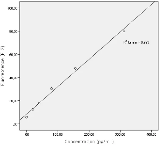
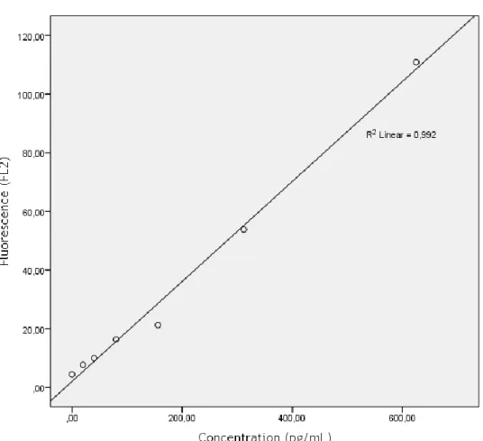
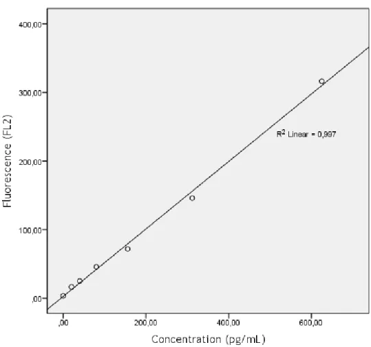
Figure 8 – Calibration curve for IL-12p70. 39
Figure 9 - Calibration curve for TNF-α. 40
Figure 10 - Calibration curve for IL-10. 41
Figure 11 - Calibration curve for IL-10. 42
Figure 12 - Calibration curve for IL-1β. 43
xiii
List of Tables
Table 1 - Characterization of the study population. 20 Table 2 – Median [max; min] levels of the studied cytokines, sorted by groups. 21 Table 3 – Summary of studies evaluating serum biomarkers of asthma. 35 Table 4 – Summary of studies evaluating serum cytokines as markers of asthma. 38 Table 5 – Fluorescence values for standard concentrations of IL-12p70. MFI values at
625pg/ml were out of the normal range of the curve. 39 Table 6 - Fluorescence values for standard concentrations of TNF-α. 40 Table 7 - Fluorescence values for standard concentrations of IL-10. 41 Table 8 - Fluorescence values for standard concentrations of IL-6 42 Table 9 - Fluorescence values for standard concentrations of IL-1β. 43 Table 10 - Fluorescence values for standard concentrations of IL-8. 44
xiv
List of Acronyms
AA – Allergic asthmatics AC – Young adult controls APC – Antigen presenting cells APP – Acute phase protein AT – After treatment
BAL – Bronchoalveolar lavage BT – Before treatment CBA – Cytometric bead array
COPD – Chronic obstructive pulmonary disease CRP – C reactive protein
EAACI – European Academy of Allergology and Clinical Immunology EC – Elderly controls
ECP – Eosinophils cationic protein
ELISA – Enzyme linked immunosorbent assay Fas – Cell death receptor
FcεRI – High affinity receptor I
FeNO – Fractionally exhaled nitric oxide
FEV1 – Forced expiratory volume at the first second HC – Healthy controls
HIV – Human immunodeficiency virus Hs-CRP – Highly sensitive C reactive protein ICS – Immunocorticosteroid
IgE – Immunoglobulin E
ISAAC – International Study of Asthma and Allergies in Childhood MMP – Membrane-type metalloproteinase
MSD – Meso Scale Discovery NA – Neutrophilic asthmatics NAA – Non-allergic asthmatics NNA – Non-neutrophilic asthmatics OAD – Obstructive airways disease PBE – Peripheral blood eosinophils RD – Respiratory diseases
SAA – Serum amyloid A sCD86 – Soluble CD86 sIgE – Specific IgE
xv SPT – Skin prick tests
tIgE – Total IgE
Treg – T regulatory lymphocytes UTI – Urinary trypsin inhibitor
xvii
Serum Biomarkers in Elderly Asthma
1
Chapter 1
Introduction
1.1 Asthma Physiopathology
Asthma is a chronic inflammatory disorder of the airways, leading to episodic shortness of breath, cough and wheezing, particularly in the night or early morning. The main physiological feature of asthma consists in episodic bronchial obstruction, which is often reversible, whereas the pathological feature is airways inflammation, including possible structural changes of the affected organs (1). Airways epithelial cells largely express chemokines, recruiting leukocytes to participate in this inflammatory process. These cells will release cytokines which will trigger a mainly Th2 immune response, increasing eosinophilia and histamine production by mast cells, among other defence mechanisms.
A subject who suffers from asthma is generally hypersensitive to certain allergens, causing an abnormal IgE-mediated immune response. Eosinophils, mast cells, neutrophils, T helper cells and dendritic cells participate in this acute inflammation (2). Dendritic cells are the main antigen-presenting cells (APCs) in the immune reaction, activating mast cells through IgE attached to their high affinity receptors (FcεRI), degranulating and releasing inflammatory components, such as histamine, which is known to be a bronchoconstrictor (3, 4). Allergens will also be presented to CD4+ T helper cells. In asthma, the activated T cells will produce a Th2 characteristic response (5), releasing IL-4 and IL-5 (among other cytokines) that will participate in the inflammation and contribute to a late chronic response (2). IL-4 will raise IgE production by B cells, while IL-5 will raise eosinophilia in affected areas. Eosinophils will also produce IL-4, further increasing IgE production. The produced IgE is released into serum and finally attaches to FcεRI receptors on mast cells, causing their degranulation and anaphylactic responses when the subject contacts with the same allergen (6).
Certain phenotypes of asthma consist in a Th1 cytokine-mediated rather than a typical Th2 response (7). Neutrophils and macrophages are characteristic of Th1 inflammatory responses. Neutrophilia is increased in patients with severe asthma and in smoking asthmatics, although it is suspected to be caused by corticosteroid therapy (1). Neutrophils seem to be recruited by IL-8 (8, 9).
Recent advances in both immunological and clinical phenotyping of asthma have raised the possibility that Th17 cells may trigger the pathology or co-exist with a Th2 type inflammation (10). Th17 cells are T helper lymphocytes that produce elevated levels of IL-17.
2 A recent study found increased numbers of Th17 cells in allergic patients, compared to non-allergic, and further showed that IL-17 could also induce class switching to IgE in human B cells, suggesting a more direct mechanism for Th17 induced atopic phenotypes (11, 12). Further studies in human patients have demonstrated that Th17 cells may also have a role in asthma pathogenesis by promoting airway smooth muscle cell migration, consequently leading to airway remodelling and possible airway narrowing (11, 13). Further proof was found using mice models, where IL-22 produced by Th17 cells also appears to have part in induction of experimental asthma, as well as suppressing the established disease (13, 14). While there is evidence about Th17 cells playing a role in asthma and atopic disease, their inflammatory mechanisms are still unclear and need further study. However, association between IL-17 and asthma is established and the possibility to use this cytokine as a marker for asthma exacerbations is being studied (15, 16).
T regulatory lymphocytes (Treg) are necessary to control the inflammatory response against allergens, releasing imunosuppressor cytokines or inducing anergy in other inflammatory cells. Tregs are known to be decreased in patients with asthma and other allergic diseases and are not as effective in suppressing allergen-stimulated effector T cells, which will consequently result in higher IL-4 serum levels and a more severe Th2 response (17).
Airway narrowing is the final common pathway leading to symptoms and physiological changes in asthma. It is caused by airway smooth muscle contraction in response to bronchoconstrictor mediators and neurotransmitters (18, 19). This occurrence is normally reversible by administration of bronchodilators. Inflammation may also produce mediators that will increase microvascular leakage and cause airway edema, especially during acute exacerbations. Furthermore, as asthma’s severity intensifies, airway remodelling will also intensify, resulting in structural changes that may cause airway thickening. This is not fully reversible by current therapy (18, 20).
The characteristic abnormality in asthma is the airway hyperresponsiveness which may lead to one or more of the airway narrowing mechanisms mentioned above. It is caused by a stimulus that would normally be innocuous in a normal person, resulting in an anaphylactic response. However, airway narrowing in asthma is not exclusively caused by hyperresponsiveness to allergens. Acute asthmatic exacerbations may occur when exposure to certain conditions are met, also called “triggers”, such as exercise, weather conditions or air pollutants (21, 22). Also, asthma tends to worsen at night. This is called nocturnal asthma and its triggers are not fully understood, but a night-time reduction in endogenous anti-inflammatory mechanisms is suspected to be the cause (1, 23).
External and temporal factors are not the only reasons for asthma exacerbations and worsening. Different phenotypes of asthma may be more difficult-to-treat, which is the case of neutrophilic asthma, severe and unresponsive to corticosteroids, with more structural changes (24). This type of asthma is generally non-allergic with a rather weak Th2 response
3 when compared to other phenotypes of the disease, but with an increased Th1 inflammatory response. It is characteristic of older patients with a late onset of asthma and it usually starts in a severe form rather than progressing from a mild/moderate form (25). Asthmatic smokers may also tend to have a more neutrophilic asthma (26).
1.2 Distinguishing Asthma from COPD
Both asthma and chronic obstructive pulmonary disease (COPD) are major chronic obstructive airways disease (OAD) that involve underlying airway inflammation and may be difficult to differentiate (1). Unlike asthma, COPD airflow limitation is not fully reversible and is usually progressive, but these symptoms may be hard to distinguish from asthma’s, especially when concerning elderly and smoking asthmatics (27). It was observed that asthmatic patients had more allergic sensitivity, greater eosinophilia in peripheral blood, bronchoalveolar lavage (BAL) and sputum, higher levels of serum IgE, greater increases in FEV1 after treatment with corticosteroids, and less common smoking history, when compared to COPD patients (28, 29). However, the levels of some markers can still overlap making it harder to diagnose. Tinkelmen et al (2006) developed a questionnaire for differentiating asthma from COPD (27). Despite being an easy and economic diagnostic method, its accuracy is questionable because the patient may not be so sure of his symptoms, among other limitations (27, 30), and it should only be used as a supplementary diagnostic tool.
1.3 Asthma in Elderly Patients
Asthma can affect all ages and it is often misdiagnosed, under diagnosed and under-treated in elderly patients (31). There is an estimated proportion of 6.5-17% sufferers of asthma in this age group, and deaths resulting from it occur mostly among these patients (32). This mortality rate is often associated with poor diagnosis and inadequate treatment. The complexity of the disease, with variable clusters of phenotypes and endotypes along with symptoms similar to those of other OAD and possible comorbidities are the principal obstacles to the diagnosis of asthma (31-34). In addition, lung capacity of individuals over 65 years of age tends to decrease by about 40% over time (35). This loss of lung function may compromise diagnostic tests, as it produces a characteristic change in the flow-volume curve which is typical of small airways disease (34). In addition, eosinophils may have an altered or decreased role in airway inflammation in elderly asthmatic patients (28), thereby leading to an increase in airways neutrophilia. Furthermore, different phenotypes of asthma may be more difficult to treat, more severe and unresponsive to corticosteroids, showing more extensive structural changes (24).
Although several distinct forms of asthma are recognised, the major focus of treatment and research has been on allergic asthma (36). This phenotype of asthma is characterised by the presence in the lungs of asthmatic patients of allergen-specific Th2 cells (37), that produce cytokines such as IL-4, IL-5, IL-9 and IL-13. Another characteristic of allergic asthma
4 is the abnormally high levels of serum allergen-specific IgE possibly due to the action of augmented IL-4 production, as well as a significant enhancement in eosinophilia in serum and bronchial mucosa, most likely due to the effects of increased IL-5 production (38).
Non-allergic asthma is a less common phenotype as it accounts for two out of every five cases of asthma in adults and constitutes both in terms of symptoms and lung function, a more severe form of the disease (39). This form of asthma tends to be more frequently associated with higher numbers of serum and sputum neutrophils and is characteristic of older patients with a late onset of the disease, with reduced response to corticosteroids and it usually has a more severe and abrupt onset rather than progressing from a mild to a moderate form (24, 25). In addition, asthmatic smokers also tend to develop neutrophilic asthma (26). This more neutrophilic cellular phenotype of asthma is characterized by a less apparent Th2-type response when compared to other phenoTh2-types, and tends to be more frequently associated with an increased Th1-type inflammatory response. This increment in a Th1-type response may be correlated with the increased neutrophilia, since IL8-mediated neutrophil influx is typically associated with activation of the innate immune system, thereby suggesting that neutrophilic asthma may be involved in its activation (9, 40, 41).
Elderly patients with non-allergic asthma may have low serum IgE levels as a consequence of immunosenescence, thus making it even harder to diagnose (42).
Apoptosis is an important procedure to depurate dysfunctional cells. As the aging process goes on, cells become more sensitive to apoptosis with an increased expression of the death receptor Fas (also called CD95), helping to keep tissue homeostasis (34, 43). Todo-Bom
et al (44) analyzed the expression of CD95 in elderly asthmatics and compared its values with
the expression of the same marker on healthy elderly controls, suspecting a decreased sensitivity to apoptosis in asthmatics. The different was non-significant, which could be caused by the Fas system reduced activity in Th2 environment, or simply because there is no relation between asthma and changes in apoptosis sensitivity (45). Concluding, apoptosis sensitivity is raised in elderly patients, which is represented by an increase in CD95 expression. Apoptosis is probably reduced in elderly asthmatics not treated with steroids, although there is no significant evidence of that incidence.
1.4 Serum Biomarkers for Asthma
Many biomarkers of asthma have been clinically studied in order to determine their diagnostic value and several are currently being used by physicians to identify the phenotype of asthma affecting their patients, thereby assisting in the choice of the most appropriate treatment. Biomarkers for asthma can be found in serum, sputum, bronchial alveolar lavage (BAL) and bronchial biopsies. Bronchial biopsies and BAL have the highest diagnostic value for asthma. However, they require extremely invasive and complicated techniques, thus becoming hard to use as a routine diagnostic tool. Fractionally Exhaled Nitric Oxide (FeNO),
5 on the other hand, is a less invasive assay that measures the exhaled nitric oxide, thereby indirectly assessing the level of inflammation in the airways. Nevertheless, the level of diagnosis based upon FeNO decreases in patients with smoking habits or other OAD (46, 47). Serum biomarkers are easily assessable and can be easily quantified, needing only blood samples to be retrieved from the patients.
In the next section we describe several clinical research studies based on various serum markers and their relevance to the diagnosis and/or monitorisation of asthma in elderly patients.
IgE
Atopic patients over express IgE, namely to common allergens, which makes them more prone to developing allergic diseases. In this regard, IgE may be a crucial factor underlying airway hyperresponsiveness in asthma. Measurement of serum total IgE (tIgE) was one of the first tests used to the study of allergic inflammation and it was thought to be a good and easily measurable marker of asthma, since asthmatic patients tended to be allergic (48). However, this was proven to be not necessarily true. It may help to exclude a diagnosis of COPD since the inflammatory response in this disease is associated with a lower concentration of serum IgE when compared with asthma (49), but despite the good sensitivity, total serum IgE has a poor specificity and negative predictive value as other diseases may also have increased IgE levels. This is the case of patients with helminthiases or other allergic diseases, such as allergic dermatitis (50-53). In addition, in elderly patients, serum tIgE levels are generally lower than in younger adults, which may decrease the sensibility of this test. Overall, determination of tIgE levels in elderly asthmatic patients has to be interpreted with caution.
In contrast, the quantification of allergen-specific IgE (sIgE) may be clinically relevant to the diagnosis of allergic diseases. It measures the circulating levels of IgE in response to a specific allergen. This can be quantified through an IgE auto-analyzer, such as Phadia® (ImmunoCAP), which measures the response of sIgE to specific allergens, thus determining the atopic status of a patient. If the test results are greater than 0.35 PAU/L the patient can be regarded as atopic. Phadiatop (ImmunoCAP) and food allergy screening panels (such as fx5 for common childhood food allergens, also for ImmunoCAP) are recommended as the IgE antibody analyses for asthma clinical trials (54). In addition, sIgE may be indirectly measured in the skin using skin prick tests (SPT). SPT measures mast cells degranulation on the skin in response to certain allergens, thus obliquely quantifying the sIgE for that allergen. The SPT method has a great diagnostic value, but the lack of full standardization, plus adverse skin topography in certain patients, reduces its value in clinical research (54, 55). An asthmatic patient can be regarded as atopic when there is positive hyperresponsiveness to at least one allergen, in a SPT (when an edema with at least 3mm in diameter is formed). King et al (2004) performed a study in elderly asthmatics (33 asthmatics, 21 healthy controls), showing that among five sIgE to cat, ragweed, German cockroach, Dermatophagoides pteronyssinus
6 and oak allergens, only IgE for Dermatophagoides pteronyssinus was significantly higher in asthmatic than in non-asthmatic patients (56).
Due to the different aspects of SPT and sIgE quantification, both tests are recommended as diagnostic tools for detecting asthma and atopy.
CRP and Hs-CRP
Acute phase proteins (APP) tend to be elevated in the serum of patients suffering from infections or other inflammatory conditions (57). C reactive protein (CRP), which is one of the most studied and easily measurable positive APP, is known to be over expressed in the presence of systemic inflammation. Since asthma is regarded as an inflammatory disease of the airways, Buyukozturk et al (2004) tested CRP as a potential marker of asthmatic inflammation, in a population of 50 adult patients with rhinitis, 20 adult asthmatics and 20 healthy controls with matching age. These authors concluded that the mean CRP levels of patients with asthma and rhinitis were not significantly different from those of healthy control groups (58). Furthermore, results obtained by Bafadhel et al (2006) showed that CRP levels did not significantly discriminate between patients with asthma and COPD (59). This study was composed of 96 adult asthmatics, 62 adult patients with pneumonia, and 161 adult patients with COPD exacerbations.
However, different results were obtained when the same marker was tested using highly sensitive CRP (hs-CRP) determination. Hs-CRP test essentially quantifies the same marker but is able to detect very small amounts of the protein in serum (0.5 to 10mg/L), whereas normal CRP quantification is only able to measure in the range from 10 to 1000 mg/L. In a study comprising 24 corticosteroid naive adult asthmatics, 26 adult asthmatics treated with corticosteroids and 15 healthy controls of similar age, Allam et al (2009) showed that hs-CRP is significantly increased in patients with asthma, even in those treated with corticosteroids, when compared to healthy controls (60). Another interesting study involving hs-CRP was performed by Wood et al (2012), showing that hs-CRP is not significantly elevated in every asthma phenotype but only in neutrophilic asthma, which is characteristically more severe, with increased systemic inflammation (7). 106 adult non-neutrophilic asthmatics, 26 adult neutrophilic asthmatics and 83 adult controls participated in this study. With these results in mind, and in face of the simplicity and accessibility of the test, hs-CRP could be a possible serum marker for further supporting a clinical diagnosis of asthma since it can indirectly detect the degree and phenotype of airways inflammation. However, this data has still to be interpreted with caution since another study, by Tilemann et al (2007), contradicted these results by showing that hs-CRP levels in asthmatic patients were not significantly different from subjects with no OAD, thus suggesting hs-CRP may only be accurate for a diagnosis of COPD (49). The population of this study consisted in 86 adult asthmatics, 36 adult patients with COPD, 15 adult patients with partial reversibility of airways alteration, and 75 healthy adult controls.
7 Altogether, hs-CRP appears to be much more clinically reliable as a marker of asthma than ordinary CRP measurement.
Urinary Trypsin Inhibitor
As discussed before, elderly asthma is characteristically more neutrophilic, which suggests that there may be increased neutrophil activity in the inflammatory process. As such, monitoring neutrophilic activity may be a good way to support a diagnosis of asthma in the elderly. In this regard, Yasui et al (2003) studied serum urinary trypsin inhibitor (UTI) in 25 asthmatic children and 15 healthy controls of matching age and showed that UTI levels were significantly greater in asthmatic children (61). These authors further suggested that serum UTI, a metabolite released by neutrophil elastase, might correlate with the patient neutrophils blood count, thus being useful for evaluating the neutrophil-mediated inflammation in childhood asthma (61). However, in spite of the good results obtained, little is known about the accuracy of UTI as a marker for asthma exacerbations since this is the only study available, to our knowledge. In addition, this study was carried out in children and the relevance of UTI levels have not yet been studied in elderly asthmatic patients.
Eosinophil Cationic Protein
Eosinophils are known to play an important role in inflammation in asthma, with increased numbers of this cell type having been reported in peripheral blood and airway secretions in asthmatic patients (for review see (62)). One of the eosinophil granule proteins, eosinophil cationic protein (ECP), has been widely characterized and researched as a marker of asthma (24, 63). In a study involving 441 adult patients with respiratory diseases and 33 healthy adult controls, Peona et al (2010) showed that ECP was significantly higher in the affected group, compared with healthy volunteers, particularly in female patients (64). In addition, the significantly high ECP levels in asthma compared to other respiratory diseases led the authors to suggest that serum ECP may be considered a marker for identifying patients with isolated asthma with an efficiency of 73%, despite not being useful for the discrimination of other respiratory disorders. However, the authors did not find any correlation between serum ECP and the absolute numbers of peripheral blood eosinophils. Studying 17 adult asthmatics before treatment with inhaled steroids, 10 adult asthmatics treated with inhaled steroids and 10 healthy adult controls, Ozseker et al (2006) found similar results in asthmatic patients not treated with inhaled steroids and further showed that there is no significant difference in the expression of ECP between healthy subjects and asthmatic patients treated with inhaled steroids (65). These findings support the theory that eosinophilic asthma is normally responsive to corticosteroids and that ECP may be a good marker for distinguishing eosinophilic asthma from other OAD and other asthma phenotypes. Yet, King et al (2004) showed that ECP levels were not significantly different between asthmatic and non-asthmatic patients in the elderly population, suggesting that ECP is elevated in the elderly population but is not related with asthma, classifying it as a potential
8 and simple test for differentiating between the allergic and non-allergic phenotypes (56). These results could compromise the usefulness of serum ECP as a marker for eosinophilic asthma in elderly. In a similar line of results, Koh et al (2006) state in their review that ECP correlates well with airway inflammation but not with airway hyper-responsiveness and it is increased not only in asthma but also in other atopic diseases, thus not being a good marker for the diagnosis of asthma (66). Nevertheless, results may support ECP as a guide to tailoring down inhaled corticosteroids.
Soluble CD86
T cells express CD28 and CTLA-4 molecules, which are important for receiving activation signals from APCs, such as monocytes and dendritic cells. These molecules interact with B7 molecules on APCs, B7-1 (CD80) and B7-2, also known as CD86 (67). Soluble CD86 (sCD86) results from an alternatively spliced transcription characterised by deletion of the transmembrane domain in resting monocytes, and is found in human serum (68).
Since monocytes and dendritic cells play an important role in antigen presentation and activation of inflammatory cells, it has been speculated that sCD86 levels may be altered in asthmatics (69, 70). Shi et al (2004) performed a study with a population consisting in 24 adult patients with stable asthma, 28 adults with aggravated asthma, and 25 healthy adult controls in which serum CD86 expression levels where measured in stable asthmatics, acute asthmatics, and healthy controls (71). Levels of serum sCD86 were significantly higher in acute asthmatics when compared to stable asthmatics and controls. Moreover, no significant difference was found between CD86 levels in stable asthmatics and controls. This significant increase in sCD86 in acute asthmatics may be correlated with the Th1-type response featured by this more severe kind of asthma, which explains increased monocyte activity when compared to the Th2-type asthma, and consequentially the increased levels of sCD86. Should these results be confirmed and sCD86 may become a possible marker of asthma severity.
Serum Amyloid A
Just like CRP, serum amyloid A (SAA) is an APP released by the liver in response to various aggressions leading to immune responses (72). In the previously mentioned study with CRP involving 20 adult patients with asthma and 50 adult patients with rhinitis, along with 20 healthy adult controls, Buyukozturk et al (2004) also measured SAA and compared the results with the same protein levels in healthy subjects (58). The results showed significantly increased serum levels of amyloid A in both patients with asthma and patients with rhinitis, compared to the healthy controls. These results suggest that SAA may be a more useful marker of asthma inflammation than CRP. However, the similar levels of SAA observed in both patients with allergic rhinitis and patients with asthma suggest that this marker does not discriminate between these two types of manifestations of the atopic syndrome (58, 73). However, it is crucial to stress that the levels of SAA in patients with asthma, both before and after treatment with FlixotideTM (fluticasone propionate) and VentolinTM (albuterol) were
9 significantly greater than in healthy controls (65), which suggests that testing for SAA levels may be sensitive enough to detect asthma inflammation even in patients on inhaled steroid treatment.
However, as far as we know, there is no study on the relevance of SAA to asthma diagnosis in elderly patients.
Neopterin
Neopterin is a pteridine derivative that is produced by activated macrophages and is a marker of immune activation (74). Mota-Pinto et al (2006) performed a study in an exclusively elderly population including 42 allergic asthmatics and 22 non-allergic asthmatics, along with 41 healthy controls, which allowed them to suggest that serum neopterin is significantly increased in non-allergic asthmatics when compared to allergic asthmatics and healthy elderly controls (75). Non-allergic asthma is generally more Th1-mediated than its allergic counterpart, and the increased neopterin levels may be related to higher macrophage activity, which could explain the results obtained. The results also suggest that serum neopterin may prove to be a useful marker for classifying asthmatic patients, including those in the elderly population (75). This may be true when classifying asthmatic patients as allergic or non-allergic, but not when attempting to discriminate between asthma and other OAD. COPD, for instance, is also characterized by Th1-type immune responses and serum neopterin levels of patients with COPD may overlap with those from non-allergic asthma. Furthermore, Frick et al (2004) showed that serum neopterin levels increase naturally with age (76), which could generate doubt about the reason for its increment in elderly patients: Are serum neopterin levels raised due to non-allergic asthma inflammation or immunosenescence? However, Mota-Pinto et al used an elderly population control group, which suggests that neopterin increase in their study was related to non-allergic asthma inflammation.
YKL-40
Chitin is commonly found in nature, as a component of the exoskeletons of crabs, the walls of fungi, or the microbial sheath of parasitic nematodes (77). Chitin cannot be synthesized by mammals, but they do synthesize chitinases, which can cleave chitin, thus playing a role in defence against chitin protected pathogens (78). YKL-40 is a chitinase like protein (also called chitinase 3-like 1) that lacks chitinase activity.
Chupp et al (2007) hypothesized that the level of expression of YKL-40 would be
increased in patients with asthma and would correlate with the severity of the disease (79). Therefore, these investigators performed a cross-sectional analysis of serum samples from three cohorts of adult patients with asthma, as well as from healthy adult controls. The results showed that serum YKL-40 levels were significantly higher in asthmatics compared to healthy patients. In addition, YKL-40 levels correlated positively with serum tIgE and
10 peripheral blood eosinophils, thereby suggesting that YKL-40 either participates in the pathogenesis of asthma, or is a marker of its severity. Similar results were obtained by Tang
et al (2009), who also reported that YKL-40 levels were significantly higher in 62 adult
asthmatics compared to 64 healthy adult controls (80). Specjalski et al (2011), showed that YKL-40 levels were significantly higher in 59 adult asthmatics compared to 29 healthy adult controls, but also stressed that no relationship was found between YKL-40 levels and asthma severity, asthma control, or serum tIgE levels, somehow contradicting the results obtained in other studies (81). Nevertheless, the authors showed that serum YKL-40 levels correlated with blood eosinophilia, thus corroborating the results from Chupp et al (79, 81).
Serum YKL-40 appeared to be a good marker for asthma, but it was not clear whether it was a biomarker of severity or a participant in the pathogenesis of asthma. However, Bara et
al (2012) showed that increased serum YKL-40 levels were positively correlated with bronchial
smooth muscle cell migration, in 40 adult asthmatics, suggesting that it may be an active player in bronchial smooth muscle remodelling in asthma (82). These results potentially link YKL-40 to the pathogenesis of asthma. Nevertheless, YKL-40 levels also appear to be increased in patients with COPD (83), probably because it involves airways remodelling as well. Other diseases such as peripheral arterial disease and diabetes or pre-diabetes also showed increased expression of YKL-40 (84, 85). It should also be stressed that none of the existing studies were focused in elderly patients with asthma, thus making it unclear whether serum YKL-40 levels are affected by aging and immunosenescence.
Leukolysin
Leukolysin is a membrane-type metalloproteinase (MMP) that appears to be expressed only in granulocytic cells of the myeloid series, mainly in neutrophils, and is released by degranulation or shedding after stimulation with pro-inflammatory cytokines, such as IL-1 and IL-8 (86, 87). Because of this limited distribution in granulocytic cells, Blumenthal et al (2010) performed a study to evaluate the role of leukolysin in chronic inflammatory conditions, including atopic asthma and COPD (88). The results showed that serum leukolysin levels correlated well with serum tIgE in patients with both atopic asthma and COPD. The population consisted in 337 patients from nuclear families with asthma (including children and adults), and 122 “married-in” individuals.
No study on the behaviour of leukolysin in elderly asthma was performed, which means there is no information about how aging affects this MMP expression in inflammatory granulocytes.
1.5 Cytokines
As inflammatory mediators, cytokines play important roles in the pathology of asthma. Therefore, there have been multiple studies measuring cytokine concentrations in asthmatics to evaluate whether these mediators may or may not be used as markers of asthma. IFN-γ, IL-1β, IL-2, IL-4, IL-5, IL-6, IL-8, IL-10, IL-17, and IL-12p70, are among the most studied serum
11 cytokines in asthma. If a different serum cytokine pattern could be established for each phenotype of asthma, it would mean increased diagnostic accuracy. Thus, measuring cytokine concentrations in serum and comparing its values between the different phenotypes may turn out to be a good diagnostic tool. In the next section we further review studies of some of the cytokines that may be related to asthma.
IL-8
One of the most studied cytokines in asthma is IL-8 since it is a major chemoattractant for neutrophils (89, 90). IL-8 is generally correlated with the extent of neutrophilic inflammation and with disease severity. This was shown in a study by Wood et al (2012), who measured serum IL-8 levels of 26 adult patients with neutrophilic asthma, 106 adult patients with non-neutrophilic asthma, and 83 healthy adult controls (7). Serum IL-8 was significantly increased in the neutrophilic asthma group when compared to the other two groups, whereas no significant difference was found between non-neutrophilic asthma and healthy controls. These results suggest that IL-8 expression in serum correlates with the levels of neutrophilic inflammation in asthma. Furthermore, they showed that neutrophilic asthma is generally more severe with increased systemic inflammation. This supports the results from a prior study performed by Silvestri et al (2006) that involved 14 adult patients with severe asthma, 22 adult patients with mild/moderate asthma, and 20 healthy adult controls (8). The results suggest that IL-8 is significantly increased in asthmatic patients when compared to the healthy group. However, serum IL-8 levels in severe asthmatics were significantly higher than in mild/moderate asthmatics. In short, the results from these two studies suggest that severe asthma is associated with IL-8 and indirectly with neutrophilia. Silvestri et al further stated that the patients with severe asthma in their study were significantly older (mean age 56.64 (8)), which also supports the notion that elderly asthma is generally more neutrophilic and more severe. Still, Hollander et al (2007) showed that serum IL-8 levels in asthma overlap with COPD (91). 13 adult asthmatics and 25 adult patients with COPD participated in this study. As we discussed before, comorbidity is a common feature in the geriatric population and asthma is often misdiagnosed as COPD, which could be a problem when using IL-8 as a marker for elderly asthma.
IL-6, IL-17 and TNF-α
IL-6, IL-17 and TNF-α are three pro-inflammatory cytokines that stimulate the production of APP by the liver, and are indirectly related to systemic inflammation during asthma exacerbations (57, 92, 93) . IL-6 is also known to be elevated in COPD patients and is associated with lung function decline and systemic inflammation (94). In the previously discussed study, Wood et al (2012) analyzed serum levels of IL-6 and TNF-α in patients with neutrophilic asthma, patients with non-neutrophilic asthma, and healthy subjects (7). Although no significant difference was found in TNF-α measurement among the three groups, IL-6 serum levels were significantly increased in asthmatics, compared to healthy controls.
12 Between the two asthmatic groups, IL-6 levels were significantly elevated in neutrophilic asthmatics, again suggesting that neutrophilic asthma is generally more severe. Furthermore, they compared these results with those of hs-CRP and concluded that IL-6 is more clinically relevant as a marker of systemic inflammation than hs-CRP. Regarding TNF-α, Silvestri et al (2006) and Agache et al (2010) showed contradictory results as serum levels of TNF-α were significantly higher in severe adult asthmatics than in mild/moderate adult asthmatics and healthy adult controls (8, 15). Thus, TNF-α may be a possible marker for systemic inflammation in asthma. Unfortunately, there are no studies that can help us to elucidate whether TNF-α levels are affected or not by neutrophilia in severe asthmatics.
In the aforementioned study, Agache et al (2010) showed that serum IL-17 is significantly increased in 36 adult patients with severe asthma, when compared with 49 adult patients with mild and moderate asthma (15). These results suggest that IL-17 may turn out to be a good serum marker for measuring asthma severity. Moreover, they concluded that TNF-α was less discriminative in predicting asthma severity, when compared to IL-17. However, serum levels of IL-17 were shown to be inversely related to smoking habits. In other studies, IL-17 in COPD was found to be considerably elevated when compared to asthma, although no association with neutrophilic inflammation was found (95).
There are no studies evaluating these cytokines levels in elderly asthma (although
Silvestri et al stated that patients in the severe asthmatics group were considerably older),
nor studies comparing the same cytokines in asthma and COPD.
IL-12p70
IL12p70 is involved in dendritic and T helper cells differentiation and it is often associated with Th1 type immune responses. Thus, IL-12p70 may play a role in non-allergic asthma. In an study including 20 Dermatophagoides pteronyssinus sensitive allergic asthmatics and 20 age-matched non-atopic controls, Huang et al (2011) showed that monocyte-derived dendritic cells obtained from the allergen sensitized asthmatics produced less 12 when compared with the cells from the non-sensitized controls, suggesting that IL-12 and consequently IL-IL-12p70 may indeed be increased in non-atopic subjects and possibly in non-allergic asthmatics (96). Contradicting these results, Vizmanos-Lamotte et al (2013) showed that IL-12p70 was significantly increased in the allergic asthmatics when compared to the non-allergic asthmatics in their study (97). However, the population of the study consisted in 60 allergic asthmatic patients, 17 non-allergic asthmatic patients and 31 healthy controls, all of them children between 7 and 15 years old, and as the authors suggest, asthma phenotypes in children may not be stable. Furthermore, IL-12p70 was measured in the sputum.
In conclusion, IL-12p70 appears to be an interesting marker to study further, as its accuracy as a marker for allergic or non-allergic asthma is yet to be determined.
13
IL-10
IL-10 is known for its regulatory and suppressive properties, inhibiting autoreactivity and regulating Th1 and Th2 immune responses. Whether IL-10 plays a distinct role in patients with asthma, who may be defective in the function of Treg cells, remains unclear. Tsai et al (2009) found no significant differences in IL-10 levels between 22 allergic asthmatic children and 16 controls of matching age (98). However, Yalcin et al (2012) showed that there are in fact differences in IL-10 circulating levels related to asthma, being significantly lower in asthmatics before anti-IgE treatment, when compared to the post-treated patients (99). This suggests that IL-10 levels are indeed altered in the pathology of asthma and may be an important key to its treatment, thus more studies regarding IL-10 are necessary.
1.6 The need for new markers
As we could tell from all the information collected, as convenient as serum markers may seem to be, a marker capable of accurately identifying asthma with sufficient specificity is yet to be found. Furthermore, all the aforementioned markers have scarcely been studied in elderly patients. For instance, studies on the accuracy of sCD86 in elderly asthma are lacking and it is also necessary to clarify whether the significant increase observed in this marker in severe asthmatics is related to the disease itself. Further studies focusing on the geriatric population and COPD are also needed to accurately classify UTI, neopterin and leukolysin as markers for evaluating neutrophil-mediated inflammation in asthma.
When evaluating IgE as a marker of asthma, sIgE appears to be more clinically relevant than tIgE for differentiating between atopic or non-atopic asthmatics. However, both specific and total IgE quantification may lose accuracy due to abnormal IgE levels produced in response to other infections. Furthermore, in elderly patients, this marker can be even less accurate since its levels are usually reduced due to natural immunosenescence (42). The same may happen in patients under pharmacotherapy, which may induce changes in serum IgE levels, thereby compromising the accuracy of the tests (100). Moreover, because elevated serum IgE levels are not an accurate discriminator of atopic asthma (especially in the geriatric population), leukolysin may indirectly share its inaccuracy as well, since changes in leukolysin levels are closely paralleled by those of tIgE in asthmatics. However, if good results are obtained in subsequent studies, leukolysin may prove to be a metabolic marker associated with atopic asthma and atopically sensitized COPD patients, that can be measured in human serum (88).
Hs-CRP and SAA appear to be more clinically reliable as markers of airway inflammation than ordinary CRP measurement, as they are able to indirectly detect asthma exacerbations. Nevertheless, they may also be increased in COPD, pneumonia and other forms of airway inflammation causing diseases. This overlap may compromise their accuracy as specific non-cytokine markers of asthma but might become important as routinely used supplemental
14 indicators of the severity of the disease. However, no studies have analyzed the impact of age on SAA and hs-CRP in elderly patients with asthma.
Currently, IL-8 seems to be a promising cytokine for measuring asthma severity, even in elderly asthma, although it cannot accurately distinguish between asthma and COPD. Still, it appears to be the more clinically reliable cytokine to be used as a severity marker of asthma.
ECP seems to be an interesting protein to be measured when researching for biomarkers of asthma. However, its accuracy is somehow questionable as each study presents slightly different results and its diagnostic value can vary with age, gender and the treatment patients are on.
YKL-40 positively correlates with blood eosinophilia and bronchial smooth muscle remodelling, thus appearing to be an emerging marker for asthma, that is not only related to asthma exacerbations, but also to the airways remodelling characteristic of the disease.
Altogether, clinically assessable markers capable of supporting a diagnosis of asthma are needed in order to counter misdiagnosis with other OAD, bearing in mind both the convenience and quality of information they provide (101). Differentiating allergic from non-allergic asthma is crucial as each phenotype can be associated with a distinct treatment. Therefore, it is essential that these novel markers are capable of diagnosing specific asthma phenotypes. This necessity becomes even more important in the case of elderly asthmatic patients, in whom morbidity is higher and symptoms often overlap with those of COPD (33). The actual process of aging may, on its own, make a diagnosis of asthma more complicated due to immunosenescence and natural changes in airways morphology, which may compromise the expression of some asthma markers. An accurate diagnosis will lead to a proper pharmacotherapy follow-up, resulting in better conditions and reduced fatality in the elderly asthmatic population.
To develop an accurate marker of asthma, one must make sure that these conditions are taken into account and controlled for, as much as possible.
1.7 Objectives of the study
Of all the serum biomarkers reviewed, we considered cytokines to be the ones that appeared the most promising, and therefore the ones with better accuracy for differentiating allergic from non-allergic asthma. Furthermore, there were no studies concerning cytokine measurement in the serum of elderly asthmatics.
The focal aim of our study was to measure six serum cytokines, including 1β, 6, IL-8, IL-10, IL-12p70 and TNF-α, in an elderly population with asthma. We also intended to:
15 1. Use flow cytometry to simultaneously quantify the cytokines, allowing us to easily
compare the resulting values and establish patterns;
2. Evaluate cytokine pattern changes related to age, using a young adult control group; 3. Investigate possible cytokine pattern differences for each phenotype of asthma
16
Chapter 2
Materials and Methods
2.1 Study Design
This study was carried out at the Faculty of Health Sciences of the University of Beira Interior, Covilhã.
The population of this cross-sectional study included 4 groups of individuals living in Cova da Beira, registered in the lists of general practitioners at the primary health care centre: one of healthy young adults (aged between 18 and 35), and three of elderly patients (aged 65 or older).
The individuals were chosen from a database of patients generated in a previously performed study at the same institution. The patients filled in a standardised questionnaire to identify symptoms of asthma and rhinitis, were submitted to skin-prick testing, and serum samples were collected and stored at -20ºC for further processing.
2.2 Population Selection
The patients were included in four different groups according to age, questionnaire answers, SPT and total and specific IgE (Phadiatop®) results. Questions used as criteria for
asthma were the ones concerning wheezing or treatment with asthma medication in the previous 12 months, as well as wheezing, dry cough or shortness of breath during or after exercise, according to the guidelines from the Global Initiative for Asthma report (1). In a similar manner, patients were checked for symptoms of rhinitis. Questions used as criteria for rhinitis were the ones concerning sneezing, “runny nose” (nasal dripping) or “stuffy nose” (nasal congestion) in the previous 12 months, in the absence of a cold or flu, as well as nasal symptoms accompanied by eye irritation manifested as itchy or watery eyes (rhinoconjunctivitis). If the patient had at least one positive answer to the previous questions, he/she was regarded as having clinical symptoms compatible with rhinitis, in accordance with the guidelines used by the European Academy of Allergology and Clinical Immunology (EAACI) for the ISAAC Study, adapted to an adult population (102).
SPT and total and specific IgE results determined whether the patient was atopic or non-atopic. Skin prick testing with a battery of common allergens adapted to local exposure was performed following EAACI allergen standardization and skin test recommendation (103). Before skin prick testing, relevant medication was discontinued. The tested aeroallergens included house dust mite (Dermatophagoides pteronyssinus), olive tree (Olea europaea),
17 grass pollen mix, weed pollen mix and Parietaria judaica. A negative control containing the diluent used to reconstitute the allergens and a positive control consisting of histamine at 10mg/ml were also applied. Allergen extracts were manufactured by LETI (Barcelona, Spain) and all belonged to the same batch. A skin test panel was considered valid if the correct outcomes of the controls were verified, including a histamine weal greater than 3mm in diameter, along with an absence of weal at the negative control site. If the test was valid, a weal diameter of at least 3mm as a reaction to any of the test allergens was regarded as a positive response. Specific and total IgE were quantified by sandwich enzyme linked immunosorbent assay (ELISA) using the Phadiatop and tIgE tests respectively, in an ImmunoCAP 250 (Phadia, Uppsala, Sweden). Any sample with sIgE quantified over 0.35kU/l was regarded as a positive Phadiatop. If the patient had a positive SPT and/or Phadiatop, he/she was regarded as atopic. In contrast, if the patient showed negative results to both SPT and Phadiatop, he/she was regarded as non-atopic. The designation “non-allergic” was applied when the SPT and serum sIgE measurements were all negative. Moreover, we only included individuals with tIgE under 150kU/l in the non-atopic elderly groups.
Using these criteria we formed four groups of individuals. The elderly allergic asthmatic group consisted in asthmatic patients aged 65 years old or over, that were regarded as atopic. Alternatively, non-atopic asthmatic patients under the same age range were included in the elderly non-allergic asthmatic group. The elderly control group consisted in non-asthmatic subjects aged 65 or over, that were also considered non-atopic. Non-asthmatic individuals aged between 18 and 35 that were also non-atopic were included in the young adult control group.
2.3 Quantification of Cytokines
IL-12p70, TNF-α, IL-10, IL-6, IL-1β and IL-8 serum cytokine levels were measured by flow cytometry, using the Cytometric Bead Array (CBA) Human Inflammatory Cytokines Kit (BD Biosciences, catalogue nº551811). The serum samples were processed according to the manufacturer instructions. The standard curve and all patient samples were acquired in the same day to guarantee similar conditions, using a dual-laser BD FACSCalibur™ flow cytometer. We conducted the cytometer setup according to the manufacturer manual, using cytometer setup beads and PE and FITC positive control detectors. Acquisition was performed using the BD Cell Quest™ Pro 5.1.1 software. Cytometric Analysis was made using the BD Cytometric Bead Array v1.4 software (BD Biosciences).
2.4 Statistical Analysis
All statistical analysis were performed using IBM SPSS Statistics 20.0 (Statistical Package for the Social Sciences Inc., Chicago, Illinois, USA).
The data was cheeked for normal distribution using the Kolmogorov-Smirnov test. All data was analysed using non-parametric tests. Results are expressed as median and range.
18 Mann-Whitney U test (continuous variables) and chi-square test (categorical variables) were used for comparisons between two groups. Kruskal-Wallis test was used for non-parametric comparison of quantitative data between the four groups.
19
Chapter 3
Results
3.1 Study Population
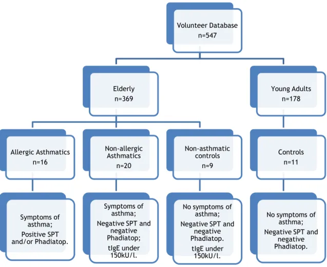
In this study, 45 elderly patients were included in three different groups with 16 being included in the allergic asthmatic group, 20 into the non-allergic asthmatic group, and finally 9 into the elderly control group. Eleven (11) young adult controls were also recruited, making a total of 56 individuals (Figure 1).
Figure 1 - Flow-chart of the methodology used for group selection.
The three elderly groups were paired according to age (p=0.574; Chi-square test), but not to sex. Twenty-three (23) individuals out of 56 were male. Furthermore, 4 out of 16 patients had allergic asthma along with allergic rhinitis, in the allergic asthmatic group.
Volunteer Database n=547 Elderly n=369 Allergic Asthmatics n=16 Symptoms of asthma; Positive SPT and/or Phadiatop. Non-allergic Asthmatics n=20 Symptoms of asthma; Negative SPT and negative Phadiatop; tIgE under 150kU/l. Non-asthmatic controls n=9 No symptoms of asthma; Negative SPT and negative Phadiatop. tIgE under 150kU/l. Young Adults n=178 Controls n=11 No symptoms of asthma; Negative SPT and negative Phadiatop.
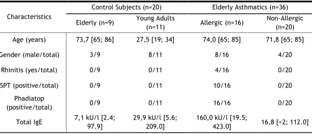
20 Moreover, in this group, all patients had positive Phadiatop values (>0.35kU), although 6 out of 16 showed negative SPT. In addition, tIgE was significantly increased in elderly allergic asthmatics in comparison with the other three groups (p<0.001). Furthermore, there was no significant difference in tIgE levels between non-allergic asthmatics and both control groups (Table 1).
Table 1 - Characterization of the study population.
Characteristics
Control Subjects (n=20) Elderly Asthmatics (n=36) Elderly (n=9) Young Adults
(n=11) Allergic (n=16) Non-Allergic (n=20) Age (years) 73,7 [65; 86] 27,5 [19; 34] 74,0 [65; 85] 71,8 [65; 85] Gender (male/total) 3/9 8/11 8/16 4/20 Rhinitis (yes/total) 0/9 0/11 4/16 0/20 SPT (positive/total) 0/9 0/11 10/16 0/20 Phadiatop (positive/total) 0/9 0/11 16/16 0/20
Total IgE 7,1 kU/l [2.4; 97.9] 29,9 kU/l [5.6; 209.0] 160,0 kU/l [19.5; 423.0] 16,8 [<2; 112.0]
3.2 Flow Cytometry
We followed the instruction manual on the kit to define the cytometer instruments settings for acquisition. The beads cluster could clearly be seen in a forward scatter - side scatter dot plot. We drew a region containing the beads, as we can see in Figure 2. In this figure example, the beads are visible inside the regions drawn in A and C, which represent forward scatter – side scatter dot plots for a 0 pg/ml standard and a sample of serum from a patient, respectively. B and D represent a gated view of the region containing the beads, in FL-2 – FL-3 dot plots, which allowed us to quantify the emitted fluorescence, thus indirectly measuring the median levels of the cytokine in the serum. It is possible to see in B and D how the dots formed separated clusters for each cytokine. Using the BD Cytometric Bead Array software we drew a standard curve using bead standards (see attachments section, page 41), which allowed us to relate the fluorescence detected in FL2 to the approximate amount of each cytokine in serum.
21 Figure 2 - Example of two acquisition cases. A – Analysis of a 0 pg/ml standard in a forward scatter - side scatter dot plot. B – A gated FL-2 – FL3 dot plot intercalated with the region drawn in A. We can see the beads clustering closely to the y axis, representing 0 pg/ml for each cytokine. C – Analysis of a sample of serum from a patient in a forward scatter - side scatter dot plot. D - A gated FL-2 – FL3 dot plot intercalated with the region drawn in B. We can see that IL-8 is increased in FL2, when compared to the 0 pg/ml standard.
Table 2 – Median [max; min] levels of the studied cytokines, sorted by groups.
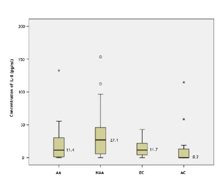
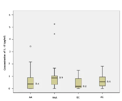
Table 2 presents a summary of the median levels of the cytokines quantified, including minimum and maximum values. IL-12p70 was not detected in any subject. TNF-α, IL-10 and IL-8 levels were increased in the non-allergic asthmatics group, although with no statistical
Cytokines Control Subjects (n=20) Elderly Asthmatics (n=36) Elderly (n=9) Young Adults (n=11) Allergic (n=16) Non-Allergic (n=20)
IL-12p70 (pg/mL) 0,0 0,0 0,0 0,0 TNF-α (pg/mL) 5,3 [0,0; 8,5] 7,6 [0,0; 10,2] 6,5 [0,0; 12,6] 7,7 [0,0; 17,6] IL-10 (pg/mL) 0,2 [0,0; 1,5] 0,6 [0,0; 1,8] 0,4 [0,0; 3,4] 0,9 [0,0; 5,2] IL-6 (pg/mL) 4,5 [0,0; 75,5] 4,9 [0,0; 7,2] 4,8 [0,0; 17,7] 6,0 [0,0;74,8] IL-1β (pg/mL) 9,7 [0,0; 11,6] 11,5 [0,0; 18,1] 10,0 [0,0; 25,7] 11,2 [0,0; 24,4] IL-8 (pg/mL) 11,7 [0,0; 43,1] 0,2 [0,0;114,7] 11,4 [0,0; 132,9] 27,1 [0,0; 153,4] A C B D
22 significance (p=0.163; 0.054; 0.092 respectively; Mann-Whitney U test). IL-6 was significantly increased in the elderly non-allergic asthmatic group, when compared to both control groups (p=0.033; Mann-Whitney U test) and when compared to the young adult controls alone (p=0.044; Mann-Whitney U test), but no significant differences were found when comparing to the elderly control group separately (p=0.167; Mann-Whitney U test). IL-1β median levels were shown to be elevated in the young adults control group, but also without statistical significant difference (p=0.322; Mann-Whitney U test). IL-8 was generally increased in the elderly, when compared to the young adults group, although without significance (p=0.079; Mann-Whitney U test).
No significant different was found when comparing the four groups altogether (p>0.05 for all cytokines; Kruskal-Wallis test).
Figures 3 to 7 show boxplots of the cytokine values measured in each group of individuals.
Figure 3 – Box plot of the measured concentrations of IL-8, sorted by groups. AA= Allergic Asthmatics; NAA= Non-allergic Asthmatics; EC= Elderly Controls; AC= Young Adult Controls. The º symbols represent the outliers and the * represent the extreme outliers. The horizontal black lines and the numbers represent the median for each group.
![Table 2 – Median [max; min] levels of the studied cytokines, sorted by groups.](https://thumb-eu.123doks.com/thumbv2/123dok_br/18942615.939765/38.893.175.756.117.620/table-median-max-levels-studied-cytokines-sorted-groups.webp)