Recent advances in smart biotechnology: hydrogels and nanocarriers for
tailored bioactive molecules depot
Gesmi Milcovich1, Paolo Marizza2, Stefania Lettieri1, Filipe E. Antunes3, Bruno Medronho4, Ana C. Fonseca5, Jorge F. J. Coelho5, Francesca Perrone6, Rossella Farra6, Barbara Dapas6, Gabriele Grassi6, Mario Grassi7, Silvia Giordani1,8*
1 Nano Carbon Materials Research Lab, Istituto Italiano di Tecnologia (IIT), Via Morego 30, 16163 Genova, Italy
2 Department of Micro- and Nanotechnology, Technical University of Denmark (DTU), Ørsteds Plads Bygning 345Ø, Kongens Lyngby 2800, Denmark
3 Coimbra Chemistry Centre, Dept. of Chemistry, University of Coimbra, Rua Larga, Coimbra, Portugal
4 Faculty of Sciences and Technology (MEDITBIO), University of Algarve, Campus de Gambelas, Ed. 8, 8005-139 Faro, Portugal
5 CEMMPRE, Department of Chemical Engineering, University of Coimbra, Polo II, Rua Silvio Lima, Coimbra, Portugal
6 Department of Life Sciences, Cattinara Hospital, University of Trieste, Strada di Fiume 447, 34100 Trieste, Italy
7 Department of Engineering and Architecture, University of Trieste, Via Valerio 6, 34127 Trieste, Italy
8 Chemistry Department, Università di Torino, Via Giuria 7, 10125, Turin, Italy
*Corresponding author. E-mail: [email protected]
Keywords: Bioactive molecule delivery, micro/nanocarriers, hydrogels, liposomes, carbon nano materials
Abstract
Over the past ten years, the global biopharmaceutical market has grown impressively, with ten over the top twenty worldwide high performance medical treatment sales being biologics. Thus, biotech R&D (research and development) sector is becoming a key leading market, with emerging revenues to progressively expand. Biotechnology offers considerable advantages compared to traditional pharmaceuticals, such as reducing side effects, targeted treatments, higher patient compliance and therefore more effective treatments leading to lower healthcare costs. Within this sector, smart nanotechnology and colloidal self-assembling systems represent pivotal tools able to modulate the delivery of therapeutics. A comprehensive understanding of the processes involved in the self-assembly of the colloidal structures discussed therein is essential for the development of relevant biomedical applications.
In this review we report the most promising and best performing platforms for specific classes of bioactive molecules and related target, spanning from siRNAs, gene/plasmids, proteins/growth factors, small synthetic therapeutics and bioimaging probes.
Introduction
The application of emerging nanotechnology to biomedical and pharmaceutical research allowed real progresses in the development of temporal and site specific drug delivery, leading to a new field of research defined as nanomedicine which nowadays is one of key fields of research [1]. Such a breakthrough was supported by the advanced scientific knowledge and technological development of different types of systems, such as carbon nanomaterials (fullerenes, nanotubes) [2], polymeric carriers (micelles, niosomes, nanoparticles, nanogels and macrogels) and lipid-based nanosystems (lipid nanovesicles, cubosomes and solid lipid nanoparticles) [3].
The use of colloidal delivery systems proved to be an efficient approach to improve the bioavailability and pharmacokinetics of small therapeutic molecules; hereby we describe the most recent advances in the field, with particular focus on the most suitable system depending on the desired bioactive molecule to be delivered.
1. Hydrogels and colloidal stuctures for drug delivery
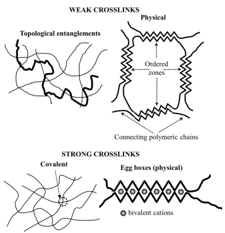
Hydrogels can be defined as coherent systems composed by a three-dimensional fibrous network, usually of polymeric origins, containing a huge amount of a continuous aqueous phase which cannot dissolve the network due to the presence of interconnections, called crosslinks [4]. Interestingly, despite the remarkable amount of the aqueous phase that these systems are able to host (the solid network volume fraction can be lower than 1%), hydrogels show rheological-mechanical properties closer to solids rather than to liquids [4], mimicking living tissues [5]. Hydrogels can be classified according to the nature of the crosslinks, their origin, composition, charge and configuration [6]. From a crosslinking point of view, hydrogels can be chemical or
physical. In chemical hydrogels, crosslinks between different chains (fibers) are strong, permanent
and punctual, due to covalent bonds. Conversely, physical hydrogels are characterized by either chains topological entanglements (spaghetti-like configuration, Fig. 1) or physical interactions (this being typical of polysaccharides such as glucans and xanthan) such as H-bonds, ionic, Coulombic, van der Waals, dipole-dipole and hydrophobic interactions.
Covalent
bivalent cations
Egg boxes (physical) WEAK CROSSLINKS
Topological entanglements
Ordered zones
Connecting polymeric chains
Physical
STRONG CROSSLINKS
Figure 1. Representative examples of chemical and physical crosslinks occurring in hydrogels. Adapted from [4].
Additional junctions can occur, with long chain segments departing from the ordered junction zones towards further chains, generating a polymeric three-dimensional network. Physical interactions are often transient, with non-strong bonding points, able to lead to a network characterized by a constant average crosslink density (i. e. moles of crosslinks per gel unit volume) and a time dependent spatial distribution of crosslinks. Thus, network meshes configure as a dynamic equilibrium, due to chains segments size and related Brownian motions, being the average mesh number and dimension constant [4,7]. The formation of ordered zones is favored/hindered by environmental conditions such as temperature and ionic strength [8]. For instance, agarose undergoes a thermo-reversible gelation process occurring when hot solutions are cooled below ∼ 40°C. In the hot state, agarose chains appear to behave as stiffened coils whereas, after cooling, a particularly extensive re-organization takes place, resulting in a hydrogel aggregation, at very low polymer concentrations (≥ 0.1% w/w) [9]. Physical crosslinks are usually associated with
mechanically weak gels, except for e.g., the case of alginates, where a strong physical hydrogel arises. Alginates are linear polymers of vegetal or bacterial origin containing β-D-mannuronic (M) and α-L-guluronic (G) acid [10], where the addition of different cations, such as Ca2+, Cu2+, Ba2+ and Sr2+, can induce gelation. These cations bind to stretches of guluronic acid residues within the polysaccharide chain, leading to the formation of junctions, which physically hold together the polysaccharide chains in a 3D continuum according to the egg-box model (see Fig. 1). Pectins are another outstanding example of polysaccharides leading to strong physical hydrogels, triggered by the presence of divalent cations, although with some differences connected to the existence of neutral sugars in the chains (that should hinder inter-chain association) and the methylation of some galactunorate residues (that do not contribute to the electrostatic ion binding).
With reference to their origin, hydrogels can be natural or synthetic. Among the plethora of natural hydrogels, those based on agar, collagen, chitosan, alginate, hyaluronic acid, gelatin, fibrin and polysaccharides (animal, vegetal and microorganisms origin [10]) are most represented [11]. Conversely, D,L-lactide-co-glycolide (PLGA), polyamidoamine (PAMAM), poly(caprolactone-co-ethylethylene phosphate) (PCLEEP) and poly(N-vinyl-2-pyrrolidone) PVP can be included in the synthetic class.
For homopolymeric hydrogels, the network is formed towards a polymer constituted by a single species of monomer, whereas in copolymeric hydrogels two or more different monomer species compose the chains of the polymeric network. Finally, interpenetrating polymeric hydrogels (IPN) are made up of two (or more) independent cross-linked synthetic and/or natural polymeric chains [12].
Hydrogels can be categorized depending on their charge features, as nonionic (neutral), ionic (anionic or cationic), amphoteric electrolyte (ampholytic) containing both acidic and basic groups,
zwitterionic (polybetaines) containing both anionic and cationic groups in each structural repeating
unit. From a configuration perspective, they can be amorphous, semicrystalline (a mixture of amorphous and crystalline phases) and crystalline [6].
Hydrogel production can be achieved by means of any technique allowing the formation of bonds among different polymeric chains, such as chemical reaction, ionizing radiations, physical interactions (e.g. entanglements and electrostatics) and crystallite formation. Moreover, hydrogels can be obtained thanks to polymerization techniques, including bulk, solution, and suspension polymerization [6]. However, when hydrogels are formed starting from a solution containing the polymer and hydrophilic drugs that can easily undergo denaturation such proteins, peptides and drugs based on nucleic acid (NABDs), an aqueous environment and room temperature are mandatory to perform a safe crosslinking procedure. For example, these requirements are perfectly accomplished by the ionic gelation of polysaccharides such as alginates and galacturonic [13]. Ionic gelation can also occur in the case of polycations with an anion as the crosslinker. Specifically, the ionic interaction between chitosan (polycation) and the trivalent negatively charged glycerol phosphate was shown to induce hydrogel formation [14].
Both macroscopic and micro/nanoscopic properties of hydrogels play an important role in biomedical applications. It has been recently demonstrated that, in three-dimensional culturing, the (macroscopic) viscoelastic properties of hydrogels used as substitutes of natural extracellular matrix (EM) can affect cells behaviour in terms of spreading, proliferation and differentiation. Chaudhuri and co-workers [15] demonstrated that the osteogenic differentiation of mesenchymal stem cells (MSCs) strictly depends on the viscoelastic properties of the alginates hydrogels used as substitute of EM. In detail, mesenchymal stem cells form mineralized, collagen-1-rich matrix similar to bone only when they are in contact with highly elastic hydrogels.
On the other hand, when hydrogels are devoted to the release of active agents, the mesh size distribution of the three-dimensional network (nanoscopic property) is a core characteristic. Indeed, it can represent the key parameter ruling the release kinetics of an embedded drug, or it can be essential to protect hydrogel load (drug, cells and so on) by external factors such as enzymes and the immune system agents as it can happen in the case of hydrogel based implantable systems. An
interesting example is represented by immunoisolant membranes, which serve to protect encapsulated pancreatic cells (aimed at the production of insulin) from antibodies [16].
In terms of release mechanisms, hydrogels drug depot can be controlled by physical, physicochemical and system related strands [17]. Swelling/shrinking processes are related to physical phenomena, whereas erosion, drug dissolution (recrystallization), drug transport (by diffusion and convection) and drug interaction throughout the matrix structure constitute the physicochemical phenomena. System related mechanisms depend on the initial drug distribution and concentration inside the hydrogel, hydrogel geometry (cylindrical, spherical, etc.) and size distribution in the case of polydispersed ensembles of hydrogels.
The swelling/shrinking process occurs upon variation of external factors (temperature and pH, more frequently), inducing a new equilibrium condition or when the dry hydrogel is in contact with an aqueous environment. The above mentioned process relies on the chemical potential difference between the water inside and outside the hydrogel [18].
Hydrogel erosion can be ruled out by chemical and/or physical factors. Erosion can be defined as peripheral or heterogeneous, when it affects only hydrogel surface. On the other hand, bulk or homogeneous erosion involves the whole hydrogel volume [19]. Chemical erosion is due to hydrolytic/enzymatic degradation of polymeric chains, while physical erosion depends on chains disentanglement due to the hydrodynamic conditions of the external aqueous environment.
Stability restrictions often require a hydrogels storage in the dry status. In such a case, drug release will begin as soon as an external aqueous fluid diffuses towards the polymeric network and a key step can be represented by the drug dissolution over the water permeating the network. When metastable bioactive molecules like polymorphs, amorphous or nano-crystalline drugs are present in the dry hydrogel, the dissolution process may correlate with recrystallization which leads to the formation of a new, more stable, drug crystallographic organization induced by the contact with the absorbed water [20].
Bioactive molecule depot and mobility towards colloidal networks can be strongly affected by the hydrogel mesh size distribution, as well as by the drug physical and chemical interactions with the 3D polymeric network [21]. For instance, drug adsorption/desorption phenomena may be due to electrostatic interactions, such as charged polypeptides and antibiotics in collagen matrices [22]. Further elements able to influence and drive bioactive molecule depot involve hydrogen bonds [23], lipophilic [24], as well as non-covalent interactions among imprinted polymeric networks and template molecules that need to be recognized in a physiological environment [25].
Colloidal and hydrogel frameworks are key structures for several bioactive molecule controlled delivery, with a specific application for Nucleic Acid based Drugs (NABDs) release.
NABDs are constituted by short sequences of either DNA or RNA, including antisense oligonucleotides, decoys oligonucleotides, aptamers, triple helix forming oligonucleotides, DNAzymes, Ribozymes, small interfering RNAs (siRNAs) and micro interfering RNAs (miRNAs) [10]. Despite their huge therapeutic potential towards different hyper-proliferative diseases [26], their daily clinic application is still very limited because of their rapid degradation by several enzymes, such as blood and cellular nucleases [27]. Moreover, as detailed in the next section (2.2), considering that both NABDs and cellular membranes are negatively charged, crossing the cellular membrane represents the core drawback, due to electrostatic repulsion. Thus, if delivered as naked NABDs, they have no chance to exploit their therapeutic activity. Delivery vectors can be divided into three classes, based on their size [28]: nano, micro and macro scales vectors. Nanoscale vectors are represented by polycationic polymers or lipids that self-assemble with NABDs to form polyelectrolyte complexes (poly- or lipo-plexes, respectively, as detailed in section 2). Microscale vectors can be outlined, for example, as hydrogels entrapping the poly- or lipo-plexes. Macroscale vectors are three dimensional matrices (such as hydrogels) that can host microvectors containing, in turn, poly- lipo-plexes to give rise to a chimeric system [29]. An outstanding example of chimeric system has been proposed by Knipe and co-workers [30], who dealt with the oral release of siRNA targeting TNF-β, an inflammatory cytokine that is a clinical target of inflammatory bowel diseases.
The chimeric delivery system consists of micro-gels (size < 30 µm) composed of poly(methacrylic acid-co-N-vinyl-2-pyrrolidone) (PMANVP) crosslinked with a trypsin – degradable peptide linker. PMANVP micro-gels contained siRNA-loaded polycationic nanogels (2-(diethylamino)ethyl metacrylate) (size ≈ 120 nm) that proved to guarantee siRNA protection and cells transfection. PMANVP matrix was designed to collapse around nanogels to protect them from degradation in the stomach (pH 2 – 4), while PMANVP swelling in the small intestine environment at pH 6 – 7.5, allowed matrix degradation, due to the uptake of intestinal fluids containing trypsin. Consequently, nano-gels could be released and internalized by cells, resulting in a considerable TNF-β knockdown in a murine macrophage model.
Chimeric systems can be used for the systemic delivery of NABDs too. Indeed, following injection administration, the NABD-vector complex is supposed to circulate towards capillaries and microvasculature structures (blood vessel diameter < 100 µm), cross the blood vessel wall and finally reach the target cells. Thus, the NABD-vector complex is required to move radially towards the vessel wall, by means of a margination mechanism. D’Apolito and co-workers [31] experimentally showed that margination is due to red blood cells and NABD-vector complexes interaction. The mentioned process is possible whether complexes size spans in diameter range > 1 μm, with 3 µm vectors better marginating than 1 µm sized particles. Accordingly, nano-sized complexes have poor chances to get the blood wall. However, nano complexes embedding into micro-vectors allows the overall structure to reach blood vessel wall. Therein, nano-vectors can be released, for example, by micro-vectors surface or bulk erosion.
2. siRNA delivery
2.1. Small interfering RNAs
In recent years the most commonly tested NABDs have been siRNAs. These short double stranded RNA molecules approximately 22 nucleotides in length, are mostly of exogenous origin, being generated from invasive nucleic acids such as viruses and transposons [32]. With reference to the
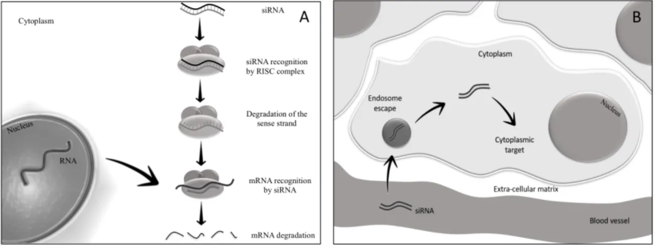
mechanism of the two siRNAs filaments (Fig. 2A), mostly the antisense strand is uptaken by the cytoplasmic RNA-induced silencing protein complex (RISC). The antisense strand drives RISC to a target RNA via a perfect sequence complementarity to the target. Following binding, RISC
mediates the degradation of the target RNA thus resulting in the downregulation of gene expression. It is possible to take advantage of this mechanism of action to generate siRNAs able to target RNAs causing disease as shown by many works [33-36].
Figure 2. Schematic representation of (A) siRNA cascade towards cytoplasmic RNA-induced silencing protein complex (RISC) and (B) siRNA metabolism throughout cellular mechanisms. 2.2. siRNA delivery problems
Despite the great siRNA therapeutic potential, their practical use is limited by their chemical nature. Following systemic administration, siRNAs encounter blood nucleases, which can rapidly degrade their nucleic acid structure (Fig. 2B).
Moreover, siRNAs tend to be removed by the reticulo-endothelial system, by kidney filtration [37] and, depending on the sequence, to activate the innate immune response [38]. Additional barriers to siRNAs cellular uptake are represented by the vessel wall and the cellular membrane, due to the electrostatic repulsion between the negatively charged phosphate groups present on siRNAs and the negatively charged surface of cellular membranes. Moreover, cell uptake is difficult by the
hydrophilic nature of siRNAs that does favor the crossing of the hydrophobic layer of the cell membranes.
The fraction of siRNAs that succeed crossing the cell membrane, further will face with cytosolic nucleases that can reduce their amount. Finally, siRNAs experience the problem of cellular
trafficking [39]. Depending on the mechanism of cellular internalization, siRNAs can be uptaken by endosomes. At this stage, when confined into these intracellular vesicles, siRNAs have no chance to reach their targets and thus to exert any biological effect. Based on the above mentioned
considerations, the administration of naked siRNA results in negligible therapeutic effects. 2.3. Strategies to minimize siRNA delivery issues
To minimize the delivery issues of exogenous siRNAs, two main strategies can be employed. The first consists of the chemical modification of siRNA structure to make these molecules more resistant to degradation. The second strategy is based on the siRNA complexation with synthetic engineered vectors to effectively bind and protect siRNAs and to allow their delivery to the target cells [40]. Frequently, the two strategies are used in combination, despite some chemical
modifications may affect siRNA effectiveness.
The choice of the optimal delivery materials is not a trivial task [10,32,40,41], with the net superficial charge of the delivery carrier/siRNA complex playing a key role. Anionic and cationic complexes usually show good solubility/stability in the physiological environment, despite they exhibit some drawbacks. Anionic complexes cannot transfect cells per se, due to the electrostatic repulsion with the negatively charged cell membrane. Conversely, cationic complexes bind to cell membrane towards strong electrostatic interactions, leading to non-specific cellular uptake and cell toxicity if the positive charge is not optimal [42]. On the other hand, neutral complexes tend to associate in the physiological environment, resulting in a limited solubility. Thus, the development of optimal delivery carriers requires a careful evaluation of different parameters such as the surface charge density, within a multidisciplinary team.
Besides providing for siRNA protection and targeting, the ideal delivery vector should be able to allow efficient extravasation of the siRNA. This feature is crucial for siRNA-vector complexes systemic administration. Therein, the size of
siRNA-vector complexes plays a relevant role, as above mentioned for NABDs.It has been recently showed that particles in the 1-3 µm diameter range [31] tend to localize closer to the endothelial layer (margination effect) of the vessel, compared to smaller particles. Thus, 1-3 µm particles localize closer to the vessel fenestration, being more susceptible to extravasation compared to smaller particles, which, alternatively, tend to localize in the middle of the vessel. Despite this advantage, it should be considered that particles bigger than 0.2 µm are readily scavenged non-specifically by monocytes and the reticuloendothelial system, thus not be efficiently uptaken by cells [43,44]. A possible solution may rely on the preparation of microparticles able to undergo a disassembly, upon extravasation, originating nano-metric particles. As previously introduced, the preparation of micro-sized delivery systems containing nano-metric particles can be included within the class of delivery strategies known as “chimeric systems” [28,45].This approach present a dual advantage: on one side, micro-particles are easy to handle, to produce on large scale and to store, whereas on the other hand, nanostructures are characterized by an extremely high surface/volume ratio, with a valuable drug payload efficiency. Polycationic polymers and lipids are most commonly employed to form nanoscale vectors. On the other hand, microscale vectors entrapping nanoscale vectors usually consist of two/three dimensional scaffolds or matrices mainly made by polymers. The following sections will focus on the presentation of strategies most commonly used to prepare nanoscale vectors, i.e. lipid and polymers nanoparticles [46-50].
2.3.1 Lipidic nanoparticles
Lipid-based nanoparticles (LNPs) have been extensively used as delivery systems for drugs and siRNAs, showing promising results both in vivo and in vitro. LNPs have appropriate delivery characteristics as their structure mimics cellular membranes, thus enhance the fusion with the target cell. Moreover, LNPs can be easily loaded with several cargo molecules.
2.3.1.1 Liposomes
Liposomes are spherical self-assembled vesicles, deriving from synthetic or natural phospholipids containing aqueous compartments (Fig. 3A).
The polar heads of phospholipids interact with the hydrophilic environment thus stabilizing lipids structure; in contrast, the long phospholipid chains interact each other, forming lipid layers in aqueous solution. Liposomes can be structured as unilamellar or multilamellar lipid bilayers. Due to the amphiphilic nature of phospholipids, these molecules can generate hydrophilic and hydrophobic compartments in the same system, thus allow for both hydrophobic and hydrophilic molecules (siRNA) hosting. Liposomes can also accumulate into tumours, present a low immunogenicity and are biodegradable [51].
2.3.1.2 Cationic liposomes
Positively charged (cationic) liposomes are most frequently used for siRNA delivery. They can electrostatically interact with the negatively charged siRNAs and allow an efficient molecule loading [41,52]. Additionally, cationic liposomes can easily interact with the negatively charged cell membrane. Sometimes, to further improve the ability to integrate with cell membrane, they are added with non-cationic lipids, such as DOPE (dioleoylphosphatidylethanolamine) and DSPC (1,2-Distearoyl-sn-glycero-3-phosphocholine). The positive surface charge is favourable for siRNA binding, but it can cause the side interaction with negatively charged serum protein such as albumin. In such a case, the negatively charged serum protein can displace siRNA from the positively charged liposome, thus significantly reducing the amount of siRNA delivered to the target tissue. The modification of liposomes with the neutral lipids such as cholesterol can contribute to overcome this limitation [53]. Moreover, cholesterol can be used also to bind other molecules such as polyethylene glycol (PEG), a polymer able to improve the delivery properties both in vitro and in vivo [51].
There are different types of cationic liposomes for siRNA delivery, such as monovalent cationic liposomes and multivalent cationic liposomes. For instance, N-[1-(2,3-dioleyloxy) propyl]-N,N,N-trimethylammonium chloride (DOTMA), 1,2-bis(oleoyloxy)-3-(trimethylammonio)-propane (DOTAP) and 3β-[N-(N'N'-dimethylaminoethane) carbamoyl]cholesterol (DC-Chol) are monovalent cationic lipids, characterized by a high in vitro transfection efficiency [54].
Multivalent cationic lipids (MCLs), synthetized from monovalent cationic lipids, exhibit an increased positive charge compared to monovalent cationic lipids. However, they tend to be more toxic than monovalent cationic lipids [55]. A widely used MCL transfection agent is Lipofectamine (2,3-dioleyloxy-N-[2(sperminecarboxamido)ethyl]-N,N-dimethyl-1-pro-paneammonium
trifluoroacetate and dioleoyl-hosphatidylethanolamine in ratio 3:1 (DOSPA/DOPE 3/1)), which contains the multivalent cationic lipid DOSPA. This mixture of lipids forms multilayers structures with the siRNA being embedded between adjacent lipid bilayers [56]. Lipofectamine can efficiently deliver siRNAs to a broad range of cells,although they exert a significant unspecific cell toxicity. For example, siRNAs directed against the mRNAs of the cell cycle promoting genes cyclin E and E2F resulted in a relevant inhibition of smooth muscle cells (SMC) proliferation. As SMC aberrant proliferation is a key event in many coronary artery diseases [44,57], the mentioned approach has the potential to minimize this pathological event. Per se, the cationic liposome-mediated delivery of siRNAs to the coronary wall is not sufficient to guarantee an effective delivery. In this case, the delivery of siRNA-cationic liposomes entrapped into gel matrix has been proposed, in order to prevent the rapid wash out of siRNA complexes due to blood flow [58].
2.3.1.3 Stable nucleic acid lipid particles (SNALPs)
Recently, stable nucleic acid lipid particles (SNALPs) have been developed for siRNAs delivery (Fig. 3B) [41]. SNAPLs are constituted by a lipid bilayer containing the ionisable cationic lipid 1,2-dilinoleyloxy-3-dimethylaminopropane (DLinDMA) or 2,2-dilinoleyl-4-(2-dimethylaminoethyl)-[1,3]-dioxolane (DLin-KC2-DMA) in the inner part, to allow the binding with siRNAs. Moreover, they contain PEG, which can stabilize the complex and a neutral lipid, like DSPC or cholesterol, which enhances the endosomal escape of the SNALP/siRNAcomplex [59].Thus, whereas the inner part of SNALPs is hydrophilic and allows the electrostatic binding with siRNAs, the surface charge is nearly neutral. Appropriate modifications in SNALPs, for example in the type and ratio of the different components, can extend the circulation time and minimize complement system activation [51].
SNALPs delivery systems were for example used to encapsulate a COP9 Signalosome Subunit 5 (CSN5) siRNA [60]. CSN5 is the catalytic center of the COP9 Signalosome that is involved in the control of proteolysis via the ubiquitin proteasoma pathway. CSN5 seems also to act as
transcriptional coactivator for MYC and TGFβ1, gene products involved in the control of
proliferation, apoptotic cell death and hepatocellular carcinoma (HCC) progression. This delivery system significantly inhibited tumor growth in an orthotopic mouse model of HCC [60].
2.3.1.4 Lipidoid nanoparticles
Lipidoid nanoparticles are made up of synthetic lipids obtained by the chemical combination of alkyl-amines with alkyl-acrylates containing carbon chain tail of variable length [61] (Fig. 3C). The particles containing the lipidoid have a polar and ionisable core, surrounded by hydrophobic carbon tails. The particles can also contain cholesterol and PEG, two types of molecules that can enhance particles stability and delivery efficiency [56]. In addition, their easy synthetic protocol allows the production of a considerable amount of different particles, which can be tailored to any different delivery purpose. For example, a siRNA embedded into lipidoid-based nanoparticles was used to downregulate β1 and αν integrin subunits in the hepatocytes of a xenograft mice model of HCC [62]. β1 and αν integrins are relevant extracellular matrix receptors involved in many cellular processes. Moreover, they play critical biological roles both in normal liver and in HCC tumor cells. Integrin silencing had, as major outcome, an extended morbidity-free survival of HCC tumor-bearing mice[62].
A) Liposome B) SNALP
C) Lipidoid
D) Solid Lipid Nanoparticle
Ionisable cationic lipid
Neutral lipid PEG Lipid core siRNA Nautral/Anionic /Cationic lipid Cholesterol Synthetic lipid
Figure 3. Schematic representation of the main nanoscale vectors: (A) liposomes, (B) stable nucleic acid lipid particles (SNALPs), (C) lipidoids and (D) solid lipid nanoparticles.
2.3.1.5 Solid-lipid nanoparticles
Solid-lipid nanoparticles (SLN) are novel siRNAs carriers derived from nano-emulsions where the oil emulsion component is replaced by a solid lipid dispersed in a surfactant solution (Fig. 3D). The loaded molecules are incorporated in the solid lipophilic matrix. SLN, solid at room temperature, are stable, non-cytotoxic, present a large surface area and can efficiently protect the encapsulated molecules [63].However, they present some disadvantages such as the low molecules loading capacity and the possible expulsion of the incorporated molecules during storage. The chemical nature of the solid-lipid matrix determines the loading capacity, as well as the type of molecule to be loaded [64]. SLN usually contain a combination of triglycerides, partial glycerides, fatty acids, steroids and waxes. To decrease cytotoxicity and immune responses, SLN can be prepared using physiological lipids present in the natural low density lipoprotein (LDL) such as cholesteryl ester, triglyceride, cholesterol, DOPE, and DC-cholesterol [65]. Lipids found in the natural high-density lipoproteins (HDL) represent an alternative. Overall, chemical modifications are required to allow the incorporation of the hydrophilic siRNA [66]. An example of siRNA delivery by SLN has been reported by Jin J. et al. [67]. Considering that c-mesenchymal-epithelial transition (c-MET) is a signaling receptor for hepatocyte growth factor, SLN were reconstituted from natural components of protein-free LDL and further conjugated to PEGylated c-Met siRNA. Inappropriate c-Met activation relates to different form of human tumours including glioblastomas (GBMs). The latter is the most frequent and malignant form of brain tumor, with limited treatment options due to the blood-brain barrier. In orthotopic U-87MG xenograft tumour model of GBM, intravenous administration of the complex significantly inhibited c-Met expression and suppressed tumour growth. Notably, no major signs of systemic toxicity were observed in mice.
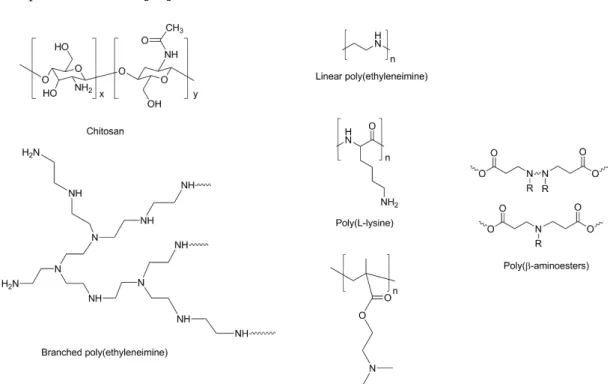
Polymers are solid and biodegradable molecules widely employed for siRNA delivery. Many different polymers have been tested so far, such as Chitosan (CH), Polyethylenimine (PEI), PEG, α,β-poly(N-2-hydroxyethyl)-D,L-aspartamide and Inulin-derived polymers.
CH is a polymer characterized by low toxicity, high biocompatibility and biodegradability [10]. CH is derived from chitin, has a carbohydrate backbone characterized by two types of repeating
residues, 2-amino-2-deoxy-glucose (glucosamine) and 2-N-acetyl-2-deoxy-glucose
(N-glucosamine), linked by (1-4)-β-glycosidic linkage. Furthermore, CH has a positive charge, due to the presence of positively charged amino groups present in its structure, thus, it can easily and efficiently bind negatively charged molecules such as siRNAs. On the other hand, CH has some disadvantages such as the low transfection efficiency and low solubility, which can be avoided via the conjugation with other molecules such as PEI, PEG, Poly (amidoamine) (PAMAM) dendrimers. As an alternative, CH physicochemical and biological properties can be improved by modulating the deacetylation rate and/or modifying the molecular weight [68].
PEI contains repeating units composed of an amine group and two aliphatic CH2-CH2 spacers. It can exist both in linear and branched forms. It is one of the most used cationic polymers, even though it tends to be more toxic than natural polymers [69]. Usually, PEI with high molecular weight has higher cytotoxicity compared to low molecular weight PEI, despite its transfection effectiveness. To decrease toxicity, PEI can undergo chemical modifications, such as the addition of hydrophilic and hydrophobic segments or cell/tissue-specific ligands [70]. As previously described for CH, PEI easily binds negatively charged molecule such as siRNAs. Moreover, PEI favours siRNA escape from endosomes, thanks to its “proton sponge effect” [71]. Finally, liposome coating with PEI results in an increased liposomes circulatory time [72], thus improving systemic delivery. PEG is a polymer of ethylene oxide monomers considered to be non-toxic and safe [73,74]. It is widely used because of its solubility in aqueous environment and organic solvents. PEG addition (PEGylation) to deliver particles reduces toxicity and stabilizes the particles, as it is the case of
PEGylated liposomes where PEG is added into the liposomal bilayer [75]. PEG is also used to bind specific ligands to be fixed on the liposome surface [76].
For instance, a copolymer based on α,β-poly(N-2-hydroxyethyl)-D,L-aspartamide (PHEA) bearing positively chargeable side oligochains, with diethylamino ethyl methacrylate (DEAEMA) as monomer has been developed [77]. The PHEA-DEAEMA polymer was able to efficiently forming complexes with siRNA, demonstrated stability in liquid fluids and protected siRNA without being significantly cytotoxic in the HCC cell line HuH-7. The copolymer was loaded with an anti E2F1 siRNA and tested in JHH6. E2F1 is a transcription factor promoting cell proliferation that plays an important role in the growth of HCC cells [26]. A significant down regulation of JHH6 cell growth was observed, in addition to a reduction in siRNA target.
As an alternative polymer for HCC cell delivery, a siRNA delivery system based on inulin (Inu) was reported. This is an abundant and natural polysaccharide functionalized for the specific requirements of siRNA delivery by conjugation with diethylenetriamine (DETA) residues (Inu-DETA) [78]. Inu-DETA copolymers can effectively bind siRNAs, are highly biocompatible and, in the HCC cell line JHH6, can effectively deliver functional siRNAs. The Inu-DETA particles loaded with a siRNA anti E2F1 [26], effectively reduced the levels of E2F1 and the proliferation of JHH6. Moreover, the uptake and trafficking mechanisms, mainly based on micropinocytosis and clatrin mediated endocytosis, allowed a triggered release of siRNA within the cytoplasm of JHH6. The above mentioned examples clearly indicate how cationic copolymers can bind siRNAs via electrostatic interaction, thus forming complexes able to promote cellular uptake and significantly improve the half-life. However, polymers can be also used in the form of micelles for delivery purposes. Micelles are spherical structures containing simple units (unimers) oriented with the hydrophobic tail towards the inner part of the micelles and the hydrophilic heads towards the external shell (Fig. 4). Unimers can be generated conjugating cationic polymers with stearic acid (SA), a saturated fatty acid with an 18-carbon chain. For example, CH polymer does not self-assembly into micelles; nevertheless, following the conjugation with stearic acid (SA), CH polymer
can form micelles. This example indicates the great plasticity of polymers and their derivatives to prepare siRNA delivery systems [79].Other example of cationic polymer able to form micelles in solution is PEG and poly(propylene sulfide) [80,81].
Hydrophilic shell
Hydrophobic core Polymeric micelle
Figure 4. Schematic representation of a polymeric micelle.
3. Plasmid, gene and probiotic delivery
Considering drug-based treatments are limited to treat symptoms, gene delivery emerged as a very promising method for the treatment (or elimination) of the causes associated to a broad range of diseases related to genetic factors [82]. This approach relies on the effective delivery and transfer of genes into specific cells to alter the expression of the existing ones. Theoretically, it should either cure the disease or, at least, slowdown its progression [82]. There are two main components in the system: the carrier (i. e. gene delivery vector) and the therapeutic agent (i. e. genetic material). The delivery system should be able to carry, protect and delivery the genes in a safe and effective mode to a wide range of different cells [83]. Although viral based vectors are extremely efficient carriers to deliver genes, several drawbacks associated to their high cost, difficulties in large scale production and safety issues (e.g immunogenicity, tumorigenic mutations) have driven the attention to other technological alternatives. In this context, during the last decades, a lot of attention has been paid to the development of efficient non-viral vectors for gene delivery. Despite the low efficiency compared to viral vectors, these systems present several important advantages, such as
low immunogenicity, absence of endogenous virus recombination, low production cost, easy implementation in large scale [84,85].
As previously discussed, within the non-viral systems class, cationic polymers have received a growing interest due to the possibility to easily tune their structure and characteristics (e.g molecular weight and composition) in order to enhance the performance of the gene delivery system [86]. The positive charges of the cationic polymers interact electrostatically with the negative charges of the gene material, leading to the formation of complexes known as polyplexes. Since the pioneer work of Vaheri and Pagamo, in 1965, that proposed the use of dextran functionalized with diethylaminoethyl groups [87], an enormous library of cationic polymers have been suggested for gene delivery, such as chitosan [88,89], poly(L-Lysine) (PLL) [90], poly(ethylenimine) (PEI) [91], poly [(2-dimethylamino) ethyl methacrylate] (PDMAEMA) [92] and poly(β-amino ester) (PβAE) [93]. Despite branched PEI [91] (Mw=25000g.mol-1) has been considered as the “gold standard” due to the high transfection efficiency, its high toxicity seriously compromises its use [94].
Generically, two main problems are associated to the polymer-based systems: minor efficiency due to the deficient capacity to overcome some extra- and intracellular gene delivery obstacles (e.g diffusion to the endothelial membrane, endosomal escape, unpacking among others); and cytotoxicity [95]. These issues have been mitigated using different approaches that include: the use of poly(ethylene glycol) (PEG); introduction of hydrophobic segments [95]; and the functionalization of polymers with moieties such as sugar molecules, antibodies, growth factors, etc. [85].
Despite the remarkable advances registered over the last decades on gene therapy, the levels of efficiency are generally very unsatisfactory to turn their clinical usage a routine. One promising approach to overcome this limitation involves the use of stimuli-responsive polymers that can change their physico-chemical characteristics upon the application of a specific stimulus. In the gene therapy area, these stimuli can be endogenous like enzyme concentration, pH, redox potential, or exogenous such as temperature, light and ultrasounds. The remarkable feature of this class of polymers relies on the possibility to rationally adapt the physical/chemical characteristics of polyplexes during their path to maximize their efficiency. The binding capability of the cationic segment should be high to afford dense and stable polyplex, while during the unpacking step weaker bindings are required [96,97]. Therefore, the use of stimuli-responsive polymers can be a very effective approach to tailor the properties of polyplexes in the different environments both outside and inside the cell. Aiming to explore the potential of these smart systems, several carriers have been used in gene therapy [98-100]. PDMAEMA is a thermoresponsive and pH-responsive polymer, being one of the most used systems in gene delivery, able to easily complex with DNA [92].
Reversible Deactivation Radical Polymerization (RDRP) methods brought an outstanding toolbox of techniques that allows to synthesize tailor-made polymers with controlled composition, architecture, molecular weight and active chain-end functional groups [101]. Advances on “click” chemistry techniques [102] as 1,3-dipolar azide-alkyne cycloaddition (CuAAC) [103] and/or
thiol-ene [104] reactions has opthiol-ened important routes to the synthesis of novel block copolymers and in the functionalization with different moieties (e.g proteins, sugars) to well-defined polymer backbone.
Nowadays, it is possible to establish precise structure/properties relationships for the different polymers due to the ability to control their composition, structure, and functionality. Atom Transfer Radical Polymerization (ATRP) [101,105] and RAFT (Reversible Addition Fragmentation Transfer) [106-108] are the most used ones to develop block copolymers for gene delivery. Several recent reviews have highlighted the important progresses achieved with these two advanced polymerization techniques in biomedical applications and gene delivery [109,110]. Controlled molecular architectures in terms of chain topology [101,106] (cycles, stars, combs, brushes), composition [101,106] (block, graft, alternating, gradient copolymers), and functionality are now easily accessible for most type of monomers used to afford stimuli-responsive structures. Star polymers, due to the higher ability of forming spherical polyplexes, which facilitates the internalization in the cell, have received particular attention [111]. Indeed, several contributions have shown promising results for stars regarding transfection efficiency when compared to their linear or randomly branched counterparts [112-114].
The simultaneous development of the polymer synthesis strategies over the next years will allow the preparation of new block copolymers with innovative sequential addition of new monomers. Consequently, it can be easily envisaged that there will be the appearance of the new cationic block copolymers rationally designed to overcome the aforementioned limitations (e.g biological barriers) leading to broad use of non-viral vectors in clinical practice.
Surfactants, such as polymers, are excellent candidates for non-viral methods in gene delivery. As previously discussed, depending on surfactant architecture, they can self-assembly into different structures and work as efficient nano-compartments for gene delivery [115]. For this purpose, many synthetic surfactants or lipids have been designed to increase the performance of cellular uptake, transfection efficiency [116] and to enhance the solubility of hydrophobic molecules, making them
suitable for parenteral administration. An extensive number of contributions encompassing the advantages of surfactant based gene delivery systems is available in the literature [117-122].
Surfactant based nanoparticles are already been used to treat genetic and acquired disorders [123] due to their demonstrated high ability to condense and deliver nucleic acid molecules [124]. As mentioned in the previous section, the stabilization of the vectors composed of surfactant or lipid aggregates can be achieved by the addition of different additives such as cholesterol to increase the packing of phospholipid [125,126], or polyethylene glycol (PEG) to protect nanoparticles from the immune system [127].
A large range of reports suggests the use of cationic surfactants as vectors for the delivery of nucleic acids [124,128,129]. Their success is related to easy synthetic pathways and to an efficient condensation and delivery of anionic nucleic acids through electrostatic interactions.
The cellular uptake of the nucleic acid-cationic surfactant complexes is driven by the disruption of the endosomal membrane due to the ion pair of the cationic surfactant and anionic lipids within the endosome membrane [130]. Regarding the structure, the complexes of the cationic surfactants and anionic phospholipids are usually arranged as inverted hexagonal phases, which apparently facilitates the release of the plasmid from the endosome into the cytoplasm [130].
Gemini are another class of very promising type surfactants that has been highly effective in delivering genetic material to cells. These systems show a very low critical micelle concentration which allows the use of less surfactant to achieve the same encapsulation capability [115].
Polymers and surfactants are able to encapsulate genes and drugs, as well as further cargo of interest for the treatment of different diseases or health problems. There have been considerable efforts in understanding and proving beneficial effects via the use of probiotic bacteria for different types of health issues ranging from gastric diseases to atopic dermatitis [131]. Probiotics are living microorganisms giving beneficial health effects to the host by replenishing natural gastrointestinal microbiota. The effectiveness of probiotics intake in some clinical cases is already recognized e.g. in acute gastroenteritis [132]. An estimation of about 1014 viable probiotic cells to potentially
harmful bacteria harbor the gastrointestinal tract of an adult and their balance is strictly depended on the diet of the host, medication intake, hygiene habits and diseased state [133]. Recent evidences highlight the remarkable role played by the gut microbiota in the predisposition to different disease phenotypes, which are often followed by dysbiosis, and are a major public health concern, e.g. obesity, diabetes and intestinal syndromes [134-136]. In this context, probiotics play a key role in the modification of the gene expressions, which are involved in immunomodulation, nutrient absorption, suppression of pathogens, energy metabolism and intestinal barrier function such as stimulation of epithelial cell proliferation or induction of mucin secretion [131,137-139].
The positive health benefits provided by these probiotic agents are essentially strain- and disease-dependent. Moreover, designing a delivery vehicle capable to overcome the physiological variations (i.e. temperature, pH, ionic strength, etc) with an efficient approach and without any harmful effect to the cells and the tissues is a challenging task. As previously discussed for the gene delivery, several (but rather similar) barriers can delay or prevent the delivery of probiotic bacteria in a safe and effective fashion. Probiotics are often administered orally and, in such case, the cells are primarily microencapsulated in order to protect them from the harsh conditions of the gastrointestinal passage, i.e. low pH, presence of bile salts and enzymes. The microcapsules are usually formed by extrusion, emulsion, spray- and freeze-drying [140,141]. Polysaccharides (alginate, chitosan, gellan and xanthan gums) and milk proteins are among the most commonly used encapsulating agents [142]. From a formulation point of view, alginate-based materials are by far the most explored due to their biocompatibility, biodegradability, very good cytocompatibility and mucoadhesive properties. Nevertheless, new systems which can exhibit a wide variety of “smart” responses according to the changes in the surrounding physiological parameters are being used for the encapsulation and delivery of probiotics [143]. As in the gene delivey, it is possible to have, for instance, pH- or thermo-sensitive systems with delivery triggered by precise changes on those parameters [144]. As mentioned in previous sections, among the many based systems developed, high focus has been given to sugar-based biopolymers for biocompatibility and availability reasons
[145]. The use of new tailor-made polymers with smart responses (for instance, using the aforementioned RDRP methods) for probiotic delivery emerges as a promising field to be explored in the future soon which will definitely generalize the use of probiotics in the treatment of different health issues.
4. Proteins and growth factor delivery
Smart biomaterial approach is one of the fastest growing segments for the treatment of high impact diseases, such as cancers, dietary-related disorders, ischemic diseases and dramatic inflammatory responses. It is set to expand over the near future, fuelled mainly by the unmet requirements from the market for less invasive and more successful treatments, in particular in the key areas of inflammatory pathological conditions. The convenience and home use appeal for patients is the main driving factor of this industrial branch.
In this context, the most high impact cause of death and/or disability in developed countries refers to enzyme and protein disorders and related consequences [146,147]. Currently, affected patients cannot rely on a wide variety of surgical and medical options; this condition involves an urgent need of possible treatment alternatives, with a main challenge lying in the decrease of side effects and specific tailoring of the therapeutic approach. In detail, a promising strategy concerns the study of key pathological signalling mechanisms and related pathways involved in the switch control between normal and aberrant conditions. The rationale behind such an innovative perspective is on stage for the development of more effective chances to therapeutically tune the inflammatory pathways. An important target disorder where inflammatory processes are actively incorporated deals with cancers: therein, biosensing approaches, such as tuning and silencing of protein and enzymatic activity, as well as regulation of small molecule signalling are clever strategies with a huge potential for impact of people’s daily life, towards the industrial nanobiotechnology sector growth. Over the years, nanotechnology has developed considerably as a result of novel
technological discoveries, in the area of smart materials and related application. One area in bionanomaterials that offers great potential is represented by self-assembly approaches, thanks to
their easy strategy for preparation and suitability for biotech engineering tailored modification. Self-assembly is the process of inter- and intra-molecular bonding through van der Waals forces, ionic interactions, H-bonding or hydrophobic interactions, which results in the formation of particularly formed structures, that can either form colloidal crystals or particle cluster among others. The intrinsic mobility of self-assembled complexes leads to ordered nanostructures upon equilibration between aggregated and non-aggregated states, thus providing a number of interesting properties such as error correction, self-healing, and high sensitivity to external stimuli. These structuring features are nowadays well understood and can be finely controlled in order to introduce and tune functional properties of self-assembled nanomaterials used for a wide range of applications. Polymer-based self-assembled nanostructures, as well as organic nanomaterials (e. g. carbon nano-onions [148]) have great potential as drug delivery vehicles for invasive cancers, mainly as a result of their good biocompatibility [149] and natural degradation/resorption pathways [150]. On the other hand, self-assembled hydrogels, composed of biocompatible and amphiphilic polymer conjugates, have been shown to exhibit prolonged circulation in blood and preferential
accumulation when administrated either in vitro or in vivo. From a targeted responsiveness point-of-view, cancers and aberrant conditions, demonstrated to be associated to low pH [151]. Thus, pH proved to be an excellent trigger for targeted cancer release carriers and further silencing of key pathological mechanisms therein involved. Another target involves small molecule changes in the cellular environment, such as Reactive Oxygen Species (ROS) [152] and formation of small amines/alcohol [153].
Besides, intercommunication between cells is mainly driven by growth factors, polypeptides which can modulate cellular behavior related to differentiation, proliferation and their ability to synthesize extra cellular matrix (ECM), by specifically targeting receptors on the cell surface [154]. Most biological processes are initiated by self-assembly reactions in the body. For example, assembly of amino acids to peptides to specific secondary, tertiary and quaternary structures that give rise to structural and functional proteins. From a material point-of-view, this naturally occurring
phenomenon can be recapitulated artificially through spontaneous and random assembly of naturally occurring proteins or with a bottom-up approach by incorporating functional sequences, which dictate a specific biological response. In this context, hydrogel systems have been
successfully employed for growth factor delivery and regenerative purposes, thanks to their ability to ensure a controlled cell migration [155,156]. Moreover, as previously discussed, hydrogels are widely applied for drug delivery purposes, as they mimic the natural ECM [157,158] and allow a dual responsive drug release, offering the advantage of incorporation of further supramolecular structure, such as liposomes and further micro/nano carriers.
For instance, the peptide sequence (Pro-Lys-Gly)4(Pro-Hyp-Gly)4(Asp-Hyp-Gly)4 simulates the collagen self-assembly into a fibrous structure, which eventually triggers a hydrogel formation [159]. Similarly, α helical peptides [160], β sheet peptides [161], β hairpin peptides [162], amphiphilic peptides [163] and multi-domain peptides [164,165] can lead to self-assembled hydrogels based on the chirality and nature of the aminoacidic side chains. As a result, the supramolecular hydrogels may vary in their physico-chemical properties such as stiffness, porosity and degradation properties.
The physico-chemical properties of hydrogel platforms play an important role in controlled delivery of bioactive factors or cells with a therapeutic potential. A recent study has shown the use of a three-dimensional, self-assembled type-I collagen microgel in modulating angiogenic responses of human mesenchymal stem cells [166]. Such ECM based microniches also help in promoting phenotypic changes by mimicking the signature matrix microenvironment of the predominant cell population in the tissue [167,168]. Design of self-assembling peptides using phage display with high affinity to growth factors have been utilized for spatio-temporal release of growth factors such as BMP-2 (YPVHPST) and TGF-B1 (LPLGNSH) for cartilage and bone regeneration [169]. Stimulus responsiveness property has also been incorporated in self-assembling hydrogels, where degradation is triggered in presence of an enzyme or reactive species as catalyst. The self-assembly of such multi-domain peptides allows greater control on fiber length and diameter, gelling and
viscoelastic properties due to its modular design. K(SL)3RG(SL)3K-GRGDS is one example of a MMP2 cleavage specific peptide which undergoes the proteolytic degradation by collagenase IV [164]. For drug delivery, peptide amphiphiles loaded with therapeutic drugs self-assemble into fibrous nanostructures that disassembles and releases the drug upon enzymatic phosphorylation of the serine residue [170]. However, in vivo it becomes increasingly challenging to predict the local concentration of reactive enzymes. Hence, peptide self-assembling strategies incorporating ester groups, which hydrolyze in a more controlled manner than enzymatic reactions are favourable [171].
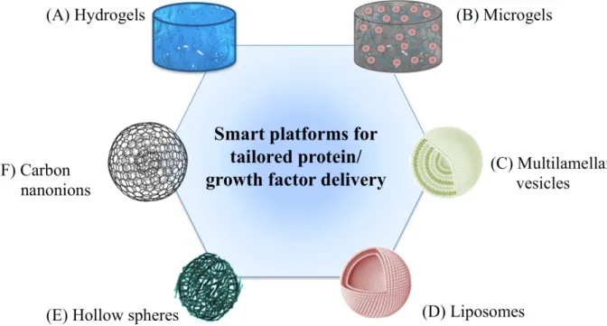
Self-assembled hydrogels made from functional extracellular matrix components have the advantage of achieving better biological recognition in a 3D tissue like environments with high fidelity. But the complexity, lack of control of the self-assembly process and variation from batch to batch may affect reproducibity. However, a semi synthetic approach in designing peptide sequences known to influence a biological function such as differentiation, maintenance of stemness or influence cellular secretome can be easily fabricated or coupled to a polymeric framework to match the physical properties of the tissue. This approach will endorse the need for designing a customised 3D hydrogel with tunable mechanical and biochemical features using functional recognition sequences. Another perspective which has been thoroughly investigated involves spherical microgels, that are an interesting bridging among macro-hydrogels and spherical microcarriers [156,172]. They can be defined as microscale hydrogels, offering a higher surface to volume ratio, compare to macroscale hydrogels. This unique feature enhances both microgels stability and their integration within the local tissue mass transport, by decreasing the bulk resistance [173,174]. In a size and structural complexity decreasing progression, soft self-assembling frameworks for protein delivery can be encompassed by catanionic vesicles, colloidal hollow structures, based on pairing of oppositely charged ionic amphiphiles. In detail, the above mentioned amphiphiles easily self-aggregate [175] into multi-walled vesicular structures [176]. Vesicles are of increasing interest in the biotechnological field [177,178], mimicking biological membranes and their
compartmentalization features [179]. Moreover, triggering factors for these structures involves salt/co-solutes addition, chain length and temperature, able to provide to multi-to-unilamellar transition and thus a controlled therapeutic release [180]. On the other side, liposomes offer another ‘soft’ alternative for tunable compartmentalization and biological responsive protein delivery. Microspheres and hollow spheres are an additional tool for tailored delivery of proteins and growth factors, which have been extensively studied. Both synthetic (i.e. polyethyetylene glycol-PEG and polydimetylacrylamide-PDMA) and natural based polymers have been employed in their fabrication [181,182] Different strategies for their production were proposed, such as emulsion, sacrifical template coating, chemical binding. Furthermore, ECM mimicking motifs allow the specifi targeting of cellular proteins: this unique feature provide these reservoir systems with excellent performances and targeting properties, for growth factors delivery [183,184].
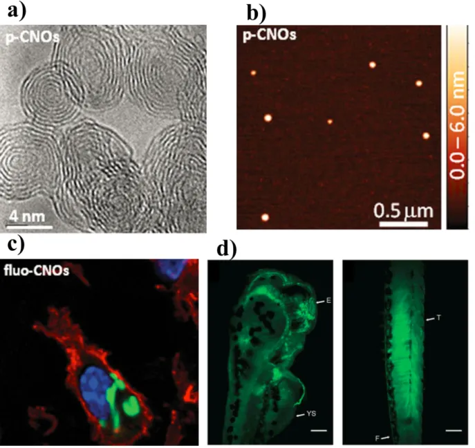
Still keep on decreasing the available protein vehicle systems, there is considerable interest for carbon nano-onion [148]. These multi-shell fullerene structures pertain unique physicochemical properties of, such as ultra-small size, large surface to mass ratio, high flexibility and capability to be complexed (either covalently or non-covalently) with bioactive molecules, including small proteins like enzyme binding moieties [148].
Figure 6. Schematic representation recapitulating platforms for protein/growth factor delivery. Scaffold size is decreasing clockwise, from (A) to (F).
5. Synthetic therapeutics delivery
5.1 Challenges of small molecules in drug delivery
Despite in the last twenty years the main trend in pharmaceutical research was driven towards a growing interest in macromolecular therapeutics [185], most of the active ingredients still belongs to the class of small molecules under 500 Da. For instance, in cancer research, almost 75% of these therapeutics derive from synthetic sources [186]. The major issue of small molecule therapeutics is represented by the toxic side effects of high doses, due to an overall poor biodistribution and rapid clearance [187]. Moreover, when a low invasive and high patient friendly administration route is chosen, such as the oral approach, additional factors can influence and reduce the therapeutic efficacy of the majority of Active Pharmaceutical Ingredients (APIs) with poor solubility and low permeability in gastro-intestinal environment [188]. Besides solubility issues of hydrophobic compunds, a larger group of drugs are susceptible to several biochemical barriers, such as enzymatic attack, hydrolysis, degradation in the low gastric pH, unspecific endocytosis of the
extended mucosa surface [189], pre-systemic metabolic pathways [190], intracellular biotrasformations [191] and plasma proteins complexation and recognition by the cells of the mononuclear phagocyte system [192].
Nowadays, the low bioavailability still represents a concrete issue: more than 90% of active compounds discovered and approved since 1995 belong either to class II or IV in the standard of Biopharmaceutical Classification System (BCS) [193]. Several strategies were developed to enhance both solubility and permeability of APIs. These approaches include the modification of both physical and chemical properties of the API [194]. The modification of physical characteristics is mainly focused on micronization techniques [195,196] reduction of crystal sizes, amorphization and stabilization of such solid dispersions [197,198]. Recently, further efforts have been paid to enhance specific organs and tissues targeting, while improving drug permeability on specific diseased cell targets.
5.2 Lipid based- nanovesicles
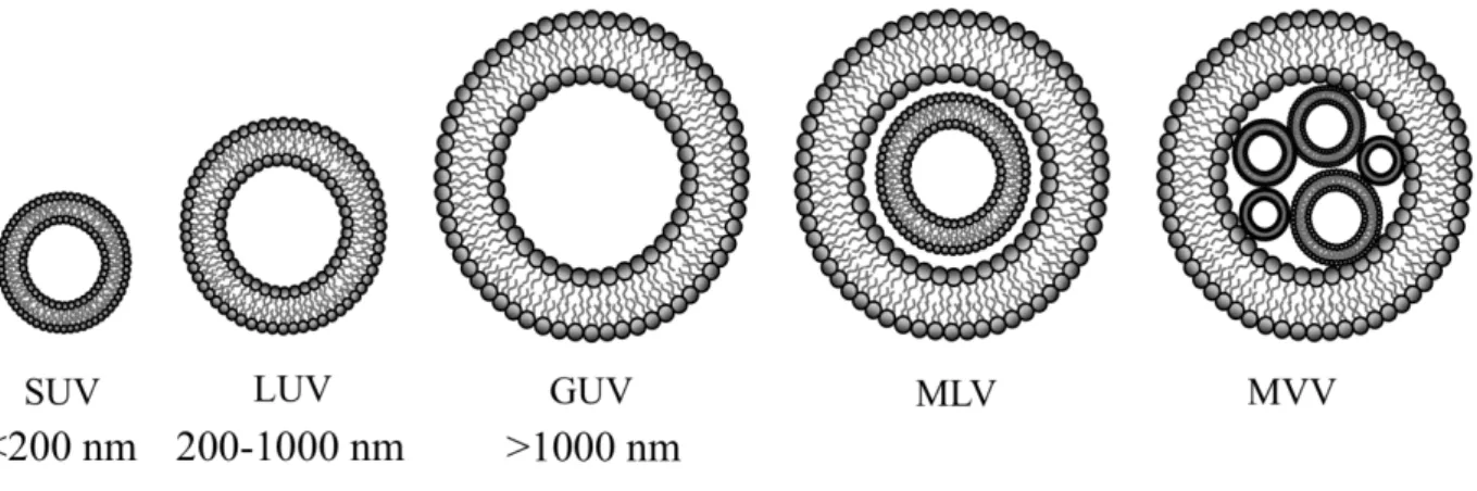
As previously mentioned, lipid based nanovesicles (LNVs) are defined as spherical vesicles constituted by a bilayered lipid membrane. In a size-driven perspective, these colloidal carriers can be divided into small unilamellar vesicles (SUVs, with diameter <200 nm and single bilayer membrane), large unilamellar vesicles (LUVs, diameter ranging 200-1000 nm, single bilayer membrane), giant unilamellar vesicles (GUVs, diameter >1000 nm, single bilayer membrane), multilamellar vesicles (MLVs composed by concentric vesicles, or multiple concentric bilayers) and multi vesicular vesicles (MVVs, composed by multiple vesicles confined inside a larger one). Figure 7 groups the different types of nanovesicular systems, with increasing size characteristics. Since the first LNV proposed by Gregoriadis in 1974 [120], a large number of nanovesicicular systems were developed. Such an impressive success is due to their ability to incorporate functional biomacromolecules on the lipid bilayer, proposing as smart, flexible, stimuli responsive systems.
Key features of these colloidal carriers involve the API encapsulation efficiency (EE), the particle size distribution and the z potential.
Figure 7. Schematic representation of the main lipid based nanovesicular systems.
Over lipidic nanovesicles class, liposomes were the first discovered structures, composed by natural phospholipids membranes in the form of SUV and MLV. Nowadays, further in-depth researches explored the use of many synthetic phospholipids like phosphatidylglycerol, phosphatidylethanolamine, phosphatidylcholine and phosphatidylserine [199]. The first method developed for the encapsulation of hydrophilic compounds refers to the thin layer hydration (TLH), characterized by a very low EE due to the highly non-favoured partition of hydrophilic molecules inside the vesicle cavity versus the surrounding aqueous medium. Further methods involved the combination of TLH with other approached to enhance the EE of hydrophilic molecules: reiterated freeze–thaw (FT), reverse phase evaporation (REV) and dehydration–rehydration of empty vesicles (DRV).
The FT method allows the spontaneous MLVs disruption by the water ice crystals produced in the freezing process, leading to the fabrication of SUVs. The EE can be modulated by tuning the FT cycle rate, the aqueous solute and lipid concentrations. Additional extrusion steps after FT have shown to improve the size distribution, such as the encapsulation of the hydrophilic drug itopride in liposomes [200]. On the other hand, multiple FT cycles can increase liposome diameter and
polydispersity, when liposome components are highly susceptible to salt concentration (i.e. egg phosphatidylcholine) [201].
Reverse phase evaporation method (REV) is another approach to encapsulate small hydrophilic drugs. Therein, the lipids are dissolved in an organic phase and further incorporated in an aqueous phase with the drug. The obtained emulsion is subsequently treated with evaporation-hydration cycles, leading to the formation of liposomes, mainly LUVs with large EE. For example, the addition of the REV step to TLH introduced an EE increase in the case of sumatriptan succinate [202]. Conversely the utilization of REV can result in a decrease of hydrophobic drugs encapsulation efficiency, compared to TFH, as in the case of acetazolamide, inside multilamellar vesicles [203] and ketoprofen-hydroxypropyl-β-cyclodextrin complexes included into large unilamellar vesicles [204]. Liposomal vesicles can be fabricated by means of a dehydration– rehydration method (DRV), adding a buffer to the thin film and further lyophilize. The solid pellet is then rehydrated with the drug solution. Compared to conventional TFH, DRV produced a high drug/lipid ratio in the case of vancomycin [205], with an impressive increase of EE (30 and 130 fold), for both non-decorated and pegylated liposomes, respectively. Recent reviews discussed innovative methods for liposomes fabrication [206-208], including microfluidics [209,210], compressed/supercritical fluids for the incorporation of both hydrophilic and lipophilic drugs into the vesicles [211,212].
A large variety of liposomal formulations was developed by incorporating one or more multitasking ligands within the vesicle membrane. Liposomes can exert a passive targeting by enhancing the permeability retention time [213] or by depletion of macrophages, as shown in the case of the hydrophobic drug amphotericin B [214]. On the other hand, active targeting features can involve the binding mechanisms of liposomes towards bioactive receptors, such as antibody conjugates [215] or permeation enhancers, like cell-penetrating peptides (CPPs) [216]. Stimuli-responsiveness is an attractive perspective for specifically triggered drug release towards liposomes, aiming to reduce the side effects of unspecific targeting through a dual action. In the first instance, by
targeting the lower pH inside the endosome; secondly, while providing for conformational changes of dioleylphosphoethanolamine (DOPE) and cholesterylhemisuccinate, the therapeutic cargo can be released in the cytoplasm [217]. At present, doxorubicin is one of the most studied drugs for liposomal triggered-release, with a thermosensitive formulaton, ThermoDox®, in clinical trials. Other liposomal formulations with small bioactive molecules are available in the market, with good encapsulation efficiency. Nevertheless targeted formulations or stimuli-responsive liposomes are still challenging at a research stage.
The main reason for such a limited transfer of liposomal formulation into the bipharmaceutical market relies on the drawback of their low physico-chemical stability, due to the intrinsic poor aqueous solubility of phospholipids, which tend to aggregate into bigger clusters [218]. As a consequence, liposome suspensions are usually lyophilized and stored as dry products [219].
5.3 Non-liposomal nanovesicles
The limited efficacy of liposomes broadens research horizons towards employing lipids in combination with alternative self-assembling materials, such as surfactants [220,221], polymers [222] and peptides [223].
Non-liposomal nanovesicles can be classified according to the alternative component versus phospholipid: niosomes, sphingosomes, pharmacosomes and quatsomes.
Niosomes are prepared by hydrating a mixture of lipids with non-ionic surfactants, such as alkyl ethers, alkylesters alkylamides, fatty acids and amino acids. Niosomes are able to encapsulate both hydrophilic and hydrophobic drugs. The use of cholesterol as lipid component leads to a more rigid and less leaky bilayers, which makes it particularly suitable for small drugs such as calcein [224] or poorly soluble beclomethasone dipropionate [221]. Niosomes exhibit an overall short-term stability, which strongly depends on the additional membrane components [225].
Sphingosomes are composed by natural or synthetic sphingolipids, which form nanovesicles in the form of SUVs and LUVs. Compared to niosomes, they show an enhanced stability, as well as an improved resistance against hydrolysis, compared to liposomes. A promising application of