S E
Biotechnol. Agron. Soc. Environ.2004 8(4), 267–273
1. INTRODUCTION
Blood products like spray-dried blood, plasma or red cells have been recognized as a source of proteins with good amino acid profile and digestibility, and therefore considered as of high nutritional value for their use as ingredients in farm animal diets for years. Spray-dried porcine plasma has been recommended during years to improve the growth and conversion index in weaning piglets, especially under high pathogen environment (Coffey, Cromwell, 1995; Van Dijk et al., 2001). Recently, efficient replacement of growth promoters by spray-dried porcine plasma in
the weaning period diets, without reduction on the productive parameters of the farm, has been demonstrated (Torrallardona et al., 2003).
However, in Europe, the Commission Regulation 1234/2003/EC temporally bans the use of processed animal proteins, including blood-derivative products, in feedstuffs for all farm animals which are fattened or bred for the production of food, with some exceptions, like the authorization of the use of non ruminant blood products and blood meal into the feed of farm fish. Authorization of the re-introduction of these proteins into animal feed formulations, especially non ruminant proteins into the feed for non ruminant farm
Identification of bovine material in porcine spray-dried
blood derivatives using the Polymerase Chain Reaction
technique
Anna Castelló
( 1 ), Olga Francino
( 1 ), Betlem Cabrera
( 1 ), Javier Polo
( 2 ),
Armand Sánchez
( 1 )(1)Servei Veterinari de Genètica Molecular. Facultat de Veterinària. Universitat Autonoma de Barcelona. E-08193 Bellaterra (Spain). E-mail: [email protected]
(2)R&D Department. APC EUROPE SA. Avda. Sant Julià 246-258. Pol. Ind. El Congost. E-08400 Granollers (Spain).
Due to the widely supported theory of bovine spongiform encephalopathy (BSE) spread in cattle by contaminated animal feeds, screening of feed products has become essential. For many years, manufacturers have used blood and plasma proteins as high quality ingredients of foods for both pets and farm animals. However, in Europe, the Commission Regulation 1234/2003/EC temporally bans the use of processed animal proteins, including blood-derivative products, in feedstuffs for all farm animals which are fattened or bred for the production of food. This regulation has some exceptions, such as the use of non ruminant blood products into the feed of farm fish. Authorization of the re-introduction of these proteins into animal feed formulations, especially non ruminant proteins into the feed for non ruminant farm animals, is expected when adequate control methods to discriminate ruminant proteins exist. Currently, the number of validated methods to differentiate the species of origin for most of the animal by-products is limited. Here we report the development of a rapid and sensitive polymerase chain reaction (PCR)-based assay, which allows detection of bovine or porcine specific mitochondrial DNAfrom spray-dried blood derivate products (plasma, whole blood and red cells), as a marker for bovine contamination in porcine products. Sample extracts, suitable for PCR, were easily and quickly obtained with the commercial PrepManTMUltrareagent
(Applied Biosystems). To confirm the porcine origin of the samples, primers targeting a specific region of 134 bp of the porcine cytochrome bcoding sequence were designed (cytbporc1-F and cytbporc2-R). Previously published PCR primers (L8129 and H8357), specific for a 271 bp fragment of the bovine mitochondrial ATPase 8-ATPase 6 genes, were chosen to accomplish amplification of bovine DNA. The limit of detection (LOD) of the bovine PCR assay was at least of 0.05% (v/v) of bovine inclusion in spray-dried porcine plasma or red cells fraction. In dried whole blood samples, sensitivity of the method was found to be at least of 0.1 % (v/v). Since the method described here exhibits high specificity and sensitivity and it is rapid, simple and consistent, it could be successfully utilized as a routine control assay to evaluate the presence of bovine materials in spray-dried blood products.
animals, is expected when adequate control methods to discriminate ruminant proteins exist.
Traditionally, species identification in animal by-products, as well as detection and identification of constituents of animal origin in feed or food, has been established through different methods, mainly based on:
a) the analysis of proteins (electrophoretic, isoelectric focusing, immunochemical and HPLC methods) (Martin et al., 1991; Zerifi et al., 1991; Cutrufelli
et al., 1993; Knuutinen, Harjula, 1998);
b) fatty acids identification (Verbeke, De Brabander, 1980);
c) DNA analysis (Meyer et al., 1994; 1995; Rehbein
et al., 1997);
d) microscopic detection of specific structure elements (Koolmees, 1999).
Successful detection of the species of origin depends on how the assay has been conducted and on the previous treatment imposed to the animal by-products.
In general, mitochondrial DNA (mtDNA) based PCR methods have given quite good results in the analysis of critical samples (submitted to moderate or severe temperature and pressure treatments) in which DNA has been partly degraded (Tartaglia et al., 1998; Wang et al., 2000; Bellagamba et al., 2001; Myers
et al., 2001; Bellagamba et al., 2003; Bottero et al., 2003; Kromar, Rencova, 2003; Rodriguez e t a l ., 2004). PCR primers designed on the basis of sequences of short interspersed repetitive elements (SINEs) have also been successfully used to detect tiny contamination levels of feed by animal material (Tajima et al., 2002). Recently, with the emergence of the real-time PCR technology, PCR designs on mitochondrial encoded targets (Lahiffet al., 2002) or in nuclear sequences (Brodmann, Moor, 2003) based on the use of Taqmanprobes, have been reported for bovine detection in meat products, meat and bone meal (MBM) and in feedstuff containing animal MBM.
In spite of the important efforts done in the last years to improve existing procedures and to develop new techniques for animal detection in feedstuffs, the only official method currently approved by EU is microscopic detection (2003/126/EC Directive). This method is laborious, requires skills and has a low throughput. It may differentiate the tissue origin of the animal by-product (hair, MBM, feather meal, fish meal, blood meal, blood products, etc.) but is unable to identify the species of origin.
As far as we know, few reports of species identification in blood-derived products are available (Etzel et al., 1997; Newgard et al., 2002; Polo et al., 2004). Nevertheless, rapid control methods, capable of
high throughput analysis of samples and providing reliable species identification of blood products, are urgently required to allow the re-introduction of these proteins into animal feed formulation.
The aim of the present work was to develop an easy, sensitive and accurate test, based in species-specific mtDNA amplification, to detect the presence of bovine contamination in spray-dried porcine blood derived products. The study was also intended to establish the limit of detection (sensitivity) of the PCR method developed. Comparison of positive detection of bovine presence in blood derived samples between the PCR method described here and the Agar Gel Immunodiffusion technique (AGID) recently reported (Polo et al., 2004) was also considered.
2. MATERIALS AND METHODS
2.1. Samples
Three kinds of blood-derived products were used: spray-dried animal plasma (SDAP), spray-dried animal whole blood (SDAWB) and spray-dried animal red cells (SDARC). Detailed description of the method to produce the spray-dried blood products will be found elsewhere (Polo et al., 2004). Whole blood prepared liquid samples, as well as plasma and red cell fractions obtained after centrifugation at 3000 rpm for 15 min (Sorvall RC-5B, New York, USA), were dried using a laboratory spray-dryer (Büchi 190 Mini Spray D ry e r, Büchi Laboratoriums-Technik AG, Flawil, Switzerland) following the conditions reported by Polo et al., 2004.
Thirteen samples of SDAWB and 20 of SDAP of porcine origin with different levels of bovine inclusion ranging from 0 to 1% (v/v) were analysed in the present study. Pure bovine SDAWB and SDAP samples (two of each) were also included as positive controls. The whole aforementioned products were part of the samples previously analyzed by AGID technique (Polo et al., 2004).
Moreover, 18 SDARC samples, including one of pure bovine origin and 17 of porcine origin (with bovine inclusion ranging from 0 to 20%, v/v), were subjected to study.
2.2. DNA extraction
The DNA preparation procedure, based on the use of
SDAWB) or a 3 ml aliquot (for SDAPand SDARC) of the obtained suspension was centrifuged at 12000 x g for 3 min and the supernatant was removed. The pellet was subsequently incubated with 100 µl (for SDAWB) or 200 µl (for SDAP and SDARC) of PrepManTM
Ultra reagent (Applied Biosystems) at 100ºC for 1 0 min and digested material was centrifuged at 12000× g for 3 min. Cleared supernatant from each preparation was recovered, placed in a fresh microcentrifuge tube and stored at –20ºC till PCR was accomplished.
To prevent DNA contamination, extractions were carried out in a dedicated laboratory room. PCR preparations and post-PCR analyses were also carried out in separate rooms.
2.3. PCR amplification
Porcine specific PCR primers were designed from porcine mtDNA sequence available in the GenBank database (AF034253). The primer sequences used were as follows: cytbporc1-F (forward; 5’-GGA GTC TTG GCC CTA G TA GC-3’) and cytbporc2-R (reverse; 5’-GTA ATG AGG TCT GCT ACT AGT TAT-3´). Previously published PCR primers (L8129 and H8357) specific for a 271 bp fragment of the bovine mitochondrial AT P a s e 8 - ATPase6 gene (Tartaglia et al., 1998) were chosen to accomplish specific amplification of bovine DNA. All primers were purchased from TIB MOLBIOL Syntheselabor (Berlin, Germany).
PCR amplifications were carried out using PCR C o re Kit Plus (Roche Diagnostics, Mannheim, Germany) in a final reaction volume of 20 µl. Each reaction contained 2,5 mM MgCl2, 1 x PCR reaction buffer, 250 nM of each appropriate species-specific forward and reverse primer, 2 units of Taq DNA polymerase, 0.5 units of Uracil DNA glycosylase heat labile, 200 µM of each dATP, dCTP, dGTP, 600 µM dUTP and 1 µl of DNA extract from spray-dried plasma, blood or red cells as template. The use of dUTP/Uracil DNAglycosylase in each reaction allows eliminating dUMP containing amplicons avoiding the risk of carryover contamination of previous PCR (Longo et al., 1990). PCR reactions were performed with GeneAmp PCR System 9700 (Perkin Elmer, Applied Biosystems). After an initial incubation step at 20ºC for 10 min (reaction of Uracil DNA glycosylase) followed by a denaturation step at 94ºC for 2 min (L8129 and H8357 primer set) or for 5 min (cytbporc1-F and cytbporc2-R primer set), cycling parameters were as follows: 35 cycles either at 94ºC for 59 s, 58ºC for 30 s and 72ºC for 30 s for bovine specific amplification or at 94ºC for 30 s, 53ºC for 3 0 s and 72ºC for 30 s for porcine specific amplification with a final extension at 72ºC for 7 min
in both cases. PCR negative and positive controls were also included in parallel with each amplification set.
PCR products were analysed on a 1.8 % high-resolution agarose (AG-500, Ecogen S.R.L, Spain) gel electrophoresis containing ethidium bromide and visualised with UV light. A molecular weight marker (φ×174 DNA/Hae-III; Promega, Madison, USA) was used to identify the PCR products sizes.
3. RESULTS AND DISCUSSION
3.1. Bovine and porcine specific mtDNA amplification test
Spray-dried blood products contain a high level of proteins, some of them being strong inhibitors of PCR (e.g. haemoglobin); therefore different protocols have been tested to prepare total DNA free of PCR inhibitors. We found that the use of the commercial
PrepManTMUltra reagent (Applied Biosystems) was
an easy, fast, effective and consistent method to obtain sample extracts suitable to be used for the analysis of DNA by PCR assays. Obtaining DNA extracts with this procedure requires only few minutes, in contrast to the overnight lysis incubations and the long ethanol precipitation steps normally used in more conventional DNA extraction protocols. The spray-drying procedure used for sample production, which involves a moderately heat-treatment (Polo e t a l ., 2002), did not compromise the quality of the DNA recovered by this method, that resulted suitable for PCR analysis in all cases (Figures 1and 2).
3.2. Detection of bovine contamination in spray-dried porcine blood derivate products
Spray-dried plasma. The porcine specific PCR test, using cytbporc1-F and cytbporc2-R primer pair, produced amplicons of the expected size from all porcine spray-dried plasma samples, confirming the porcine origin and the suitability in PCR of DNA extracts obtained from the plasma samples (data not shown). Of the 22 samples of spray-dried animal plasma (SDAP) analyzed in our study, only two, corresponding to pure porcine SDAP, showed negative results of PCR amplification with bovine specific primer pair (L8129 and H8357). The remaining ones, including two pure bovine spray-dried plasmas and 18 porcine plasmas with levels of bovine inclusion ranging from 0.05 to 1% (v/v), gave positive amplification with bovine species-specific PCR assay (Table 1).
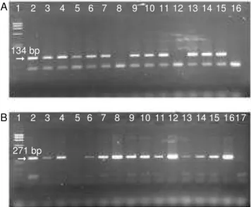
Typical results of some SDAP samples of porcine origin with bovine inclusion levels ranging from 0 to 1% (v/v) are illustrated in f i g u re 1. Porcine and bovine specific amplicons of 134 bp and 271 bp are showed in figure 1Aand figure 1B, respectively. The detection limit (lower percentage producing visible
DNA amplification) of the bovine specific PCR test was at least of 0.05% of bovine plasma in porcine plasma (Figure 1B, lanes 6 and 13).
As mentioned above, all SDAP samples analyzed by bovine specific PCR test in the present study were part of those analyzed by the AGID method previously reported (Polo et al., 2004). Comparison of the results obtained with the PCR method described here and with the AGID technique is showed in table 1. The detection of bovine material in SDAP porcine samples by AGID shows a LOD of 0.5% (v/v) of bovine plasma in porcine plasma in 15/16 of the samples analyzed and of 0.7 % (v/v) in all samples studied (16/16). The sensitivity of the bovine PCR assay increases the detection of bovine material in SDAP samples to at least 0.05% (v/v).
Figure 1.PCR amplification of porcine (A) or bovine (B) specific mitochondrial DNA using DNA extracts from spray-dried porcine plasma with different spiking levels of bovine material (from 0 to 1%, v/v).
Lanes: (1) molecular weight marker; (2-7, 9-11, 13-14) dried porcine plasma containing 1, 0.1, 0.3, 0, 0.05, 0.5, 0.7, 0.9, 0.5, 0.05 and 0.3 % of bovine material, respectively; (8 and 12) dried pure bovine plasma; (15) porcine DNA used as positive PCR control in A and dried porcine plasma containing 0.7% of bovine material in B; (16) bovine DNA used as negative PCR control in A and as positive PCR control in B and (17) porcine DNA used as negative PCR control in B.
A
B
1 2 3 4 5 6 7 8 9 10 11 12 13 14 15 16
1 2 3 4 5 6 7 8 9 10 11 12 13 14 15 1617 ➞
134 bp
➞ 271 bp
1 2 3 4 5 6 7 8 9 10 11 12
➞ 271 bp
Figure 2. Sensitivity screening of PCR method to detect bovine presence in porcine spray-dried red cell samples. Bovine species-specific amplicon of 271 bp was detected with L8129 and H8357 primer pair.
Lanes: (1 to 8) spray-dried porcine red cells containing 0, 0.05, 0.1, 0.3, 0.5, 0.7, 0.9, and 1% of bovine material; (9) pure bovine dried red cells; (10) spray-dried porcine red cells containing 5% of bovine material; (11) bovine DNA used as positive PCR control and (12) PCR negative control.
Ta b l e 1 . Frequency of positive detections of diff e r e n t spiking levels of bovine material in spray-dried porcine plasma or whole blood, depending on the method used to detect contamination (AGID or PCR).
Bovine inclusion Porcine blood product analytical level (% v/v) sample source
Spray-dried Spray-dried
plasma whole blood
PCR AGID PCR AGID
0.00 0/2 0/16 0/1 0/13
0.05 2/2 0/16 -
-0.10 2/2 1/16 2/2 0/13
0.30 3/3 11/16 2/2 8/13
0.50 3/3 15/16 2/2 10/13
0.70 3/3 16/16 2/2 13/13
0.90 3/3 16/16 2/2 13/13
1.00 2/2 16/16 2/2 13/13
Spray-dried whole blood.Fifteen spray-dried animal whole blood (SDAWB) samples were analyzed by PCR with the porcine and bovine specific primers. Porcine origin of 13 samples was confirmed using cytbporc1-F and cytbporc2-R primer pair (data not shown). Positive detection of bovine was obtained in 12 porcine SDAWB samples with bovine inclusion levels ranging from 0.1 to 1 % (v/v) and also in two pure bovine dried whole blood samples by bovine specific PCR test (Table 1). The sensitivity of the PCR method to detect bovine contamination was found to be at least 0.1% of bovine whole blood in porcine whole blood (Table 1).
The SDAWB samples included in this study were also analyzed by Polo et al.(2004), so a comparison of positive detection of different levels of bovine inclusion in whole blood samples depending on the method used was possible. PCR is certainly more sensitive in SDAWB samples than the AGID method; at least seven fold higher (LOD of 0.1% versus0.7%, if we consider 100% of success, respectively).
Spray-dried red cells. A total of 18 s p r a y - d r i e d animal red cells (SDARC) samples were analyzed by the PCR method described here to detect bovine contamination. One sample, corresponding to pure bovine SDARC, and 15 of porcine origin with different levels of bovine inclusion (from 0.05 to 20%) produced a bovine specific amplicon of the expected size, the remaining two, corresponding to pure porcine SDARC, as expected, failed to produce specific bovine amplification (data not shown). Confirmation of porcine origin of the aforementioned samples was accomplished by porcine specific PCR assay (data not shown).
Sensitivity of bovine specific PCR test in SDARC samples is illustrated in figure 2. The LOD was at least of 0.05% of bovine red cells in porcine red cells (v/v). It was observed that the lower the percentage of bovine inclusion in the porcine red cells, the fainter the band obtained in PCR; nevertheless saturated signal was quickly observed (between 0.5 and 0.7% of bovine presence in porcine red cells). Most of the analytical methods available are based on immune identification of blood proteins mainly present in the plasma fraction, like IgG and others. Therefore, these techniques are unable to identify the presence of bovine contamination in the red cells fraction due to the lack of immuno-reactivity. The use of the specific PCR to identify the presence of bovine contamination in blood products derived from the red cells fraction, like SDARC, provides a strong tool to guarantee the identification of bovine contamination in the different range of blood products that could be obtained following the processing conditions established in the Regulation 1774/2002/EC.
4. CONCLUSIONS
Here we report a method that, coupling the specificity and sensitivity of the PCR and the efficiency of
Prepman Ultrafor nucleic acid preparation, allows rapid detection of bovine or porcine specific mtDNA from spray-dried animal blood products, as a marker for bovine contamination in porcine products.
Despite the low level of DNAmolecules recovered and the considerable degradation of nucleic acids, that is expected to occur in the rendering process of samples, the method developed was able to amplify the mtDNA molecules extracted from spray-dried blood derived products. The amplification of the mtDNA regions selected fits our purposes because of: a) the high number of copies per cell of mtDNA
(conferring sensitivity);
b) its mutation rate, which induces genetic interspecies variation (confers specificity); and
c) the length of amplicons (that are short enough to allow amplification from mildly degraded DNA).
The LOD of the bovine specific PCR assay was at least of 0.05% (v/v) of bovine inclusion levels in spray-dried animal plasma or red cells fraction. In spray-dried whole blood samples, sensitivity of the method was found to be at least of 0.1 % (v/v), lower inclusion levels where not tested.
Our method is more sensitive than the automated turbidimetric immunoassay technique of bovine IgG quantification (Etzel et al., 1997), which is accurate to 2.5% (v/v), and even more than the AGID assay (Polo
et al., 2004), which detect the presence of bovine IgG in porcine SDAP or SDAWB at inclusion levels of 0.5% (v/v). In a European Commission report on cross-contamination of ruminant feed with mammalian MBM, dated September 24, 1998, Commission stated that contamination levels >0.5% MBM in ruminant feedstuffs should be prevented. If this level should be considered as a guideline for the European Commission’s level of acceptability, the method described here is suitable for determining that spray-dried blood products meet the guideline.
Acknowledgements
This research was part of Eureka Project (E!2452 EUROAGRI IMMUCON) and was funded by a Spanish grant (PROFIT FIT-060000-2000-188).
List of abbreviations
AGID: agar gel immunodiffusion technique LOD: limit of detection
MBM: meat and bone meal mtDNA: mitochondrial DNA PCR: polymerase chain reaction SDAP: spray-dried animal plasma SDARC: spray-dried animal red cells SDAWB: spray-dried animal whole blood SINEs: short interspersed repetitive elements
Bibliography
B e l l a g a m b a F., Moretti VM., Comincini S., Va l f r è F. (2001). Identification of species in animal feedstuffs by polymerase chain reaction-restriction fragment length polymorphism analysis of mitochondrial DNA. J . Agric. Food Chem.49, p. 3775–3781.
B e l l a g a m b a F., Va l f r è F., Panseri S., Moretti V. (2003). Polymerase chain reaction-based analysis to detect terrestrial animal protein in fish meal. J. Food Prot.66 (4), p. 682–685.
Bottero MT., Dalmasso A., Nucera D., Turi RM., Rosati S., S q u a d r o n e S., Goria M., Civera T. (2003). Development of a PCR assay for the detection of animal tissues in ruminant feeds. J. Food Prot. 6 6 ( 1 2 ) , p. 2307–2312.
B r o d m a n n PD., Moor D. (2003). Sensitive and semi-quantitative Taqman real-time polymerase chain reaction systems for the detection of beef (Bos taurus) and the detection of the family Mammalia in food and feed. Meat Sci.65, p. 599–607.
C o ff e y RD., Cromwell GL. (1995). The impact of environment and antimicrobial agents on the growth response of early-weaned pigs to spray-dried porcine plasma. J. Anim. Sci.73, p. 2532–2539.
C u t r u f e l l i ME., Mageau R P., Schwab B., Johnston RW. (1993). Development of a multispecies identification field test by modified agar-gel immunodiffusion. J. AOAC Int.76, p.1022–1026.
Etzel LR., Strohbehn RE., Mc Vicker JK. (1997). Develop-ment of an automated turbidimetric immunoassay for quantification of bovine serum immunoglobulin G. Am. J. Vet. Res.58(11), p. 1201–1205.
K n u u t i n e n J., Harjula P. (1998). Identification of fish species by reversed-phase high-performance liquid chromatography with photodiode-array detection. J. Chromatogr.705, p. 11–21.
Koolmees PA. (1999). Histology as an additional technique for species identification in meat products. I n Bergwerff AA. (Ed). Species Identification in Meat P ro d u c t s. Utrecht, The Netherlands: ECCEAMST, p. 35–41.
Kromar P., Rencova E. (2003). Identification of species-specific DNA in feedstuffs. J. Agric. Food Chem.51, p. 7655–7658.
L a h i ff S., Glennon M., Ly n g J., Smith T., Shilton N . , Maher M. (2002). Real-Time polymerase chain reaction detection of bovine DNA in meat and bone meal samples. J. Food Prot. 65(7), p. 1158–1165.
L o n g o MC., Beringer MS., Hartley JL. (1990). Use of uracil DNA glycosylase to control carry-over contamination in polymerase chain reactions. Gene93, p. 125–128. M a r t i n R., Wa r d a l e RJ., Jones SL., Hernández P E . ,
P a t t e r s s o n RLS. (1991). Monoclonal antibody sandwich ELISA for the potential detection of chicken meat mixtures of raw beef and pork. Meat Sci. 30, p. 23–31.
Meyer R., Candrian U., Luthy J. (1994). Detection of pork in heated meat products by Polymerase Chain Reaction. J. AOAC Int.77(3), p. 617–622.
M e y e r R., Hofelein C., Luthy J., Candrian U. (1995). Polymerase Chain Reaction-restriction fragment length polymorphism analysis: a simple method for species identification in food. J. AOAC Int. 7 8 ( 6 ) , p. 1542–1551.
M y e r s MJ., Friedmann S., Farrel D., Dove-Pettit D A . , B u c k e r M F., Kelly S., Madzo S., Campbell W. , Wang RF., Paine D., Cerniglia CE. (2001). Validation of a polymerase chain reaction method for the detection of rendered bovine-derived materials in feedstuffs. J. Food Prot. 64(4), p. 564–566.
Newgard JR., Rouse GC., McVicker JK. (2002). A novel method for detecting bovine immunoglobulin G in dried porcine plasma as an indicator of bovine plasma contamination. J. Agric. Food Chem. 5 0 ( 11 ) , p. 3094–3097.
P o l o J., Rodríguez C., Ródenas J., Saborido N. (2002). Bactericidal effect of the spray-drying system for animal plasma on two different E. colianimal strains. P roceedings of 48th ICoMST, vol 1, Rome, Italy, p. 194–195.
P o l o J., Saborido N., Ródenas J., Rodríguez C. (2004). Determination of the presence of bovine immunoglobulin G in liquid or spray-dried porcine plasma and whole blood by agar gel immunodiffusion. J. AOAC Int.87(1), p. 78–82.
Rehbein H., Kress G., Schmidt T. (1997). Application of P C R - S S C P to species identification of fishery products. J. Sci. Food Agric.74, p. 35–41.
Ta j i m a K., Enishi O., A m a r i M., Mitsumori M . , K a j i k a w a H., Kurihara M., Ya n a i S., Matsui H . , Yasue H., Mitsuhashi T., Kawashima T., Matsumoto M. (2002). PCR detection of DNAs of animal origin in feed by primers based on sequences of short and long interspersed repetitive elements. Biosci. Biotechnol. Biochem.66(10), p. 2247–2250.
Ta r t a g l i a M., Saulle E., Pestalozza S., Morelli L . , Antonucci G., Battaglia PA. (1998). Detection of bovine mitochondrial DNA in ruminant feeds: a molecular approach to test for the presence of bovine-derived materials. J. Food Prot.61, p. 513–518.
Torrallardona D., Conde MR., Badiola I., Polo J., Brufau J. (2003). Effect of fishmeal replacement with spray-dried animal plasma and colistin on intestinal structure, intestinal microbiology, and performance of weanling pigs challenged with Escherichia coliK99. J. Anim. Sci. 81, p. 1220–1226.
Va n D i j k AJ., Everts H., Nabuurs MJA., Marg r y R J C F. , Beynen AC. (2001). Growth performance of weanling pigs fed Spray-Dried Animal Plasma: A Review.Livest. Prod. Sci. 68, p. 263–274.
Ve r b e k e R., De B r a b a n d e r H F. (1980). Identification of animal fat species. Proc. Eur. Meet. Meat Res. Workers 26, p.150–153.
Wang RF., Myers MJ., Campbell W., Cao WW., Paine D., Cerniglia CE. (2000). A rapid method for PCR detection of bovine materials in animal feedstuffs. Mol. Cell. Probes14, p. 1–5.
Z e r i f i C., Lanie C., Benard G. (1991). SDS-PA G E technique for species identification of cooked meat. Fleischwirtschaft71, p. 1060–1062.