RESUMO.- [Perfil de resistência a antimicrobianos de grupos clonais de Staphylococcus aureus isolados de
pequenas propriedades leiteiras do sul do Brasil.]A
in-fecção intramamária persistente em bovinos leiteiros tem sido associada com estirpes de Staphylococcus (S.)aureus específicos, os quais podem ser resistentes a antimicrobia
-nos. Os objetivos deste estudo foram avaliar os fenótipos
de resistência aos antimicrobianos de isolados de S. au-reus e a distribuição e persistência de grupos clonais em pequenos rebanhos leiteiros do sul do Brasil. As amostras
de leite foram coletadas de todas as vacas em lactação de
21 propriedades leiteiras, ao longo de um período de dois anos, perfazendo um total de 1.060 amostras. Isolados de S. aureus foram testados quanto à resistência frente a treze antimicrobianos, pelo método de disco-difusão. O DNA to
-tal dos isolados foi clivado com a enzima Smal e submetido a eletroforese em gel de campo pulsado (PFGE). Padrões
Antimicrobial resistance profiles of
Staphylococcus aureus
clusters on small dairy farms in southern Brazil
1Lilian K. Girardini2, Daniel S. Paim3, Thais C. Ausani3, Graciela V. Lopes3, Debora C.P. Pellegrini4, Maria Aparecida V.P. Brito5 and Marisa Cardoso3*
ABSTRACT.- Girardini L.K., Paim D.S., Ausani T.C., Lopes G.V., Pellegrini D.C.P., Brito M.A.V.P. & Cardoso M. 2016. Antimicrobial resistance profiles of Staphylococcus aureus clus -ters on small dairy farms in southern Brazil. Pesquisa Vetrinária Brasileira 36(10):951-956. Departamento de Medicina Veterinária Preventiva, Faculdade de Veterinária, Universi
-dade Federal do Rio Grande do Sul, Av. Bento Gonçalves 9090, Porto Alegre, RS 91540-000, Brazil. E-mail: [email protected]
In intensive dairy farming, persistent intramammary infection has been associated with specific Staphylococcus (S.) aureus strains, and these strains may be resistant to antimicrobials. The objective of this study was to evaluate the antimicrobial resistan
-ce phenotypes of S. aureus isolates and to assess the distribution and the persistence of clonal groups in small dairy herds of southern Brazil. Milk samples were collected from all lactating cows from 21 dairy farms over a two-year period, totaling 1,060 sam -ples. S.aureus isolates were tested for susceptibility to thirteen antimicrobials using the disk diffusion method. The total DNA of the isolates was subjected to SmaI digestion followed by pulsed-field gel electrophoresis (PFGE). Banding patterns differing by ≤4 bands were considered members of a single PFGE cluster. The frequency of S. aureus isolation ranged from 3.45% to 70.59% among the 17 S. aureus-positive herds. Most S. aureus isolates (87.1%) were susceptible to all antimicrobials; resistance to penicillin (18.2%) was the most frequently observed. The 122 isolates subjected to macrorestric
-tion analysis were classified into 30 PFGE-clusters. Among them, only 10 clusters were intermittent or persistent over the two-year period. The majority (93.6%) of isolates belonging to persistent and intermittent clusters were susceptible to all tested antimi -crobials. S. aureus intramammary colonization in small dairy farms of southern Brazil is most frequently caused by sporadic PFGE clusters, although some persistent clusters can arise over time. Both sporadic and persistent isolates were highly susceptible to
antimicrobials.
INDEX TERMS:Mastitis, PFGE clusters, Methicillin-resistant Staphylococcus aureus, MRSA.
1 Received on January 26, 2016.
Accepted for publication on June 24, 2016.
2 Faculdade de Medicina Veterinária, Universidade do Oeste de Santa Catarina, Xanxerê, SC 89820-000, Brazil.
3 Departamento de Medicina Veterinária Preventiva, Faculdade de Ve -terinária, Universidade Federal do Rio Grande do Sul (UFRGS), Avenida Bento Gonçalves 9090, Agronomia, Porto Alegre, RS 91540-000, Brazil. *Corresponding author: [email protected]
4 UniversidadeFederal do Pampa, BR-472 Km 592, Uruguaiana, RS 97500-970, Brazil.
de bandas diferentes por ≤4 bandas foram considerados como pertencentes ao mesmo grupo clonal. A freqüência
de S. aureus variou de 3,45% até 70,59%, entre os 17 reba
-nhos com isolamento positivo de S. aureus. A maioria dos isolados de S. aureus (87,1%) foi suscetível a todos os an
-timicrobianos; resistência à penicilina (18,2%) foi a mais freqüentemente observada. Os 122 isolados submetidos à análise de macrorestrição foram classificados em 30 gru
-pos clonais de PFGE. Entre eles, apenas dez gru-pos clonais foram intermitentes ou persistentes ao longo do período de dois anos. A maioria (93,6%) dos isolados pertencentes a grupos clonais persistentes e intermitentes foram susce
-tíveis a todos os antimicrobianos testados. Concluiu-se que a colonização intramamária em bovinos de pequenas pro
-priedades leiteiras do Sul do Brasil é mais frequentemente causada por grupos clonais esporádicos de S. aureus, em
-bora alguns grupos clonais persistentes possam ocorrer ao longo do tempo. Em ambos os grupos clonais os isolados foram majoritariamente suscetíveis a antimicrobianos. TERMOS DE INDEXAÇÃO:Mastite, grupos clonais, PFGE, Staphylo-coccus aureus resistente à meticilina, MRSA.
INTRODUCTION
Staphylococcus (S.) aureus is the main coagulase-positive species of Staphylococci associated with intramammary in
-fections in dairy cattle and can lead to clinical or subclinical mastitis (Young et al. 2001). Although S. aureus can
colo-nize the skin and mucous membranes of cows, the udder is the most important reservoir of these bacteria (Capurro et al. 2010). It was demonstrated that S. aureus
intramam-mary infection can be widespread among cows within the same herd, and genotyping has shown that common clonal groups can be persistent in a herd over time (Mork et al. 2012, Piccinini et al. 2012). Among the genotyping techni
-ques, pulsed-field gel electrophoresis has been frequently used to determine whether S.aureus isolates are close
rela-ted and associarela-ted with persistent intramammary infection (Middleton et al. 2002, Haveri et al. 2008, Mork et al. 2012).
Antimicrobial resistance in S. aureus is a concern for
both human and animal health, because multi-resistant strains represent a challenge to effective treatment. Parti
-cularly, methicillin-resistant S. aureus (MRSA), which are resistant to almost all types of β-lactam antimicrobials, have been a concern worldwide (Lee 2003, Van Duijkeren et al. 2004). The presence of MRSA in dairy cows has been investigated because it was demonstrated that cattle may serve as a source of emergence of new MRSA strains in hu
-mans (Juhász-Kaszanyitzky et al. 2007, Vanderhaeghen et al. 2010). Therefore, colonization of dairy cattle with anti -microbial-resistant S. aureus impacts milk production and may additionally represent an infection hazard to people who work in close contact with cows or consume raw milk (Juhász-Kaszanyitzky et al. 2007).
In southern Brazil, small dairy farms based on family la
-bor contribute a large amount of the total milk production. These independent farmers usually deliver the milk pro
-duction to processing plants; however, some of the milk is also consumed by the family. One major challenge for small
dairy farm production is the lack of technical assistan
-ce, leading to failures in animal management and control measures to prevent udder infection (Stumpf et al. 2000). Often, farmers treat animals without any previous know
-ledge of antimicrobial susceptibility. These practices may influence pathogen transmission and resistance, resulting in an epidemiological scenario quite different from that of dairy farms with large-scale production. Therefore, the aim of this study was to assess the antimicrobial-resistance profiles and pulsotypes of S. aureus isolates causing intra
-mammary infection in small dairy herds.
MATERIALS AND METHODS
Herds and sampling. The study population consisted of 1,185 small dairy herds located in a region of approximately 4,821 square kilometers of the state of Rio Grande do Sul, Bra
-zil (UTM zone 22S, 6.69.000N, 350.000E, 6.830.000N, 450.000E). First, farms were stratified according to the average number of cows in lactation (≤10, 11-15, 16-20, 21-25 and ≥25). Next, the sample size was calculated, taking into account an expected S. aureus isolation frequency of 20% (Brito et al. 2001), with a pre
-cision of 10% at the 95% confidence level. The number of farms of each stratum to be included in the study was determined such that their percent representation in the total target population was maintained (80% of farms had ≤15 cows in lactation). In each stratum, farms were randomly selected for sampling using Micro
-soft Excel 2010 -software. In the 21 farms included in the study, an average of 13 cows (minimum 3 and maximum 50) was in lacta
-tion. All sampled farms used mechanical milking and washed the udder before milking. Most farms applied post-dipping treatment with iodine, although no pre-dipping protocol was used. On all farms, clinical mastitis was treated without prior antimicrobial resistance testing.
Samples were collected every six months over a two-year pe
-riod on all farms. During each sampling event, milk samples were collected from all lactating cows in the herd. Animals that were receiving antimicrobial treatment were excluded from sampling. The procedures for the collection and transport of milk samples followed the recommendations of the National Mastitis Council (2004). Milk samples were collected before the milking process began. The teats were washed and dried individually with dis
-posable paper towels before sampling. The first three streams of milk were discarded, and the teats were then disinfected with 70% alcohol. A composite sample of milk from all teats of each cow was collected in one sterile screw-cap flask. Samples were transported under refrigeration to the laboratory for processing.
Bacterial isolation and identification. Ten microliters of each milk sample was streaked onto 5% sheep blood agar and in
-cubated at 37°C for 24 - 48 hours. After incubation, predominant colonies (at least three colonies present on the agar plate) with si
-milar morphology were isolated and identified as described pre
-viously by the National Mastitis Council (2004) and Markey et al. (2013). Gram positive cocci with positive catalase and coagulase test results that displayed acetoin production (Voges-Proskauer test) were classified as Staphylococcus aureus.
Antimicrobial susceptibility testing and detection of MRSA. Isolates of S. aureus were tested for antimicrobial suscep -tibility using the disk diffusion test on Müller-Hinton agar (Oxoid, Thermo Scientific, UK) according to the guidelines of the Clinical and Laboratory Standards Institute (2008). The following antimi
-crobial disks (Oxoid, Thermo Scientific, UK) were tested: cepha
-fonamide (300µg), sulfa + trimethoprim (25µg) and tetracycline (30µg). S. aureus ATCC 25923 was used as the quality control. Iso
-lates displaying inhibition zones ≥29mm to penicillin were sub
-jected to the zone edge evaluation of the penicillin disk-diffusion test as recommended (CLSI 2009). Fuzzy zone edges were consi
-dered as indicative of no beta-lactamase production.
Isolates that were found to be resistant to oxacillin by the disk diffusion test were subjected to minimum inhibitory concentra
-tion (MIC) determina-tion, screened by the Etest® (bioMérieux,
Marcy l’Etoile, Lyon, France), and checked for the presence of the
mecA gene by PCR, as described by Murakami et al. (1991). DNA
was extracted after disrupting the bacterial cell wall with Guanidi
-ne EDTA-Sarkosyl (Rademaker & De Bruijn 1997). The PCR reac
-tions were performed in a total volume of 50 µl composed of 45µL of PCR MasterMix, 1 µL of each primer (mecA F: AAA TCG ATG GTA
AAG GTT GGC; mecA R:AGT TCT GCA GTA CCG GAT TTG C) and 3
μL of template DNA. Amplification was performed for 40 cycles as follows: initial denaturation at 95°C for 5 min, denaturation at 95°C for 30 s, annealing at 55°C for 30 s, and extension at 72°C for 3 min with a final extension at 72°C for 5 min. Ten microliters of PCR product was analyzed using 2% agarose gel electropho
-resis. The reactions were performed in a GeneAmp® PCR System
9700 thermal cycler (Applied Biosystems, Foster City, CA, USA). S. aureus ATCC 25923 was used as a negative control for the mecA
gene. A mecA-positive strain of S. epidermidis, which was previou
-sly confirmed by sequencing of the mecA gene, was used as a posi
-tive control (Santos et al. 2015).
Macrorestriction of the total DNA and pulsed-field gel electrophoresis (PFGE). One isolate of S. aureus from each S. aureus-positive cow was submitted to macrorestriction and PFGE
analysis, according to a protocol previously proposed by McDou
-gal et al. (2003). Initially, a single colony of the isolates was inocu
-lated into 5mL of brain heart infusion broth and incubated at 37°C for 24h. The concentrations of the cells were adjusted to an ab
-sorbance at 610nm of 0.9 to 1.1 using a spectrophotometer. Two hundred-microliter aliquots of the adjusted cell suspension were centrifuged. The pellet was resuspended in 300µl of Tris-EDTA (TE) buffer (10 mM Tris HCl, 1 mM EDTA [pH 8.0]) and incubated in a water bath at 37°C for 10 min. Three hundred-microliter ali
-quots of 1.8% of agarose were added to the cell suspension, gen
-tly mixed, and dispensed into wells of a plug mold. DNA-soaked agarose plugs were submitted to treatment with 4µL lysostaphin (no. L-7386: Sigma, 1mg / mL). The solidified plugs were trans
-ferred to 3 mL of buffer lysis solution (6mM Tris-HCl, 1 M NaCl, 100mM EDTA, 0.5% Brij-58, 0.2% sodium deoxycholate, and 0.5% sodium-lauryl-sarcosine) and incubated at 37°C in a water bath with stirring for 4h or overnight. The plugs were washed four ti
-mes with 4mL of TE buffer for 10-15 min each and stored at 4°C. Next, a DNA-plug portion was subjected to digestion with 20 U of the enzyme SmaI(Invitrogen, Carlsbad, CA, USA) for 2-3 hours at 25°C. Electrophoresis was performed with a 1% agarose gel using 0.5X Tris-borate-EDTA buffer with a CHEF DR-II system (BioRad Laboratories, Hercules, CA) at 6 V/cm for 20h at 14oC,
with an initial switching time of 5 seconds and a final switching time of 40 seconds. Salmonella Braenderup (ATCC# BAA-664)
was included as a size reference. After PFGE, the gel was stained with ethidium bromide (2µg/mL, Sigma, St. Louis, USA) for 20
min in a covered container and destained in distilled water for 45
min. The gels were photographed and documented using a Kodak 2200 system (Rochester, New York, USA).
Banding patterns were compared using the Gel-Compar II software package (Applied Maths, Kortrijk, Belgium). Percent si
-milarities between isolate fingerprints were determined on the basis of the Dice correlation coefficient. A band position tolerance of 1% was used for the analysis of PFGE patterns. Dendrograms
were generated by unweighted pairwise grouping with mathema
-tical averaging (UPGMA). Isolates were considered as belonging to a common cluster when the PFGE pattern differed by ≤ 4 bands. A cluster was considered transient when isolated only once over a two-year period in a dairy farm; intermittent when it was isolated in more than one sampling event; and persistent when isolated at two or more subsequent samplings.
RESULTS
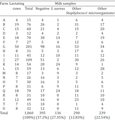
From 1,060 milk samples collected, 395 (37.2%) tested negative for bacterial growth. Staphylococcus aureus was
isolated from 136 samples (12.8%), and other Staphylococ-cus spp. were present in 290 samples (27.3%) (Table 1). Other microorganisms (Corynebacterium sp., Streptococcus sp., Enterococcus sp., Nocardia sp., Trueperella pyogenes, Es-cherichia coli, Klebsiella sp. and yeast) were found in 239 samples (22.5%). On 17 farms, S. aureus was isolated at
fre-quencies ranging from 2.6 to 52.9% of the samples; on 14
farms S. aureus isolates were obtained in two or more
sam-plings; and five farms were positive in all four samplings. Among the 132 isolates of S. aureus tested by the disk diffusion method, the majority (87.1%; 115/132) were susceptible to all antimicrobial agents. Resistance was de
-tected to penicillin (18.2%; 16/132), tetracycline (2.3%; 3/132), and enrofloxacin (0.7%; 1/132). All penicillin sus
-ceptible isolates (n=116) displayed a fuzzy edge zone on the disk diffusion test and were considered as no beta-lactama
-se producers. All strains were susceptible to cephalothin, ceftiofur, clindamycin, gentamicin, sulfonamide, and sulfa + trimethoprim. Regarding methicillin-resistance, three isolates (2.3%) were resistant in the disk diffusion test to oxacillin. The MICs presented by these strains ranged from
Table 1. Bacteriological test results from milk samples collected on small dairy farms in southern Brazil
Farm Lactating Milk samples
cows Total Negative S. aureus Other Other
Staphylococci microorganisms*
A 4 15 4 1 6 4
B 19 76 26 2 31 17
C 15 60 21 4 15 20
D 3 12 4 2 2 4
E 18 70 30 14 7 19
F 7 27 5 4 12 6
G 50 201 98 16 53 34
H 8 31 5 3 17 6
I 11 45 12 10 11 12
J 27 109 51 2 30 26
K 14 54 20 24 9 1
L 15 59 13 6 12 28
M 8 17 3 9 3 2
N 7 26 16 3 2 5
O 7 30 16 3 5 6
P 8 31 6 9 11 5
Q 18 70 17 24 18 11
R 8 31 10 0 11 10
S 12 49 16 0 23 10
T 7 15 10 0 1 4
U 8 32 12 0 11 9
Total 1,060 395 136 290 239
(100%) (37.2%) (27.35%) (12.83%) (22.54%)
0.19 to 0.75µg.mL-1 in the Etest (resistance ≥4µg.mL-1). All three isolates were also negative for mecA detection and, therefore, were considered methicillin-susceptible.
The most prevalent resistance profile included resis
-tance only to penicillin and was observed in 14 isolates (10.6%). Only one isolate was resistant to antimicrobials belonging to three different classes of antimicrobial agents (penicillin, enrofloxacin and tetracycline) and was thus considered multi-resistant.
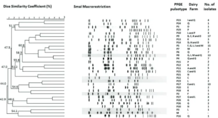
Among the 122 S. aureus isolates digested by SmaI, 102 different PFGE banding patterns were obtained. The pat
-terns presented between 6 and 16 bands ranging from 33.3 to ~1140 kb. Fourteen patterns were found in more than one isolate (from two to five); the remaining patterns were represented by single isolates. The 102 PFGE banding pat
-terns were grouped into 30 clusters of pat-terns that diffe
-red by ≤4 bands (Fig.1). Four clusters (P5, P8, P9 and P17) encompassed almost the half of the S. aureus isolates. Ten clusters (P4, P5, P6, P8, P9, P14, P17, P19, P26 and P27) were found in more than one sampling event at the same respective farms and were classified as intermittent or per
-sistent (Table 2). These clusters encompassed 63 (51.6%) of the S. aureus isolates submitted to PFGE profiling. On one farm (K), the same cluster (P9) was persistent over the two-year period of testing. On two farms (Q and G), up to three persistent or intermittent clusters were found con
-comitantly infecting different cows. Among the isolates belonging to persistent and intermittent clusters, the great
majority (59/63; 93.6%) were susceptible to all tested an -timicrobials.
DISCUSSION
In this study, milk of lactating cows with and without symp
-toms of mastitis was sampled during a two-year period in order to assess the presence of persistent Staphylococcus aureus strains causing intramammary infection in small dairy herds in southern Brazil. In 62.3% (665/1060) of the samples, at least one bacterial species was isolated, and among them the genus Staphylococci was the most frequent (426/665, 64.1%). The colonization of the udder with coagulase-negative staphylococci is frequently repor
-ted in dairy herds, and it may cause mild subclinical mas
-titis (Mendonça et al. 2012, Mork et al. 2012). In our study, the cows were not examined for alterations in the mamma
-ry gland or the milk somatic cell counts were assessed; the
-refore, it is not possible to draw any conclusion about the implications of the coagulase-negative staphylococci isola
-tion for the udder’s health of the sampled animals. On the contrary, the isolation of S. aureus from 12.8% (136/1060) of the milk samples from lactating cows, belonging to 17 (17/21, 80.9%) of those farms, demonstrates the relevan
-ce of this pathogen in the region. Among the 17 positive herds, in 14 (82.3%) S. aureus was isolated in two or more
sampling events indicating that S. aureus strains were
cir-culating among the animals over time. Staphylococcus au-reus is a frequent cause of clinical and subclinical mastitis, and chronically infected cows in a herd are an important reservoir of this pathogen (Ericsson et al. 2009, Mork et al. 2012). Considering the highly contagious nature of the infection, the presence of infected cows may constitute a serious risk of the perpetuation and spread of S. aureus to
susceptible animals and the contamination of milk. Althou
-gh the inability to eradicate S. aureus from dairy herds may be related to failures in standard milking time hygiene as well as in dry cow mastitis therapy (Hutton et al. 1990), other studies raised the hypothesis that certain strains could be more difficult to eliminate from the udder (Smith et al. 1998).
Early attempts for discrimination of S. aureus strains
were based on typing methods, such as ribotyping and phagotyping, and resulted in the identification of common strains in different herds and geographical areas (Mathews et al. 1994, Aarestrup & Jensen 1997). Later on, PFGE star
-ted to be considered the most discriminatory tool to resol
-ve clonal relationship and pro-ved to be superior to other tested techniques (Tenover et al. 1994, Olive & Bean 1999). Using PFGE, it was demonstrated that S. aureus strains are
more likely to be unique to a herd than to be found in multi
-ple herds, and once introduced in a herd these strains may become persistent (Joo et al. 2001, Mork et al. 2012). In our study, the 122 S. aureus isolates digested by SmaI could be grouped into 30 PFGE clusters, among which three (P5, P8 and P9) encompassed strains originated from four to five different herds. Seven clusters included strains present in two herds and the other 20 clusters were found in only one herd. In this sense, our results are in accordance with the conclusion of Joo et al. (2001) that S. aureus strains are
Fig.1. Dendrogram representing the similarity grouping of Sta-phylococcus aureus clusters. The total DNA of isolates was
digested withSmaI and separated by pulsed-field gel electro
-phoresis (PFGE).
Table 2. Distribution of persistent and intermittent S. aureus PFGE-pulsotypes among dairy farms sampled in a two-year
period in southern Brazil
Dairy Pulsotype [# Sampling event (number of isolates)] farm
C P9 [I(1); III(1)]
E P17 [I(2); II(7); IV(3)]
F P5 [I(1); II(2)]
G P5 [I(1); II(2)]; P26 [II(1); III(1)]; P27 [I(2); IV(1)]
H P14 [I(2)*; II(1)*]
K P9 [I(4); II(2); III(1); IV(6)]
L P19 [I(1); IV(1)]
Q P4 [I(1)*; III(5)]; P6 [II(4); III(3)]
more likely to be associated with the herd than to be wides
-pread among multiple herds in a region.
Regarding the persistence of clusters, ten were consi
-dered persistent or intermittent in the farms over the two
--year period. Among them, seven clusters were found at least once in up to five herds and two of them (P5 and P9) proved to be intermittent or persistent in two herds each. Although those clonal groups represent one third of the total of clusters, they are distributed among herds as well as persistent. Other studies also pointed out that some clo
-nal groups can cause persistent intramammary infections over months to years (Aarestrup et al. 1995, Buzzola et al. 2001). Since they persist in the udder, they are secreted over time and have a higher potential for spreading. Per
-sistent strains most likely have properties that make them particularly fit to survive in the udder. Genes encoding vi
-rulence factors, or those required for biofilm formation, have been reported in persistent strains isolated from the udders of cows (Cucarella et al. 2004, Haveri et al. 2008, Piccinini et al. 2012). Another possible explanation for per
-sistence of clonal groups could be the repeated treatment with antimicrobials, which would lead to the elimination of susceptible strains but allow for the persistence of resis
-tant strains (Rajala-Schultz et al. 2009).
In our study, antimicrobial resistance was not frequent among the S. aureus strains, and the majority (87.1%) of the S. aureus isolates, including the persistent and intermit
-tent isolates, was susceptible to all tested antimicrobials. Even the resistance to penicillin, which is highly prevalent among S. aureus strains in Brazilian herds (Medeiros et al. 2009, Silva et al. 2012), was found in only 18.2% of strains. Among the sampled farms, only clinical mastitis cases were treated with antimicrobials and no susceptibility test was performed. In Brazilian herds, a higher chance of S. aureus resistant to penicillin and ampicillin was found in farms that did not perform microbiological cultures and suscepti
-bility tests (Beuron et al. 2014). In the aforementioned stu
-dy, herds with a higher number of lactating cows (from 10 to >60) were sampled, while in our study 80% of the sam
-pled farms had less than 15 cows in lactation. The size of herds was found to influence the adoption of culture tests, and large herds were found to perform test more often than small herds. In fact, most herds of our study were small and didn’t perform culture tests, which may explain the lack of
association of antimicrobial resistance and none
antimi-crobial susceptibility testing. Another study (Pol & Ruegg 2007) pointed out that the higher resistance to penicillin
in S. aureus strains was attributed to the adoption of dry cow treatment and the long-term exposure to antimicro
-bials. However, in our study the low frequency of resistant strains cannot be related to the dry cow therapy, which, ac
-cording to the farmers, was seldom adopted in those herds. The costs of laboratory diagnosis and treatment of chroni
-cally infected animals are often not affordable in small her
-ds, and antimicrobial treatment is usually performed only in clinically affected animals. On the other hand, the lack of subclinical mastitis diagnosis may contribute to the per
-sistence of udder infection with susceptible isolates, which could have been eliminated by antimicrobial treatment.
Even considering the penicillin susceptible profile of the tested S. aureus strains, we screened MRSA strains by the agar diffusion test using oxacillin discs and submitted the positive strains to mecA detection and E-test. Althou
-gh three isolates were positive in the screening test, none of them carried mecA and the MIC values were much lo
-wer than the CLSI-breakpoint for susceptibility (≤2µg/ mL), which may suggest that the strains were in fact not
resistant. A variant of mecA has been described (Cuny et al. 2011) and identified in bovine mastitis and in humans (García-Álvarez et al. 2011). However, our isolates presen
-ted a different phenotypic resistance profile of strains car
-rying variant mecA, which were resistant to oxacillin in the screening test and displayed high MIC values.
In summary, although persistent clusters were present, a characteristic typically reported in intensive production systems, the predominance of sporadic PFGE clusters de
-monstrates the wide variability of S. aureus found on small dairy farms. This scenario might be related to several fac
-tors. Most dairy farmers in the region are not aware of the risks of S. aureus colonization to milk production and human health. Furthermore, most small farmers failed to maintain hygiene measures before and after milking, which may facilitate the presence of S. aureus on the skin of the ud
-der and its access to the mammary gland. The challenges of S. aureus body colonization of lactating cows brought on by their immediate environment have already been demons
-trated (Capurro et al. 2010), highlighting the importance of hygiene measures and grouping or culling practices of infected animals in control programs. The lack of these me
-asures on the sampled farms may have been responsible for the new S. aureus clusters that were introduced into the mammary gland, as well as for their persistence and conti
-nuous circulation among animals or between animals and their environment.
CONCLUSIONS
In summary, the results suggest that Staphylococcus aureus intramammary colonization on small dairy farms in southern Brazil is mostly caused by sporadic PFGE-clus
-ters, although some persistent clusters can be present over
time.
In both groups, isolates showed to be highly susceptible
to antimicrobials.
Acknowledgements.- This project was supported by the Conselho Na
-cional de Desenvolvimento Científico e Tecnológico (CNPq, Proc. 578430/ 2008-8). D. Paim and T. Campos received scholarships from CNPq.
Conflict of interest statement. The authors have no competing interests.
REFERENCES
Aarestrup F.M., Wegener H.C. & Rosdahl V.T. 1995. A comparative study of
Staphylococcus aureus strains isolated from bovine subclinical mastitis
during 1952-1956 and 1992. Acta Vet. Scand. 36:237-243.
Aarestrup F.M. & Jensen N.E. 1997. Prevalence and duration of intramam -mary infection in Danish heifers during the peripartum period. J. Dairy Sci. 80:307-312.
-bial resistance of Staphylococcusaureus isolated from bovine mastitis. Pesq. Vet. Bras. 34(10):947-952.
Brito M.A.V.P., Brito J.R.F., Silva M.A.S. & Carmo R.A. 2001. Concentração mínima inibitória de dez antimicrobianos para amostras de Staphylo-coccus aureus isoladas de infecção intramamária bovina. Arq. Bras. Med.
Vet. Zootec. 53 (5):10-17.
Buzzola F.R., Quelle L., Gomez M.I., Catala M., Steele-Moore L., Berg D., Gentilini E., Denamiel G. & Sordelli D.O. 2001. Genotypic analysis of Sta-phylococcus aureus from milk of dairy cows with mastitis in Argentina.
Epidemiol. Infect. 126:445-452.
Capurro A., Aspán A., Unnerstad E.H., Waller K.P. & Artursson K. 2010. Identification of potential sources of Staphylococcus aureus in herds
with mastitis problems. J. Dairy Sci.93:180-191.
Clinical and Laboratory Standards Institute. 2008. Performance Standards for Antimicrobial Disk and Dilution Susceptibility Tests for Bacteria Iso -lated from Animals; Approved Standard. 3rd ed. CLSI, Wayne.
Cucarella C., Tormo A.T., Úbeda C., Trotonda M.P., Monzón M., Peris C., Amorena B., Lasa I. & Penadés J.R. 2004. Role of biofilm-associated pro -tein Bap in the pathogenesis of bovine Staphylococcusaureus. Infect.
Immun. 72:2177-2185.
Cuny C., Layer F., Strommenger B. & Witte W. 2011. Rare occurrence of methicillin-resistant Staphylococcusaureus CC130 with a novel mecA
homologue in humans in Germany. PLos ONE 6(9):e24360.
Ericsson U.H., Lindberg A., Person W.K., Ekman T., Artursson K., Nilsson --Ost M. & Bengtsson B. 2009. Microbial aetiology of acute clinical masti -tis and agent-specific risk factors. Vet. Microbiol. 137:90-97.
García-Álvarez L., Holden M.T., Lindsay H., Webb C.R., Brown D.F., Curran M.D., Walpole E., Brooks K., Pickard D.J., Teale C., Parkhill J., Bentley S.D., Edwards G.F., Girvan E.K., Kearns A.M., Pichon B., Hill R.L., Larsen A.R., Skov R.L., Peacock S.J., Maskell D.J. & Holmes M.A. 2011. Methicillin-re -sistant Staphylococcusaureus with a novel mecA homologue in human
and bovine populations in the UK and Denmark: a descriptive study. Lancet Infect. Dis. 11(8):595-603.
Haveri M., Hovinen M., Roslöf A. & Pyörälä S. 2008.Molecular types and genetic profiles of Staphylococcus aureus strains isolated from bovine intramammary infections and extramammary sites. J. Clin. Microbiol. 46:3728-3735.
Hutton D.T., Fox L.K. & Hancock D.D. 1990. Mastitis control practices: differen -ce between herds with high and low milk somatic -cell counts. J. Dairy Sci. 73:1135-1143.
Joo Y.S., Fox L.K., Davis W.C., Bohach G.A. & Park Y.H. 2001. Staphylococcusaureus
associated with mammary glands of cows: genotyping to distinguish different strains among herds. Vet. Microbiol. 80:131-138.
Juhász-Kaszanyitzky E., Jánosi S., Somogyi P., Dán A., Bloois L.G., Van Dui -jkeren E. & Wagenaar J.A. 2007. MRSA transmission between cows and humans. Emerg. Infect. Dis. 13:630-632.
Lee J.H. 2003. Methicillin (oxacillin)-resistant Staphylococcus aureus
strains isolates from major food animals and their potential transmis -sion to humans. Appl. Environ. Microbiol. 69:6489-6494.
Markey B., Leonard F., Archambault M., Cullinane A. & Maguire D. 2013. Clinical Veterinary Microbiology. 2nd ed. Mosby Elsevier, London. Mathews K.R., Kumar S.J., O’Conner S.A., Harmon R.J., Pankey J.W., Fox L.K.
& Oliver S.P. 1994. Genomic fingerprints of Staphylococcusaureus of bovine origin by polymerase chain reaction-based DNA fingerprinting. Epidemiol. Infect. 107:177-185.
McDougal L.K., Steward C.D., Killgore G.E., Chaitram J.M., McAllister S.K. & Tenover F.C. 2003. Pulsed-field gel electrophoresis typing of oxacillin --resistant Staphylococcus aureus isolates from the United States: esta -blishing a national database. J. Clin. Microbiol. 41:5113-5120.
Medeiros E.S., Mota A.M., Santos M.V., Freitas M.F.L., Pinheiro Júnior J.W. & Teles J.A.A. 2009. Perfil de sensibilidade microbiana in vitro de linha -gens de Staphylococcus spp. isoladas de vacas com mastite subclínica.
Pesq. Vet. Bras. 29(7):569-574.
Mendonça E.C.L., Marques V.F., Melo D.A., Alencar T.A., Coelho I.S., Coelho S.M.O. & Souza M.M.S. 2012. Caracterização fenotípica da resistência an -timicrobiana em Staphylococcus spp. isolados de mastite bovina. Pesq.
Vet. Bras. 32(9):859-864.
Middleton J., Fox L.K., Gay J.M., Tyler J.W. & Besser T.E. 2002. Use of pulsed --field gel electrophoresis for detecting differences in Staphylococcus aureus strain populations between dairy herds with different cattle im -portation practices. Epidemiol. Infect. 129:387-395.
Mork T., Jorgensen H.J., Sunde M., Kvitle B., Sviland S., Waage S. & Tollers -rud T. 2012. Persistence of staphylococcal species and genotypes in the bovine udder. Vet. Microbiol. 159:171-180.
Murakami K., Minamide W., Wada K., Nakamura E., Teraoka H. & Watanabe S. 1991. Identification of methicillin-resistant strains of staphylococci by polymerase chain reaction. J. Clin. Microbiol. 29: 2240-2244. National Mastitis Council 2004. Microbiological procedures for the diag
-nosis of bovine udder infection and determination of milk quality. 4th ed. NMC, Verona.
Olive D.M. & Bean P. 1999. Principles and applications of methods for DNA --based typing of microbial organisms. J. Clin. Microb. 37:1661-1669. Piccinini R., Tassi R., Daprà V., Pilla R., Fenner J., Carter B. & Anjum M. 2012.
Study of Staphylococcusaureus collected at slaughter from dairy cows
with chronic mastitis. J. Dairy Res. 79:249-255.
Pol M. & Ruegg P.L. 2007. Relationship between antimicrobial drug usage and antimicrobial susceptibility of Gram-positive mastitis pathogens. J. Dairy Sci. 90:262-273.
Rademaker J.L.W. & De Bruijn F.J. 1997. Characterization and classification of microbes by REP-PCR genomic fingerprinting and computer-assisted pattern analysis, p.151-171. In: Caetano-Anollés G. & Gresshoff P.M. (Eds), DNA Markers: protocols, applications, and overviews. J. Wileyand Sons, New York.
Rajala-Schultz P.J., Torres A.H., DeGraves F.J., Gebreyes W.A. & Patchanee P. 2009. Antimicrobial resistance and genotypic characterization of coagulase-negative staphylococci over dry period. Vet. Microbiol. 134: 55-64.
Santos F., Mendonça L., Reis D., Guimarães A., Lange C., Ribeiro J., Macha -do M. & Brito M. 2015. Presence of mecA-positive multidrug-resistant Staphylococcusepidermidis in bovine milk samples in Brazil. J. Dairy Sci.
(In publication)
Silva E.R., Pereira A.M.G., Moraes W.S., Santoro K.R. & Silva T.R.M. 2012. Perfil de sensibilidade antimicrobiana invitro de Staphylococcusaureus
isolado de mastite subclínica bovina. Revta Bras. Saúde Prod. Anim. 13(3):701-711.
Smith T.H., Fox L.K. & Middleton J.R. 1998. Outbreak of mastitis caused by one strain of Staphylococcusaureus in closed dairy herd. J. Am. Vet. Med.
Assoc. 212:553-556.
Stumpf W.J., Bittencourt D. & Gomes J.F. 2000. Sistemas de pecuária de lei -te: uma visão na região de clima temperado. Embrapa Clima Temperado, Pelotas, RS.
Tenover F.C., Arbeit R., Archer G., Biddle J., Byrne S., Goering R., Hancock G., Herbert G.A., Hill B., Hollis R., Jarvis W.R., Kreiswirth B., Eisner W., Maslow J., McDougal L.K., Miller J.M., Mulligan M. & Pfaller M.A. 1994. Comparison of traditional and molecular methods of typing isolates of
Staphylococcusaureus. J. Clin. Microbiol. 32:407-415.
Vanderhaeghen W., Cerpentier T., Adriaensen C., Vicca J., Hermans K. & Bu -taye P.2010. Methicillin-resistant Staphylococcusaureus (MRSA) ST398
associated with clinical and subclinical mastitits in Belgian cows. Vet. Microbiol. 144:166-171.
Van Duijkeren E., Box A.T.A., Heck M.E.O.C., Wannet W.J.B. & Fluit A.C. 2004. Methicillin-resistant staphylococci isolated from animals. Vet. Micro -biol. 103:91-97.