Short Communication
ACE inhibition by astilbin isolated from
Erythroxylum gonocladum
(Mart.)
O.E. Schulz
M.D. Lucas-Filho
a, G.C. Silva
b, S.F. Cortes
b, T.R. Mares-Guia
c, V. Perpe´tua Ferraz
d, C.P. Serra
a,
F.C. Braga
e,aSchool of Pharmacy, Universidade Federal de Ouro Preto, Ouro Preto, Brazil
bDepartament of Pharmacology, ICB, Universidade Federal de Minas Gerais, Belo Horizonte, Brazil
cDepartment of Biochemistry and NUCEL – Cell and Molecular Therapy Center, IQ, Universidade de Sao Paulo, S~ ao Paulo, Brazil~ dDepartment of Chemistry, ICEX, Universidade Federal de Minas Gerais, Belo Horizonte, Brazil
eFaculty of Pharmacy, Universidade Federal de Minas Gerais, Av. Antˆonio Carlos 6627, campus Pampulha, Belo Horizonte, Brazil
a r t i c l e
i n f o
Keywords:
In vitroACE inhibitory activity Astilbin
Erythroxylum gonocladum
a b s t r a c t
Erythroxylum species have several traditional uses in different countries, including the treatment of hypertension. The ethanol extract fromE. gonocladumaerial parts, a species endemic to the Brazilian cerrado, elicited a concentration-dependent inhibition of angiotensin converting enzyme (ACE) (pIC50= 4.5370.05). Extract fractionation led to the isolation of two compounds, whose structures were assigned by spectrometric data as astilbin and b-sitosterol, along with a mixture of palmitic, stearic and linolenic acids. This is the first report on the occurrence of these compounds on E. gonocladum. Astilbin promoted significant ACE inhibitionin vitro(pIC50=5.8670.33) and its activity did not differ from captopril, when both compounds were assayed at 10mM concentration.
&2009 Elsevier GmbH. All rights reserved.
Introduction
Erythroxylum (Erythroxylaceae) species are distributed in tropical regions of South America, Africa, and the island of Madagascar, being Brazil considered a center of diversity and endemism of the genus. A total of 114 out of 187Erythroxylum species found in the tropical America occur in the country (Plowman and Hensold 2004). Despite its wide distribution in Brazil, data on chemical composition and biological activities are scarce forErythroxylumspecies, even though several ethnophar-macological uses have been described for them, such as anti-inflammatory, antibacterial, diuretic, tonic, stimulant, for treating liver, kidney and vesicular diseases, as well as for rheumatism, arthritis and respiratory disorders (Adsersen and Adsersen 1997; Bohm et al. 1988;Cano and Volpato 2004;Gonza´lez-Guevara et al. 2006).
The chemistry ofErythroxylumis characterized by the presence of tropane alkaloids (Chin et al. 2006;Griffin and Lin 2000) and flavonoids (Cha´vez et al. 1996; Johnson et al. 2003), commonly found as 3-O-monoglycosides of glucose, galactose, arabinose, xylose and rhamnose (Hegnauer 1981). Kampferol and quercetin, along with their corresponding 3-O-glycosides, are frequently
found inErythroxylum, being considered chemical markers for the genus (Inigo and Pomilio 1985).
Erythroxylum gonocladum(Mart.) O. E. Schulz is a shrub found in the Braziliancerrado, a savannah like vegetation. As far as we know, the chemistry of the species has never been investigated and no ethnomedical use has been reported so far. On the other hand, in a preliminary screening carried out by our group directed towards the search of anti-hypertensive plants, the ethanol extract from E. gonocladum aerial parts promoted significantin vitro inhibition of angiotensin converting enzyme (ACE). There-fore, the main goal of the present study was to investigate the chemical composition of the species and to isolate the potential anti-hypertensive constituents.
Material and methods
General
1H-NMR (400 MHz),13C-NMR (100 MHz), NOESY, HSQC,
HSQC-TOCSY and HMBC spectra were obtained in CD3OD+D2O or CDCl3
with TMS as internal standard and were recorded on a Bruker Avance DRX-400 equipment. Silica gel (70-320 mesh, Merck, Germany) was used for CC separation while silica gel 60 (Merck) was used for analytical (0.25 mm) TLC. Preparative HPLC was carried out on a Shimadzu system (Japan) composed of pump LC-8A, UV-VIS detector SPD-GAV, controller system SCL-8A and Contents lists available atScienceDirect
journal homepage:www.elsevier.de/phymed
Phytomedicine
0944-7113/$ - see front matter&2009 Elsevier GmbH. All rights reserved. doi:10.1016/j.phymed.2009.09.008
Corresponding author. Tel.: +55 31 34096951; fax: +55 31 34096935.
integrator C-R4A. A Zorbax SB-C18 column (25020 mm i.d.,
Agilent, USA) was employed.
Plant material
The aerial parts ofE. gonocladumwere collected in Serra da Piedade, in the municipality of Caete´, Minas Gerais state, Brazil, in July 2007. The species was identified by Dr. J. R. Stehmann, Institute of Biological Sciences, UFMG, Belo Horizonte, Brazil, where a voucher specimen (BHCB 118.812) is deposited. The fresh vegetal material was washed under current water and dried in a ventilated oven at 401C for 72 h. The plant material (264.5 g) was ground and percolated with 96% EtOH at room temperature. The solvent was removed in a rotatory evaporator under vacuum at 401C, leaving a dark residue (EGE 62.5 g), which was kept in a desiccator until constant weight.
RP-HPLC analysis of EGE, fractions and compounds
RP-HPLC analyses were carried out on a Waters alliance 2695 HPLC system composed of a quaternary pump, an auto sampler, a photodiode array detector (DAD) 2996 and a Waters Empower pro data handling system (Waters Corporation, Milford, USA). The analyses were performed on a LiChrospher 100 RP-18 column (1254 mm i.d., 5
m
m; Merck, Darmstadt, Germany), incombina-tion with a LiChrospher 100 RP-18 guard column (44 mm i.d.,
5
m
m; Merck, Darmstadt, Germany). The chromatograms were obtained employing a linear gradient of H2O (A) and CH3CN (B):0 min 95% A, 5% B; 60 min 5% A, 95% B; followed by 10 min of isocratic elution, at a temperature of 401C and flow rate of 1.0 ml/ min. Samples were dissolved in MeOH, in ultrasonic bath for 15 min, to concentration of 10 mg/ml for extract, 5 mg/ml for fractions and 1 mg/ml for the isolate substance. The chromato-grams were obtained after centrifugation at 10,000 rpm (10 min) and automatic injection of 10
m
l of supernatants onto the apparatus. The chromatograms were obtained at 210 nm and UV spectra from 190 to 400 nm were recorded on line.GC-FID analysis
The composition of the fatty acids mixture (solid 3) was analyzed by gas chromatography as methylated derivatives (FAME). The sample (1 mg) was derivatized by adding 100
m
l of BF3/MeOH (14% w/v) to the free fatty acids mixture in amicrocentrifuge tube. The closed tube was placed in boiling water for 5 min. The methyl esters formed were diluted in MeOH to a final concentration of 100 ppm. A Varian gas chromatography model 3380 with flame-ionization detector was used for analysis, employing hydrogen as carrier gas at 2 ml/min. A DB-Wax capillary column (30 m0.25 mm i.d.; J&W Scientific, USA) was
used with gradient temperature program: 1001C (1 min) to 2401C at 71C/min. The injection volume was 1
m
l. The detector (FID) and injector (split 1:100) temperatures were kept at 2601C. The FAMEs were identified by comparing retention times to a standard mixture (Supelco 37, Supelco, USA).Fractionation and isolation
A portion of EGE (2.0 g) was suspended in H2O-MeOH (11:1;
120 ml) and sequentially partitioned with equal volumes (340 ml) ofn-hexane, CH2Cl2 and EtOAc. MeOH was removed
in a rotavapor before partitioning the extract suspension with CH2Cl2 and EtOAc. Solvents were eliminated in a rotatory
evaporator, at maximum temperature of 501C. The process was repeated ten times (20 g of EGE), yielding then-hexane (2.08 g),
CH2Cl2 (80 mg), EtOAc (870 mg) and aqueous fractions (8.26 g),
along with a precipitate (PPT, 7.70 g) formed during partition with n-hexane. PPT (3.0 g) was further subjected to silica gel column chromatography eluted with n-hexane, CH2Cl2, CH2Cl2 -EtOAc
(9:1, 8:2, 6:4, 4:6, 2:8, 1:9), EtOAc, EtOAc-MeOH (9:1, 8:2, 6:4, 4:6, 2:8), MeOH and water. The EtOAc fraction (700 mg) was additionally chromatographed over a silica gel column eluted with CH2Cl2-EtOAc (4:6, 3:7, 1:9), EtOAc, EtOAc-MeOH (9:1, 8:2,
6:4, 2:8) and MeOH. The eluate CH2Cl2-EtOAc (3:7) (225.0 mg)
was further purified by HPLC on an ODS column using MeOH-H2O
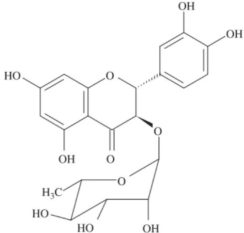
(1:1) as eluent to give astilbin (1) (Fig. 1) (11.0 mg).
The n-hexane fraction resulting from the extract partition between immiscible solvents was fractionated by silica gel column chromatography using gradient elution ofn-hexane and CH2Cl2resulting in the isolation of
b
-sitosterol (2, 10.0 mg) and amixture of fatty acids (3, 132.7 mg) composed of palmitic, stearic and linolenic acids.
Astilbin (dihydroquercetin 3-a-L-rhamnopyranoside) (1) 1H NMR (400 MHz, CD
3OD+D2O): 1.18 (d, J 6.2 Hz, CH3-600),
3.31 (m, H-400), 3.54 (dd,J3.3 and 1.5 Hz, H-200), 3.67 (dd,J9.6 and
3.3 Hz, H-300), 4.06 (d, J 1.5 Hz, H-100), 4.25 (m, H-500), 4.58 (d, J
10.7 Hz, H-3), 5.08 (d,J10.7 Hz, H-2), 5.90 (d,J2.2 Hz, H-8), 5.92 (d,J2.2 Hz, H-6), 6.81 (d,J8.3 Hz, H-50), 6.84 (dd,J8.3 and 1.9 Hz,
H-60), 6.95 (d,J1.9 Hz, H-20).13C NMR (100 MHz, CD
3OD+D2O):
17.99 (C-600), 70.66 (C-500), 71.93 (C-200), 72.33 (C-300), 73.97 (C-400),
78.72 (C-3), 84.10 (C-2), 96.43 (C-8), 97.55 (C-6), 102.29 (C-100),
102.64 (C-4a), 115.64 (C-20), 116.48 (C-50), 120.63 (C-60), 129.35
(C-10), 146.69 (C-30), 147.52 (C-40), 164.25 (C-8a), 165.66 (C-5),
168.82 (C-7), 196.11 (C-4).
Determination of the absolute configuration of 1
Absolute configuration was determined by circular dichroism spectra (Fig. 2) recorded on a JASCO J-715 spectropolarimeter (Jasco Inc., Easton, USA), equipped with a Peltier type temperature controller. The instrument was calibrated using (+)-10-camphorsulfonic acid (Sigma-Aldrich, St. Louis, USA) (Chen and Yang 1977). Spectra were obtained from 220 to 300 nm at 251C using 0.1 cm path length cells. Compound 1 was dissolved in methanol (Mallinckrodt Baker Inc., Phillipsburg, USA) to a final concentration of 0.125 mg/ml. The solution was stirred and incubated at room temperature for 10 min before each spectrum was recorded. The spectra were an average of eight scans recorded at a speed of 10 nm/min with a bandwidth of 1.0 at 0.5 nm step size and a 2 s time constant. After background subtraction and smoothing, CD data was expressed in molar circular dichroism
O
OH HO
HO H3C
O
O HO
OH
OH
OH
O
units. The software Spectra Manager (Jasco) was used for data collection and analysis.
ACE inhibition assay
ACE inhibitory activity was determined using a method described bySerra et al. (2005), modified to employ rat plasma as enzyme source. Briefly, an aliquot (25
m
l) of rat plasma was added to a microtitre plate containing 5m
l of the sample solution to be tested in different concentrations. Phosphate buffer (pH 8.3), ethanol and DMSO were employed as negative controls. Captopril (10m
M) was employed as positive control. The enzymatic reaction was started by adding the assay buffer and the substrate solution Hip-Gly-Gly (100m
M) (Sigma, USA). After homogenization, the mixture was incubated for 35 min, at 371C. The reaction was stopped by the addition of sodium tungstate (100 g/l) and sulfuric acid (0.33 mM). The system was mixed with the color reagent TNBS (Sigma, USA). After 20 min in dark, the plate absorbance was read in a microtitre plate reader (BioRad, Model 550) at 415 nm against a blank solution similarly prepared, except by adding the sodium tungstate and the sulfuric acid solutions before enzyme solution.Statistical analysis
The experimental data are expressed as mean7standard error mean (S.E.M.) of at least six experiments. Statistical analyses were performed using one-way ANOVA plus Tukey’s multiple compar-ison post-test. The value of pIC50 represents the log of
concentration values for the ethanol extract fromE. gonocladum aerial parts (g/ml) and astilbin (M) that induce 50% inhibition of ACE activity.
Results and discussion
The RP-HPLC fingerprint recorded for E. gonocladum aerial parts (EEG) indicates the predominance of a major peak with retention time of 13.5 min (compound 1), whose UV spectra showed a
l
maxat 289.9 nm and a shoulder around 340 nm, beingthese values compatible with a dihydroflavonol (Justesen et al. 1998;Bohm 1975). RP-HPLC analysis of PPT, a precipitate formed during the partition of the hydromethanolic suspension of EGE withn-hexane, indicated that compound1has been concentrated in this precipitate. Its further purification by silica gel column
chromatography and preparative RP-HPLC afforded the yellowish solid 1. Analysis of 1D and 2D NMR data, along with acid microhydrolysis on TLC and comparison with literature records for dihydroflavonoids suggested that compound1is dihydroquerce-tin-3-O-
a
-rhamnoside (Lu and Foo 1999). However, the molecule possesses two chiral centers at C-2 and C-3 resulting in four possible diastereoisomers (astilbin, neoastilbin, isoastilbin and neoisoastilbin). The 1H NMR spectrum showed that1 hastransconfiguration since it shows coupling constantJ2,3of 10.7 Hz, thus
limiting the possible isomers to astilbin and neoastilbin. In order to identify compound1unequivocally, its absolute configuration was investigated by circular dichroism. The chiroptical properties of dihydroflavonoids has been thoroughly investigated and the sign of the Cotton effect of n-
p
* origin can be used forestablishing the absolute configuration at C-2. Therefore, the CD measurement of1was carried out in the UV absorption region in methanol (Fig. 2). The absolute configuration at C-2 was concluded to beRbased on a positive Cotton effect observed in the CD spectrum of1, ascribed to the absorption band at 329.0 nm (
D
e
= +7.88) (Slade et al. 2005; Gaffield et al. 1975). Therefore, compound 1 was identified to be the 3-O-a
-L-rhamnoside of ( )(2R,3R)-5,7,30,40-tetrahydroxydihydroflavonol or ( )(2R,3R)-astilbin. Despite its presence as a major peak in the extract from E. gonocladumaerial parts, only a small mass of pure astilbin (1) was obtained. The interconversion of isomeric astilbins is well documented (Gaffield et al. 1975) and we experienced it during the chromatographic procedures carried out for the isolation and purification of1.
Two solids (2and3) were obtained from then-hexane fraction ofE. gonocladum. Analysis of13C and1H NMR data obtained for2
and comparison with literature records for triterpenes and steroids allowed identifying it as
b
-sitosterol (Forgo and K ¨ove´r 2004). Solid3was submitted to GC-FID analysis and comparison with reference compounds revealed a mixture of fatty acids composed of palmitic (30.0%), stearic (21.3%) and linolenic (4.6%) acids, along with a non-identified compound. It is worth mentioning that astilbin 1,b
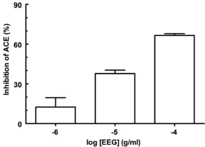
-sitosterol and the fatty acids were obtained for the first time fromE. gonocladum. On the other hand, the occurrence of the aglycone in the genus seems to be common, since several glycosides of dihydroquercetin have been described forErythroxylum rufum,E. ulei(Bohm et al. 1981),E. cocavar.ipadu andE. novogranatensevar.truxillense(Johnson et al. 1998). Fig. 2.CD curve of ( )(2R,3R)-astilbin (1) in MeOH.Fig. 3.Effect of the ethanol extract fromE. gonocladumaerial parts (EEG) on the ACEin vitroinhibition assay. Each bar represents the mean7SEM of at least 6 replicates. EEG was incubated with the serum 60 min before substrate addition. The results for each concentration of EEG are significantly different between each other (po0.01). The vehicle used for dilution of EEG (ethanol) did not inhibit ACE
EEG elicited concentration-dependent ACE inhibition (pIC50 4.5370.05; Fig. 3), employing a colorimetric in vitro
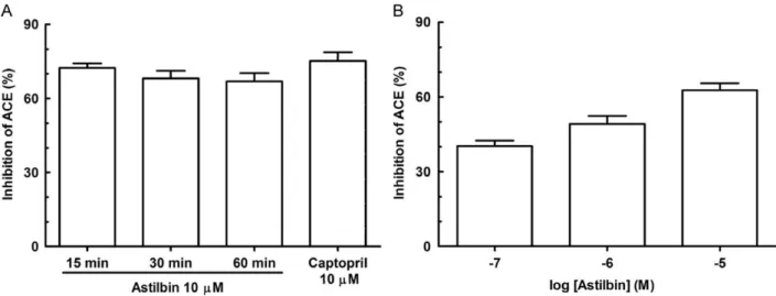
assay previously described by our group (Serra et al. 2005). Keeping in mind that flavonoids have been identified as ACE inhibiting constituents of different plant species (Lacaille-Dubois et al. 2001; Oh et al. 2004; Loizzo et al. 2007), the activity of compound1was investigated in the present study. Astilbin was evaluated at different time intervals (15, 30 and 60 min) before substrate addition, in order to determine the optimal time of reaction (Fig. 4A). It promoted ACE inhibition over 65% at each time interval assayed, statistically different to the negative control (po0.001; not shown). Besides, the ACE inhibition showed by
compound 1 (10
m
M) at 15, 30 and 60 min before substrate addition did not differ from captopril (10m
M;Fig. 4A).The ACE inhibitory activity observed for astilbin (pIC50=5.8670.33; Fig. 4B) is considerably higher than
that reported for other flavonoids, such as quercetin -3-O-
a
-6-caffeoyl-glycosyl-b
-1,2-rhamnoside (pIC50=3.9),querce-tin-3-O-
a
-6-p-coumaryl-glycosyl-b
-1,2-rhamnoside (pIC50=3.4),quercetin-3-O-
b
-glucopyranoside (pIC50=3.1) (Oh et al. 2004)and quercetin-3-O-
a
-arabinopyranoside (pIC50=3.5) (Loizzoet al. 2007). Astilbin shows superior ACE inhibition activity even in comparison to proanthocyanidins, a class of natural products regarded as the most potent ACE inhibitors from plants, including epicatechin dimer (pIC50=3.6), trimer (pIC50=3.9), tetramer
(pIC50=4.9), pentamer (pIC50=4.6) and hexamer (pIC50=5.0)
(Actis-Goretta et al. 2003).
In conclusion, the results described in here point out astilbin as one of the constituents responsible for the ACE inhibitory activity observed for the ethanol extract ofE. gonocladumaerial parts and disclose1as a promising molecule with potential anti-hyperten-sive activity, via ACE inhibition.
Acknowledgments
This work was supported by funds from CNPq – Conselho Nacional de Desenvolvimento Cientı´fico e Tecnolo´gico (Brazil). CAPES (Brazil) and CNPq (Brazil) are also acknowledged for M.Sc. (MDLF) and research (FCB and SFC) fellowships.
References
Actis-Goretta, L., Ottaviani, J.I., Keen, C.L., Fraga, C.G., 2003. Inhibition of angiotensin converting enzyme (ACE) activity by flavan-3-ols and procyani-dins. FEBS Lett. 555, 597–600.
Adsersen, A., Adsersen, H., 1997. Plants from Re´union Island with alleged antihypertensive and diuretic effects an experimental and ethnobotanical evaluation. J. Ethnopharmacol. 58, 189–206.
Bohm, B.A., 1975. In: Harborne, J.B., Mabry, T.J., Mabry, H. (Eds.), The Flavonoids. Chapman and Hall, London, pp. 68.
Bohm, B.A., Phillips, D.W., Ganders, F.R., 1981. Flavonoids ofErythroxylum rufum
andE. ulei. J. Nat. Prod. 44, 676–679.
Bohm, B.A., Loo, T., Nicholls, K.W., Plowman, T., 1988. Flavonoid variation in
Erythroxylum. Phytochemistry 27, 833–837.
Cano, J.H., Volpato, G., 2004. Herbal mixtures in the traditional medicine of Eastern Cuba. J. Ethnopharmacol. 90, 293–316.
Cha´vez, J.P., Santos, I.D.D., Cruz, F.G., David, J.M., 1996. Flavonoids and triter penes ester derivatives from Erythroxylum leal costae. Phytochemistry 41, 941–943.
Chen, G.C., Yang, J.T., 1977. Two-point calibration of circular dichrometer with d-10-camphorsulfonic acid. Anal. Lett. 10, 1195–1207.
Chin, Y.W., Jones, W.P., Waybright, T.J., Mccloud, T.G., Rasoanaivo, P., Cragg, G.M., Cassady, J.M., Kinghorn, A.D., 2006. Tropane aromatic ester alkaloids from a large-scale re-collection of Erythroxylum pervillei stem bark obtained in Madagascar. J. Nat. Prod. 69, 414–417.
Forgo, P., K ¨ove´r, K.E., 2004. Gradient enhanced selective experiments in the1H
NMR chemical shift assignment of the skeleton and side-chain resonances of stigmasterol, a phytosterol derivative. Steroids 69, 43–50.
Gaffield, W., Waiss, A.C., Tominaga, T., 1975. Structural relationships and interconversions of isomeric asilbins. J. Org. Chem. 40, 1057–1061.
Gonza´lez-Guevara, J.L., Herman, V.C., Gonza´lez-Garcia, K.L., Payo-Hill, A.L., Gonza´lez-Lavaut, J.A., Molina-Torres, J., Prieto-Gonza´lez, S., 2006. Flavonoid glycosides from Cuban Erythroxylum species. Biochem. Syst. Ecol. 34, 539–542.
Griffin, W.J., Lin, G.D., 2000. Chemotaxonomy and geographical distribution of tropane alkaloids. Phytochemistry 53, 623–637.
Hegnauer, R., 1981. Chemotaxonomy of Erythroxylaceae (including some ethno-botanical notes on old world species). J. Ethnopharmacol. 3, 279–292. Inigo, R.P.A., Pomilio, A.B., 1985. Flavonoids from Erythroxylum argentinum.
Phytochemistry 24, 347–349.
Johnson, E.L., Schmidt, W.F., Norman, H.A., 1998. Flavonoids as markers for
Erythroxylumtaxa:E. cocavar. ipaduandE. novogranatensevar.truxillense. Biochem. Syst. Ecol. 26, 743–759.
Johnson, E.L., Schmidt, W.F., Emche, S.S., Mossoba, M.M., Musser, S.M., 2003. Kaempferol (rhamnosyl) glucoside, a new flavonol fromErythroxylum cocavar.
ipadu. Biochem. Syst. Ecol. 31, 59–67.
Justesen, U., Knuthsen, P., Leith, T., 1998. Quantitative analysis of flavonols, flavones, and flavanones in fruits, vegetables and beverages by high-performance liquid chromatography with photo-diode array and mass spectrometric detection. J. Chromatogr. A 799, 101–110.
Lacaille-Dubois, M.A., Franck, U., Wagner, H., 2001. Search for potential angiotensin converting enzyme (ACE)-inhibitors from plants. Phytomedicine 8, 47–52.
Fig. 4.Effect of astilbin on the ACEin vitroinhibition assay (iACE). (A) Incubation intervals for iACE with astilbin. (B) Concentration-dependent effect of astilbin on iACE. Each bar represents the mean7SEM of at least 6 replicates. Captopril was incubated with the serum 60 min before substrate addition. Astilbin was incubated with serum 15 min before substrate addition for concentration-dependent iACE. The results illustrated in (A) are not significantly different when incubation intervals or the effect of captopril was compared with those of astilbin. The results on (B) are significantly different between each other (po0.05). The vehicles used for dilution of captopril (saline)
Loizzo, M.R., Said, A., Tundis, R., Rashed, K., Statti, G.A., Hufner, A., Menichini, F., 2007. Inhibition of angiotensin converting enzyme (ACE) by flavonoids isolated fromAilanthus excelsa(Roxb) (Simaroubaceae). Phytother. Res. 21, 32–36. Lu, Y., Foo, L.Y., 1999. The polyphenol constituents of grape pomace. Food Chem. 65,
1–8.
Oh, H., Kang, D.G., Kwon, J.W., Kwon, T.O., Lee, S.Y., Lee, D.B., Lee, H.S., 2004. Isolation of angiotensin converting enzyme (ACE) inhibitory flavonoids from
Sedum sarmentosum. Biol. Pharm. Bull. 27, 2035–2037.
Plowman, T., Hensold, N., 2004. Names, types, and distribution of neotropical species ofErythroxylum(Erythroxylaceae). Brittonia 56, 1–53.
Serra, C.P., Cˆortes, S.F., Lombradi, J.A., Oliveira, A.B., Braga, F.C., 2005. Validation of a colorimetric assay for the in vitroscreening of inhibitors of angiotensin-converting enzyme (ACE) from plants extracts. Phytomedicine 12, 424–432. Slade, D., Ferreira, D., Marais, J.P.J., 2005. Circular dichroism, a powerfull tool for