(Annals of the Brazilian Academy of Sciences)
Printed version ISSN 0001-3765 / Online version ISSN 1678-2690 www.scielo.br/aabc
http://dx.doi.org/10.1590/0001-3765201520140156
Nicotine-enhanced oxidation of low-density lipoprotein and its components by myeloperoxidase/H2O2/Cl- system
OLGA M.M.F. OLIVEIRA1, IGUATEMY L. BRUNETTI2 and NAJEH M. KHALIL3
1
Instituto de Química, UNESP, Departamento de Bioquímica e Tecnologia Química, Rua Prof. Francisco Degni, 55, Jardim Quitandinha, 14800-900 Araraquara, SP, Brasil
2
Faculdade de Ciências Farmacêuticas de Araraquara, UNESP, Rodovia Araraquara-Jaú, Km 01, 14801-902 Araraquara, SP, Brasil 3Departamento de Farmácia, UNICENTRO, Rua Simeão Camargo Varela de Sá, 03,
Vila Carli, 85040-080 Guarapuava, PR, Brasil
Manuscript received on April 8, 2014; accepted for publication on July 28, 2014
ABSTRACT
In this study, the effect of nicotine on the LDL oxidation by the MPO/H2O2/Cl- system and the effect of HOCl on LDL and some of its components, such as methyl linoleate, vitamin E and the amino acid tryptophan were explored. Nicotine, in micromolar concentrations, enhanced the tryptophan oxidation, either present in LDL or free, in solution. Nicotine also decreased the formation of conjugated dienes and oxygen consumption in a methyl linoleate / HOCl system, and there was evidence to suggest an increase in chlorohydrin formation. Acceleration of the vitamin E oxidation by HOCl was also observed in the presence of nicotine. These data show that the interaction of nicotine and HOCl can promote significant biochemical modifications in LDL particle and some of its components involved in the pathogenesis of cardiovascular and other diseases.
Key words: nicotine, myeloperoxidase, hypochlorous acid, low-density lipoprotein, methyl linoleate, vitamin E, tryptophan.
Correspondence to: Najeh Maissar Khalil E-mail: [email protected]
INTRODUCTION
Hypochlorous acid (HOCl) is a major oxidant produced by neutrophils, in a reaction catalyzed by myeloperoxidase (MPO; EC 1.11.1.7), a heme enzyme present in high concentrations in the granules of leukocytes (Winterbourn and Kettle 2013). MPO uses hydrogen peroxide (H2O2), produced during
the oxidative burst, to oxidize Cl- to HOCl, by the
following reactions (Vlasova et al. 2012):
native MPO + H2O2 → MPO I + H2O
MPO I + Cl- → native MPO + HOCl
and quinine, react with HOCl to form reactive Cl+ adducts (R3N+ - Cl, reactive quaternary
chlorammonium ions) that readily release Cl+,
enhancing the formation of chlorinated/oxidized products (Prutz 1998, Suzuki and Ohshima 2002). It is also suggested that the interaction between
significant concentrations of nicotine (in smokers)
and HOCl may promote DNA (Masuda et al. 2001) and tissue damage (CV-1 mammalian kidney cells; Salama et al. 2014).
Cardiovascular disease is currently the leading cause of morbidity and mortality worldwide and its incidence is likely to increase. Many studies
have indicated that MPO is an inflammatory
marker in coronary artery disease (Zhang et al. 2001); in particular, due to its ability to generate reactive oxygen species, which promote oxidative damage to lipoproteins and leading to progression of atherosclerosis (Haraguchi et al. 2014).
In light of such reports on the properties of R3N+ - Cl and the possibility of its in vivo
generation with nicotine, and considering their relation to oxidative stress, we explored the effect of nicotine on the LDL oxidation by the MPO/ H2O2/Cl- system and the effect of HOCl on LDL
lipid components, methyl linoleate (ML), vitamin E and tryptophan.
MATERIALS AND METHODS
REAGENTS
MPO of a purity index (A430 / A280) of at least
0.85 was purchased from Planta Naturstoffe Vertriebs GmbH. H2O2 (stocked as a 30%
solution) and NaOCl, both from Merck, were diluted shortly before use, the concentration of H2O2 being determined by measuring absorbance at 230 nm (Є = 80 M-1 cm-1; Brestel
1985) and that of OCl- at 292 nm (Є = 350 M-1 cm-1; Zgliczynski et al. 1971). Nicotine tartrate
was purchased from Sigma (St. Louis, MO, USA). All other reagents were analytical grade, obtained from Sigma or Aldrich.
ISOLATION OF LDL
Venous blood from “healthy” volunteers (non-fasting, age range 23–50 years, non-smokers) was collected into tubes containing ethylenediaminetetraacetic acid (1.0 mg/mL of blood). Plasma was separated by centrifugation at 1000 × g for 10 min, at 4 °C,
and 5.0 μM phenylmethylsulfonyl fluoride, 10.0 μM benzamidine, 10.0 μg/mL aprotinine and 100.0 μM
butylated hydroxytoluene were added to prevent protease activity and oxidative reactions. The study was approved by the research ethics committee (FCF/UNESP: protocol CEP/FCF/Car. N 02/2005 and document 022/2005). Firstly, very-low-density lipoprotein (VLDL) was separated: the plasma was placed in ultracentrifuge tubes with saline solution (about 60% of the total volume) and rotated at 55,000 g, for 7h at 4 °C. At the end of this period,
the VLDL floated and was drawn off with great
care. The remaining plasma was transferred to a test tube, where its density was adjusted to 1.063 g/L with solid KBr. It was then centrifuged again at 55.000 g for 7h, at 4 °C, bringing to the surface the LDL, which was delicately removed and dialyzed in phosphate-buffered saline in 20 mM sodium phosphate, pH 7.4 and 0.14 M NaCl, for 4h at 4 °C, the buffer being renewed every hour. The LDL was stored at 4 °C in the dark, for a maximum of 2 weeks, until used (Bagheri et al. 2013). Protein content was determined by the Lowry method (Lowry et al. 1951).
LDLOXIDATION BY MPO/H2O2/Cl-SYSTEM
LDL (80 µg protein/ml) was oxidized by the system MPO (1 nM) / H2O2 (0.5 mM) in 50 mM
sodium phosphate buffer (pH 7.4) with 0.14 M Cl-, at 25 °C. This reaction was monitored by
the fall in fluorescence (excitation at 282 nm and emission at 331 nm). The LDL α-tocopherol fluorescence (excitation at 290 nm and emission
at 323 nm) does not interfere with the tryptophan
fluorescence even at high concentrations (Jerlich
TRYPTOPHAN OXIDATION BY MPO/H2O2/Cl-SYSTEM
Tryptophan (0.1 mM) was oxidized by the system MPO (1.0 nM) / H2O2 (0.25 mM) in 50 mM sodium
acetate buffer (pH 5.5) with 0.14 M Cl-, at 30 °C. The reaction was monitored by following the decay
in fluorescence of the tryptophan (excitation at 290
nm and emission at 360 nm; Jerlich et al. 1998).
METHYL LINOLEATE (ML)OXIDATION BY HOCl
A 50 mM solution of sodium dodecyl sulphate (SDS) was made in 50 mM sodium phosphate buffer (pH 7.4). 20 µL of ML (density 0.9 g/mL) was added and the mixture shaken until it had all dissolved (to
a transparent solution of final concentration 1.2 mM).
These solutions were prepared with and without nicotine, and the ML was oxidized at 37 °C by adding HOCl. The reaction was monitored by following (i) the formation of conjugated dienes at 234 nm and (ii) the consumption of oxygen; both assays were conducted in a thermostatic cuvette (Noguchi et al. 2002).
VITAMIN EOXIDATION BY HOCI
The reactions of HOCl with vitamin E, in the absence and presence of nicotine, were carried out in 50 mM sodium phosphate buffer (pH 7.4) with SDS 1%, at 37 °C. The absorbance at 255 nm and spectral variation were monitored, at intervals of 1 second, using a diode-array spectrophotometer. The reactions were started by adding HOCl (Pattison et al. 2003).
HOCIDETERMINATION
A solution containing 14 mM 3,3’,5,5’-tetra-methylbenzidine (TMB) dissolved in 50% dimethylformamide, 100 mM potassium iodide and 400 mM acetic acid was used to measure HOCl. Under these conditions HOCl oxidizes TMB to a blue product with an absorbance maximum at 655 nm. A standard curve was generated by adding pure HOCl (Dypbukt et al. 2005).
TARTRATE BLANK
All assays performed with nicotine tartrate were repeated with sodium potassium tartrate and with
buffer alone. No difference was observed between these tartrate and buffer solutions.
STATISTICS
Statistical analysis of the data was performed via one-way analysis of variance (ANOVA). The results
were considered statistically significant if p < 0.05.
RESULTS
EFFECT OF NICOTINE ON THE OXIDATION OF FREE AND LDL TRYPTOPHAN BY MPO/H2O2/Cl-SYSTEM
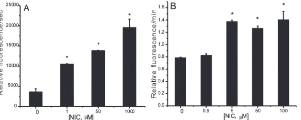
Using this model system the oxidation of free tryptophan was observed as a gradual fall in the
fluorescence intensity. In Figure 1A, it can be seen
that the MPO/H2O2/Cl- system promoted tryptophan
oxidation and that increased concentrations of nicotine led to a marked progressive rise in the
fluorescence decay, in which represents an increase
in the oxidation of tryptophan (evaluated by rate constant for the oxidation of tryptophan, measured
by tangent of fluorescence decay curve). A similar effect was observed in the fluorescence of LDL
particles exposed to the MPO/H2O2/Cl- system, which fluorescence decay was also enhanced by
nicotine in the reaction medium (Fig. 1B).
In both the above experiments, the effect of the enzymatic reaction on the LDL or free tryptophan, was only seen with the complete system, MPO/ H2O2/Cl-, indicating that the HOCl is responsible for the loss of fluorescence, as previously postulated
by others (Jerlich et al. 2000a).
EFFECT OF NICOTINE ON THE OXIDATION OF THE ML BY HOCl
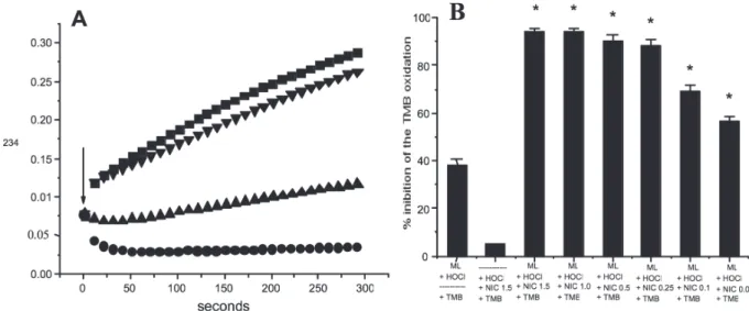
such a fall in absorbance. This fall cannot be due to a suppression of HOCl by nicotine, since, up to 1.5 µM, nicotine exhibited virtually no scavenging effect on HOCl (Fig. 2B).
In parallel to the formation of conjugated dienes, the reaction between HOCl and polyunsaturated fatty acids (PUFA) generates chlorohydrin. This reaction is well characterized for both PUFA and cholesterol (Winterbourn et al. 1992). The mechanism by which chlorohydrin is formed involves two electrophilic addition reactions: the Cl + ion is added at the
double bond and then OH- is added at the other
carbon atom of the same bond. If the loss of double bonds outstrips the formation of new conjugated dienes, a reduced rate of change will be observed in the absorbance at 234 nm. Arnhold et al. (1995), showed that there was a direct relation between the amount of HOCl consumed and the loss of double bounds of linoleic acid, which resulted in the formation of chlorohydrins. The production of these compounds may occur in certain biological events and, as they are more polar than their lipid precursors, their appearance in cell membranes might cause changes in the membrane structure and consequently in its functionality (Winterbourn et
al. 1992). In the case of cholesterol chlorohydrin, the possibility has been discussed that it may cause harmful effects in artery walls (Heinecke et al. 1994); futhermore, it has been shown to promote the lysis of red blood cells (Vissers et al. 1994).
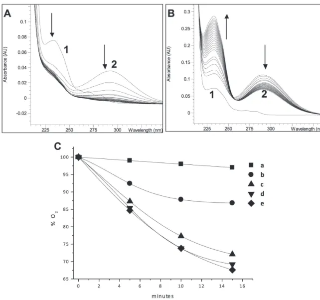
Our results show that there may be an increased formation of chlorohydrin in the interaction of the HOCl/ML system with nicotine. Thus, an experiment was done to determine the variation in the concentration of HOCl (in presence of ML) in the reaction medium, via oxidation of TMB. Nicotine, in micromolar concentrations, accentuated the depletion of the HOCl, compared to the control: 0.25 - 1.5 µM nicotine led to 80-100% decreased oxidation of TMB, while the assay without nicotine showed a fall of only 38% (Fig. 2B). In order to visualize this effect directly, the ML/HOCl reaction was followed by measuring absorbance in the region 200-300 nm; an effective fall can be seen in absorbance at 234 nm (conjugated dienes) and at 292 nm (HOCl absorption peak) in the presence of nicotine (Fig. 3A) relative to the assay without nicotine, with a fall at 292 nm and rise at 234 nm (Fig. 3B).
Figure 1 - A) Effect of nicotine on the rate of free tryptophan oxidation by the MPO/H2O2/Cl
-system. Tryptophan (0.1 mM) was incubated with MPO (1.0 nM) in 50 mM sodium acetate buffer (pH 5.5), containing 0.14 M NaCl, at 30 °C. The reaction initiated adding 0.25 mM H2O2, in the absence or presence of various concentrations of nicotine (Mean ± SD; n=5) . (B) Rates of
The oxidation of PUFA leads to the consumption of the equivalent amount of O2 from the reaction
medium, as hydroperoxides are formed. The consumption of oxygen has been used to monitor the oxidation of ML in micelles (Rossetto et al. 2002).
The oxygen depletion profile of the HOCl/
ML in SDS micelles, in the absence and presence of nicotine, is displayed in Figure 3C. The HOCl
promotes the consumption of O2 from the reaction mixture with ML and it can be seen that nicotine
at 1 and 2 µM significantly inhibits the O2
consumption. Nicotine, at these low concentrations,
has an insignificant scavenging effect on HOCl, and
therefore it seems likely that another mechanism, different from conjugated-diene formation, is responsible for this effect.
Figure 2 - A) Effect of nicotine on the ML oxidation by HOCl. 1.3 mM ML, 1.0 mM HOCl and 0 (■), 0.1 (●), 0.5 (▲) or 1.0 (▼)
µM nicotine in 50 mM sodium phosphate buffer, (pH 7.4), with 50 mM SDS at 37 °C. The arrow shows the initial absorbance,
before addition of HOCl. B) Effect of nicotine on the TMB oxidation by HOCl in the presence of ML. Nicotine (NIC, μM) and
ML (1.3 mM ) were incubated for 1 min and 0.5 mM HOCl was added, in a medium containing 50 mM SDS in phosphate buffer (pH 7.4). After 5 min, the remaining HOCl was estimated by adding 2.8 mM TMB and monitoring absorbance at 652 nm (Mean ±
SD; n=5). * Significantly different from control (the sum of % inhibition of TMB oxidation by the systems ML/HOCl and nicotine/ HOCl), with p<0.05.
EFFECT OF NICOTINE ON THE VITAMIN E/HOCISYSTEM IN SDSMICELLES
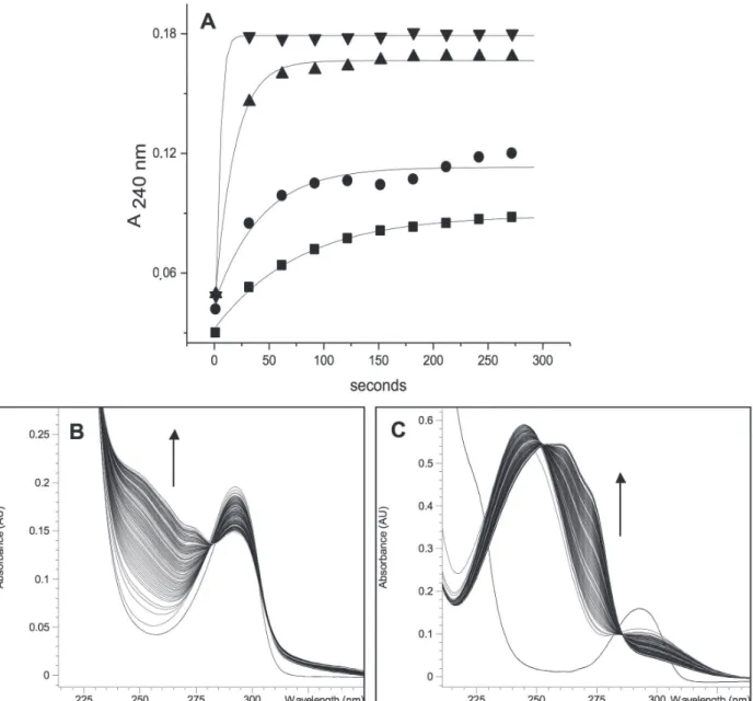
In Figure 4A, it can be seen that in the presence of nicotine there is a rise in A240nm, relative to
the vitamin E/HOCl reaction without nicotine. Concentrations of 1 and 5 µM of nicotine caused a strong and fast rise in A240nm. Figure 4B and C
also show the absorption spectrum from 220 to 350 nm, and how it develops during the reaction, in the absence or presence of nicotine. The accentuated increase in absorption between 220 and 280 nm may correspond mainly to the formation of a vitamin E dichloroquinone (Ho et al. 2000).
DISCUSSION
The great majority of experiments on LDL oxidation are conduced “in vitro”. In this case, the oxidation reaction can be followed by observing changes in the physicochemical and biological properties of the LDL (Jerlich et al. 1998, 2000a).
It is well established that the apoproteins (apo B-100 being the main one in LDL) contain the aromatic residues of tyrosine and tryptophan which contribute
visibly to the intrinsic fluorescence of the LDL particle. During LDL oxidation, there is a significant fall in the fluorescence of tryptophan and this is used
consequence of the oxidation of tryptophan residues,
the structure of LDL suffers modification, affecting its
biological properties, such as its binding to receptors on cells (Jerlich et al. 2000a).
Oxidative reactions convert tryptophan to kynurenine, N-formylkynurenine, oxindolylalanine and hydroxytryptophan, altering both the structure and function of the protein (Thomas and Stocker 1999). Tryptophan is oxidized by HOCl, and
its fluorescence significantly decreases when
lipoproteins are exposed to the MPO/H2O2/Cl
-system. The indole ring of tryptophan initially reacts with HOCl to yield a chlorinated species, which is likely to be 3-chloroindolenine or perhaps the N-chloroindole species (Fu et al. 2006).
The presence of PUFA makes the membrane susceptible to oxidation processes known as lipid peroxidation (Gasparovic et al. 2013).
The radicals produced, undergo rearrangements to form conjugated dienes. These molecules react with O2, generating peroxide radicals that, in turn,
can abstract another H atom to form hydroperoxides, thus propagating the chain of reactions. This process leads to the formation of secondary products such as alcohols, ketones and aldehydes. The formation of conjugated dienes has been widely studied by
UV absorption spectroscopy, to be used as a marker for lipid peroxidation (Noguchi et al. 2002, Rekdal and Melo 1995).
LDL contains PUFA in its structure, and these molecules may be a target for oxidative damage during atherosclerosis; thus, the effects of HOCl on these moieties have been investigated in a number of studies (Spiteller 2005).
Figure 4 - Effect of nicotine on the oxidation of vitamin E (5 mM) by HOCl (0.05 mM) in 50 mM sodium phosphate buffer (pH
7.4) with 50 mM SDS at 37 °C. (A) Reaction time-course: without nicotine (■); with 0.1 (●), 1.0(▲) or 5.0 (▼) µM nicotine.
One such mechanism might be chlorohydrin formation in the ML/HOCl/ nicotine system. As can be seen, nicotine leads to a fall in absorbance at 234 nm, which must be due to the loss of one or more double bonds. Chlorohydrin formation in this system would result in a smaller proportion of conjugated dienes, which in turn would diminish the rate of consumption of dissolved oxygen. It is worth remembering that this effect could occur anywhere in the body where nicotine is found, together with PUFA and HOCl generation. Chlorinated lipoproteins have been located in human atherosclerotic lesions (Malle et al. 2006), so a relevant factor might be the effect of nicotine on the oxidation of LDL, which is rich in PUFA, by HOCl or the MPO/H2O2/Cl- system, making that
oxidation potentially more atherogenic. It is important to note that chlorohydrins are the main products of the reaction between HOCl and lipids in relation to the formation of hydroperoxides (Jerlich et al. 2000b).
Vitamin E (tocopherol) is an important antioxidant in the diet, the main lipophilic antioxidant in plasma, membranes and tissues and also an important inhibitor of lipid peroxidation that blocks the propagation of this chain reaction in biological membranes and lipoproteins (Leichtle et al. 2006).
Vitamin E is also the main antioxidant present in the LDL particle, making it important to investigate the interaction of nicotine and the vitamin E / HOCl system, given that HOCl is one of the factors leading to LDL oxidation. HOCl reacts primarily with the chromanol ring system of this vitamin (as described using Trolox, a water soluble analogue), leading to formation of a quinone, with a gradual absorbance rise in ultraviolet region (Ho et al. 2000, Pattison et al. 2003).
According to published reports, the reaction of tertiary amines with hypochlorous acid leads to formation of quaternary chlorammonium ions that dramatically enhance the chlorination of free (2’-deoxy) nucleosides (Masuda et al. 2001), salicylate and sorbate (Prutz 1998).
A recent study demonstrated that nicotine increase CD36 expression in macrophages (with consequent enhanced oxLDL uptake), contributing to the development of atherosclerosis (Zhou et al. 2013). This study demonstrates the important role of nicotine on LDL in reactive oxygen species generation systems, and their deleterious effect on the development of atherogenesis.
CONCLUSIONS
In conclusion, we have shown that nicotine enhances the oxidation of tryptophan (free or in LDL) by the MPO/H2O2/Cl- system and the oxidation/
chlorination of its lipid components by HOCl directly. Several studies support the hypothesis that the risk of atherosclerosis is associated with
the oxidative modification of LDL and there is
diverse evidence that MPO and HOCl play a part in the development of atherosclerosis. The results presented here afford evidence on the biochemical effects of nicotine on components involved in the pathogenesis of cardiovascular diseases.
Abbreviations: LDL, low-density lipoprotein; ML, methyl linoleate; MPO, myeloperoxidase; PUFA, Polyunsaturated Fatty Acid; ROS, reactive oxygen species; TMB, 3,3’,5,5’-tetramethylbenzidine.
ACKNOWLEDGMENTS
This work was sponsored by the Fundação de Amparo à Pesquisa do Estado de São Paulo (FAPESP) (Process 99/10229-6). The authors report
no conflict of interest.
RESUMO
consumo de oxigênio no sistema metil linoleato/HOCl, e houve evidência que sugere aumento na formação de clorohidrinas. O aumento da oxidação da vitamina E pelo HOCl também foi observada na presença de nicotina. Esses dados mostram que a interação da nicotina e HOCl pode promover alterações bioquímicas significativas na partícula de LDL e alguns de seus componentes envolvidos na patogênese de doenças cardiovasculares e de outras doenças.
Palavras-chave: nicotina, mieloperoxidase, ácido hipocloroso, lipoproteína de baixa densidade, metil linoleato, vitamina E, triptofano.
REFERENCES
ALIPOUR A, RIBALTA J, NJO TL, JANSSEN HW, BIRNIE E, VANMILTENBURG AJ AND ELTE JW. 2013. Trans-vessel gradient of myeloperoxidase in coronary artery disease. Eur J Clin Invest 43: 920-925.
ARNHOLD J, PANASENKO OM, SCHILLER J, VLADIMIROV A AND ARNOLD K. 1995. The action of hypochlorous acid on phosphatidylcholine liposomes in dependence on the content of double bonds. Stoichiometry and NMR analysis. Chem Phys Lipids 78: 55-64.
BAGHERI S, AHMADVAND H, KHOSROWBEYGI A, GHAZANFARI F, JAFARI N, NAZEM H AND HOSSEINI RH. 2013. Antioxidant properties and inhibitory effects of Satureja khozestanica essential oil on LDL oxidation induced-CuSO4 in vitro. Asian Pac J Trop Biomed 3: 22-27.
BRESTEL EP. 1985. Co-oxidation of luminol by hypochlorite and hydrogen peroxide implications for neutrophil chemiluminescence. Biochem Biophys Res Commun 126: 482-488.
CLAYTON PM, VAS CA, BUI TT, DRAKE AF AND MCADAM K. 2013. Spectroscopic studies on nicotine and nornicotine in the UV region. Chirality 25: 288-293.
DYPBUKT JM, BISHOP C, BROOKS WM, THONG B, ERIKSSON H AND KETTLE AJ. 2005. Sensitive and selective assay for chloramine production by myeloperoxidase. Free Radic Biol Med 39: 1468-1477.
FU X, WANG Y, KAO J, IRWIN A, D'AVIGNON A AND MECHAM
RP. 2006. Specific sequence motifs direct the oxygenation
and chlorination of tryptophan by myeloperoxidase. Biochemistry 45: 3961-3971.
GASPAROVIC AC, JAGANJAC M, MIHALJEVIC B, SUNJIC SB AND ZARKOVIC N. 2013. Assays for the measurement of lipid peroxidation. Methods Mol Biol 965: 283-296. HARAGUCHI Y, TOHB R, HASOKAWAA M, NAKAJIMAA H,
HONJOA T AND OTSUI K. 2014. Serum myeloperoxidase/ paraoxonase 1 ratio as potential indicator of dysfunctional
high-density lipoprotein and risk stratification in coronary
artery disease. Atherosclerosis 234: 288-294.
HEINECKE JW, LI W, MUELLER DM, BOHRER A AND TURK J. 1994. Cholesterol chlorohydrin synthesis by the myeloperoxidase-hydrogen peroxide-chloride system: Potential markers for lipoproteins oxidatively damaged by phagocytes. Biochemistry 33: 10127-10136.
HO H, SOLDEVILLA J, HOOK JM AND SOUTH-WELL-KEELY PT. 2000. Oxidation of 2,2,7,8-tetramethyl-6-chromanol, the model compound of gamma-tocopherol, by hypochlorous acid. Redox Rep 5: 60-62.
JERLICH A ET AL. 1998. Human low density lipoprotein as a target of hypochlorite generated by myeloperoxidase. Free Radical Biol Med 24: 1139-1148.
JERLICH A, FRITZ G, KHARRAZI H, HAMMEL M, TSCHABUSCHNIG S AND GLATTER O. 2000a. Comparison of HOCl traps with myeloperoxidase inhibitors in prevention of low density lipoprotein oxidation. Biochim Biophys Acta 31: 109-118.
JERLICH A, PITT AR, SCHAUR RJ AND SPICKETT CM. 2000b. Pathways of phospholipid oxidation by HOCl in human LDL detected by LC-MS. Free Radic Biol Med 28: 673-682. LEICHTLE A, TEUPSER D AND THIERY J. 2006. Alpha-tocopherol
distribution in lipoproteins and anti-inflammatory effects
differ between CHD-patients and healthy subjects. J Am Coll Nutr 25: 420-428.
LOWRY OH, ROSEBROUGH NJ, FARR AL AND RANDALL RJ. 1951. Protein measurement with the Folin phenol reagent. J Biol Chem 193: 265-275.
MALLE E, MARSCHE G, ARNHOLD J AND DAVIES MJ. 2006.
Modification of low-density lipoprotein by
myeloperoxidase-derived oxidants and reagent hypochlorous acid. Biochim Biophys Acta 1761: 392-415.
MASUDA M, SUZUKI V, FRIESEN MD, RAVANAT JL, CADET J AND PIGNATELLI B. 2001. Chlorination of guanosine and others nucleosides by hypochlorous acid and myeloperoxidase of actived human neutrophils: catalysis by nicotine and trimethylamine. J Biol Chem 276: 40486-40496.
NEWMAN MB, ARENDASH GW, SHYTLE RD, BICKFORD PC, TIGHE TandSANBERG PR. 2002. Nicotine’s oxidative and antioxidant properties in CNS. Life Sci 71: 2807-2820. NOGUCHI N, NAKADA A, ITOH Y, WATANABE A AND NIKI
E. 2002. Formation of active oxygen species and lipid peroxidation induced by hypochlorite. Arch Biochem Biophys 397: 440-447.
PATTISON DI, HAWKINS CL AND DAVIES MJ. 2003. Hypochlorous acid-mediated oxidation of lipid components and antioxidants present in low-density lipoproteins: absolute rate constants, product analysis, and computational modeling. Chem Res Toxicol 16: 439-449.
PRUTZ WA. 1998. Reactions of hypochlorous acid with biological substrates are activated catalytically by tertiary amines. Arch Biochem Biophys 357: 265-273.
ROSSETTO M, VANZANI P, MATTIVI F, LUNELLI M, SCARPA Mand RIGO A. 2002. Synergistic antioxidant effect of catechin and malvidin 3-glucoside on free radical-initiated peroxidation of linoleic acid in micelles. Arch Biochem Biophys 15: 239-245.
SALAMA SA, ARAB HH, OMAR HA, MAGHRABI IA AND SNAPKA RM. 2014. Nicotine Mediates Hypochlorous Acid-Induced Nuclear Protein Damage in Mammalian
Cells. Inflammation 37: 785-792.
SALONEN I, HUTTUNEN K, HIRVONEN MR, DUFVA J, GROUNDSTROEM K AND DUFVA H. 2012. Serum myelo-peroxidase is independent of the risk factors of atherosclerosis. Coron Artery Dis 23: 251-258.
SASSA A, KAMOSHITA N, MATSUDA T, ISHII Y, KURAOKA I, NOHMI T, OHTA T, HONMA M AND YASUI M. 2013. Miscoding properties of 8-chloro-2'-deoxyguanosine, a hypochlorous acid-induced DNA adduct, catalysed by human DNA polymerases. Mutagenesis 28: 81-88. SPITELLER G. 2005. Is atherosclerosis a multifactorial disease
or is it induced by a sequence of lipid peroxidation reactions? Ann N Y Acad Sci 1043: 355-366.
SUZUKI T AND OHSHIMA V. 2002. Nicotine-modulated formation of spiroiminodihydantoin nucleoside via 8-oxo-7,8-dihydro-2’-deoxyguanosine in 2’-deoxyguanosine-hypochlorous acid reaction. FEBS Letters 516: 67-70. THOMAS SR AND STOCKER R. 1999. Redox reactions related to
indoleamine 2,3-dioxygenase and tryptophan metabolism along the kynurenine pathway. Redox Rep 4: 199-220.
VISSERS MC, STERN A, KUYPERS F,VAN DEN BERG JJ AND WINTERBOURN CC. 1994. Membrane changes associated with lysis of red blood cells by hypochlorous acid. Free Radical Biol Med 16: 703-712.
VLASOVA II, SOKOLOV AV AND ARNHOLD J. 2012. The free amino acid tyrosine enhances the chlorinating activity of human myeloperoxidase. J Inorg Biochem 106: 76-83. WINTERBOURN CC, KETTLE AJ. 2013. Redox reactions and
microbial killing in the neutrophil phagosome. Antioxid Redox Signal 18: 642-660.
WINTERBOURN CC, VAN DEN BERG JJ, ROITMAN E AND KUYPERS FA. 1992. Chlorohydrin formation from unsaturated fatty acids reacted with hypochlorous acid. Arch Biochem Biophys 296: 547-555.
ZGLICZYNSKI TJM, STELMASZYNSKA T, DOMANSKA J and OSTROWISKI W. 1971. Chloramines as intermediates of oxidation reaction of amino acids by myeloperoxidase. Biochim Biophys Acta 235: 419-424.
ZHANG V, BRENNAN ML, FU X, AVILES RJ, PEARCE GL AND PENN MS. 2001. Association between myeloperoxidase levels and risk of coronary artery disease. JAMA 286: 2136-2142.