Carrying a DsRed-Monomer Reporter Gene
Chih-Jen Chou1., Shao-Yu Peng1., Mei-Han Wu2,7
, Cho-Chen Yang3, Yu-Sheng Lin4, Winston Teng-Kui Cheng4,5, Shinn-Chih Wu1,4*, Yao-Ping Lin6,7*
1Institute of Biotechnology, National Taiwan University, Taipei, Taiwan,2Department of Radiology, Taipei Veterans General Hospital, Taipei, Taiwan,3Department of Animal Science, National Chiayi University, Chiayi, Taiwan,4Department of Animal Science and Technology, National Taiwan University, Taipei, Taiwan,5Department of Animal Science and Biotechnology, Tunghai University, Taichung, Taiwan,6Division of Nephrology, Department of Medicine, Taipei Veterans General Hospital, Taipei, Taiwan,7Department of Medicine, National Yang-Ming University, Taipei, Taiwan
Abstract
Background:Pigs are an optimal animal for conducting biomedical research because of their anatomical and physiological resemblance to humans. In contrast to the abundant resources available in the study of mice, few fluorescent protein-harboring porcine models are available for preclinical studies. In this paper, we report the successful generation and characterization of a transgenic DsRed-Monomer porcine model.
Methods:The transgene comprised a CMV enhancer/chicken-beta actin promoter and DsRed monomeric cDNA. Transgenic pigs were produced by using pronuclear microinjection. PCR and Southern blot analyses were applied for identification of the transgene. Histology, blood examinations and computed tomography were performed to study the health conditions. The pig amniotic fluid progenitor/stem cells were also isolated to examine the existence of red fluorescence and differentiation ability.
Results: Transgenic pigs were successfully generated and transmitted to offspring at a germ-line transmission rate of 43.59% (17/39). Ubiquitous expression of red fluorescence was detected in the brain, eye, tongue, heart, lung, liver, pancreas, spleen, stomach, small intestine, large intestine, kidney, testis, and muscle; this was confirmed by histology and western blot analyses. In addition, we confirmed the differentiation potential of amniotic fluid progenitor stem cells isolated from the transgenic pig.
Conclusions: This red fluorescent pig can serve as a host for other fluorescent-labeled cells in order to study cell-microenvironment interactions, and can provide optimal red-fluorescent-labeled cells and tissues for research in developmental biology, regenerative medicine, and xenotransplantation.
Citation:Chou C-J, Peng S-Y, Wu M-H, Yang C-C, Lin Y-S, et al. (2014) Generation and Characterization of a Transgenic Pig Carrying a DsRed-Monomer Reporter Gene. PLoS ONE 9(9): e106864. doi:10.1371/journal.pone.0106864
Editor:David Warburton, University of Southern California, United States of America
ReceivedApril 8, 2014;AcceptedMay 7, 2014;PublishedSeptember 4, 2014
Copyright:ß2014 Chou et al. This is an open-access article distributed under the terms of the Creative Commons Attribution License, which permits unrestricted use, distribution, and reproduction in any medium, provided the original author and source are credited.
Data Availability:The authors confirm that all data underlying the findings are fully available without restriction. All data are included within the manuscript.
Funding:This work was supported by a grant from National Taiwan University (Grant 97R-99R0066-41) and the Council of Agriculture, Executive Yuan, R.O.C. (Taiwan) (Grant 99AS-1.2.3-ST-aM). The funders had no role in study design, data collection and analysis, decision to publish, or preparation of the manuscript.
Competing Interests:The authors have declared that no competing interests exist. * Email: [email protected] (YPL); [email protected] (SCW)
.These authors contributed equally to this work.
Introduction
Rodents are the main laboratory animals used in establishing human disease models. However, research using rodents frequent-ly yields findings that are not applicable to humans. Because of their anatomical and physiological resemblance to humans, pigs are an optimal model for preclinical research. Porcine models have been used in the pharmaceutical industry for the production of insulin, heparin, and coagulation factors. The completion of the swine genome [1] further facilitated genetic modifications for biomedical research and applications [2]. Genetically modified pigs can serve as donors to overcome the severe shortage of human organs for transplantation [3]. Several human clinical trials evaluating the therapeutic effects of using porcine
xenotransplan-tation to treat type I diabetes mellitus (ClinicalTrials.gov NCT00940173), corneal disease (NCT01443559), and Parkinson’s disease (NCT00226460) are ongoing. Pigs can also provide excellent sources of pluripotent stem cells for cell therapy.
Stra8-enhanced yellow fluorescent protein. Matsunari et al. [7] successfully created Kusabira-Orange-expressing transgenic pigs. Recently, Lu et al. [8] created red-fluorescent transgenic Wuzhishan miniature pigs. However, reports of red-fluorescence-expressing large pigs with comprehensive phenotype character-ization remain scarce. In this study, we investigated monomeric red fluorescent pigs as a potential model for biomedical research on human disease.
Materials and Methods
The experiments were conducted in accordance with the Guide for the Care and Use of Agricultural Animals in Research and Teaching (Federation of Animal Science Societies 2010) and were approved by the Institutional Animal Care and Use Committee of National Taiwan University (NTU-96-EL-20).
Construction of the DsRed transgene
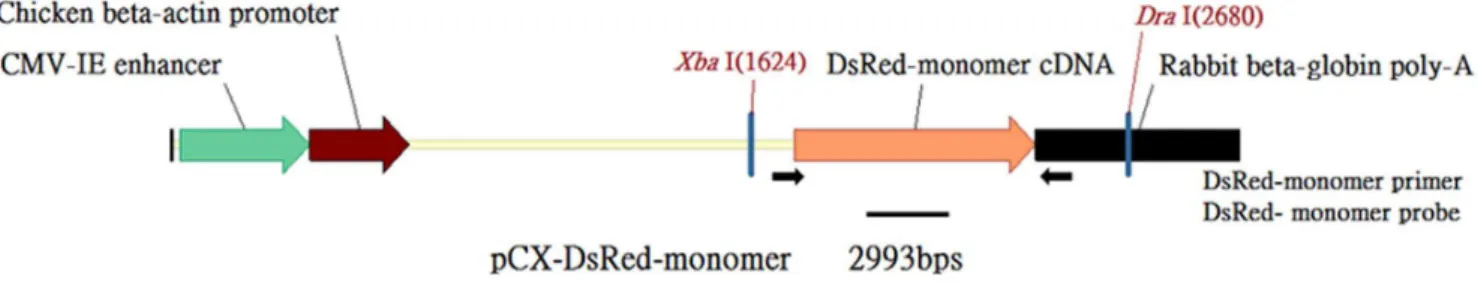
The DsRed monomer coding sequence was cut from the pDsRed-Monomer-N1 Vector (Clontech Laboratories Inc., Mountain View, CA, USA) by using EcoRI digestion, treated with klenow, and used to replace the EGFP coding sequence between 2 EcoRI sites in pCX-EGFP [9]. The resulting plasmid was designated as the pCX-DsRed-Monomer, and contains a CMV enhancer, a chicken-beta actin promoter, a DsRed-Monomer cDNA fragment, and a rabbit beta-globin polyA site (Fig. 1).
Production of transgenic pigs by using pronuclear microinjection
The transgenic pigs were generated by using procedures described previously [4]. Sexually mature crossbred gilts (Duroc
6 Landrace 6Yorkshire ) aged between 240 to 300 d and
weighing greater than 120 kg were used as embryo donors or recipients. The animals’ estruses were synchronized through daily oral administration of 20 mg of Regumate (Intervet, Millsboro, DE, USA) to the gilts for a period of 18 d. Donor animals were administered 2000 IU of pregnant mare’s serum gonadotropin (PMSG) through injection (China Chemical and Pharmaceutical, Taipei, Taiwan) 24 h after the final dose of Regumate, and were injected with 1500 IU of hCG 80 h after being injected with the PMSG. Animals exhibiting standing estrus were artificially inseminated. Pronuclear embryos were collected from the oviduct 55–62 h after hCG injection.
The linearized transgene was purified from the Sal I and Hind III fragments of the pCX-DsRed-Monomer. The injected DNA was dissolved in a microinjection buffer (10 mM Tris, 0.1 mMEDTA, pH 7.4) at a concentration of 4 ng/uL. After microinjection, the pronuclear embryos were surgically transferred into recipient gilts on the same day.
Identification of the transgene
Genomic DNA was extracted as described previously [4]. A PCR analysis of the genomic DNA was performed by primers specific for DsRed (forward, 59 -ATGGACAACACCGAG-GACGTCATC-39, and reverse, 59- TCAGTGGTATTTGT-GAGCCAGGGC-39). Each reaction mixture contained 200 ng of genomic DNA extract, 0.1mM of each primer, 1.25 units of
Taq DNA polymerase (Invitrogen, Brazil) and 2.5 mM MgCl2,
constituting a final volume of 20 uL. A PCR was conducted at 94uC for 3 min for 35 cycles (94uC, 30 s; 65uC, 30 s; and 72uC, 30 s), with a final extension at 72uC for 7 min. The amplified PCR product was separated on 1% agarose gel and exhibited the expected bands of 780 bp.
A Southern blot analysis was performed to confirm the transgene. The DNA were digested using XbaI and DraI (New England Biolabs, Beverly, MA, USA), separated in 1% agarose gel, and then transferred to a Hybond-N+membrane (Amersham
Biosciences) by using capillary diffusion. The blots were probed with the 232-bp PCR fragment of DsRed (forward, 59 -CGACATCCCCGACTACATGA-39, and reverse, 59 -TCCTGGGGGTACAGCTTCTC-39), which was [p32 ]dCTP-labeled by using the random primed labeling method. Hybridiza-tion was conducted under high stringency condiHybridiza-tions (20% dextran sulfate, 26SSC, 0.5% fat free milk powder, and 1%
SDS) in the presence of 750mg/mL of heat-denatured salmon sperm DNA. After a final wash with 26SSC/0.1% SDS for 5 min at 65uC, the nylon membrane was sealed and exposed to an exposure cassette (Amersham Biosciences) with an intensifying screen at 4uC for 7 d.
Histology and hematoxylin/eosin staining
Tissues were fixed in a 10% neutral formaldehyde solution overnight at 4uC, dehydrated through a series of graded ethanol baths, and then embedded in paraffin. The samples were sectioned at a thickness of 3mm and stained with hematoxylin/eosin for
analysis.
Blood sample collection and measurements
Blood samples were collected in plastic vacutainer spray-coated K2EDTA tubes (Becton Dickinson, Franklin Lakes, NJ, USA) and lithium heparin vacuum tubes (Vacuette, Greiner Labortechnik, Kremsmunster, Austria) by using ear venipuncture. Routine blood measurements, including measurements of the red blood cell count, hemoglobin, hematocrit, white blood cell count, and platelet counts, were executed using the CELL-DYNE 1400 (Abbot Laboratories, Abbott Park, IL, USA). Blood biochemical parameters were analyzed using a Hitachi 7600 auto-analyzer (Hitachi Ltd, Tokyo, Japan), which also measured nutritional indices (total protein and albumin), renal function indices (blood urea nitrogen and creatinine), liver function indices (alanine
Figure 1. The DsRed-Monomer transgenic construct pCX-DsRed-Monomer.Arrows indicate the positions of the PCR DsRed primers. XbaI and DraI were restriction enzyme digestion sites. The thick black line indicates the position of the Southern blot probe.
aminotransferase, aspartate aminotransferase, and alkaline phos-phatase), and metabolic indices (glucose, cholesterol, and triglyc-eride).
Computed tomography
Whole-body computed tomography (CT) was performed to confirm the structural and functional integrity of the transgenic pigs. The pigs were anesthetized through an intramuscular injection of ketamine (12.5 mg/kg) and fentanyl (0.04 mg/kg). A 24-gauze catheter was subsequently inserted into the peripheral vein for contrast media infusion.
All images were acquired using a 256-slice multidetector CT scanner (Brilliance iCT; Philips Healthcare, Cleveland, OH, USA). Scanning was conducted at a tube voltage of 120 kV, a tube current of 120 mAs, a detector collimation of 12860.625 mm, a 0.601 pitch, a gantry rotation of 270 ms, a field of view set to 20–25 cm, and a matrix size of 5126512. An iodinated contrast medium (2 ml/kg; Ultravist 370, Iopromide, Schering, Germany) followed by 5 mL of saline were infused through the venous catheter at 0.5 mL/sec. The contrast media and saline were infused, and after ensuring the opacification of the vasculature, scanning was immediately initiated from head to hoof, with simultaneous electrocardiographic recording. After scanning, the images were reconstructed with a slice thickness of 0.9 mm and a slice interval of 0.45 mm by using a standard algorithm (medium soft-tissue convolution kernel).
Detection of red fluorescence
A portable light (excitation = 520–540 nm, emission = 580– 650 nm) was used to detect red fluorescence in the pig. Paraffin-embedded sections of tissues were observed using a fluorescence microscope (Nikon Eclipse i80, Japan).
Western blot analysis
Total proteins were extracted from snap-frozen tissues and separated on 10% SDS-PAGE gels. After being transferred to a PVDF membrane, the blots were probed with rabbit anti-DsRed polyclonal antibodies (1:1500; BD Clontech, Palo Alto, CA, USA) and detected using HRP-conjugated goat anti-rabbit antibodies (1:15,000; Bio-Rad) by using the ECL detection system (Amer-sham Bioscience).
Isolation of pig amniotic fluid progenitor/stem cells
Pig amniotic fluid progenitor/stem cells (pAFPCs) were isolated from the Dsred pig at 70 d of pregnancy. The collected amniotic fluid was filtered through a 70-mm mesh (Miltenyi Biotec Inc., Auburn, CA, USA) and centrifuged at 12 000 rpm for 10 min. The pellets were resuspended and cultured in a 10-cm dish (TPP, Trasadingen, Switzerland) at a density of 26105cells/cm2.
Fluorescence-activated cell-sorting analysis
Ten to 12 passages of pAFPCs were treated using 0.25% trypsin EDTA for 5 min at 37uC, washed with a fluorescence-activated
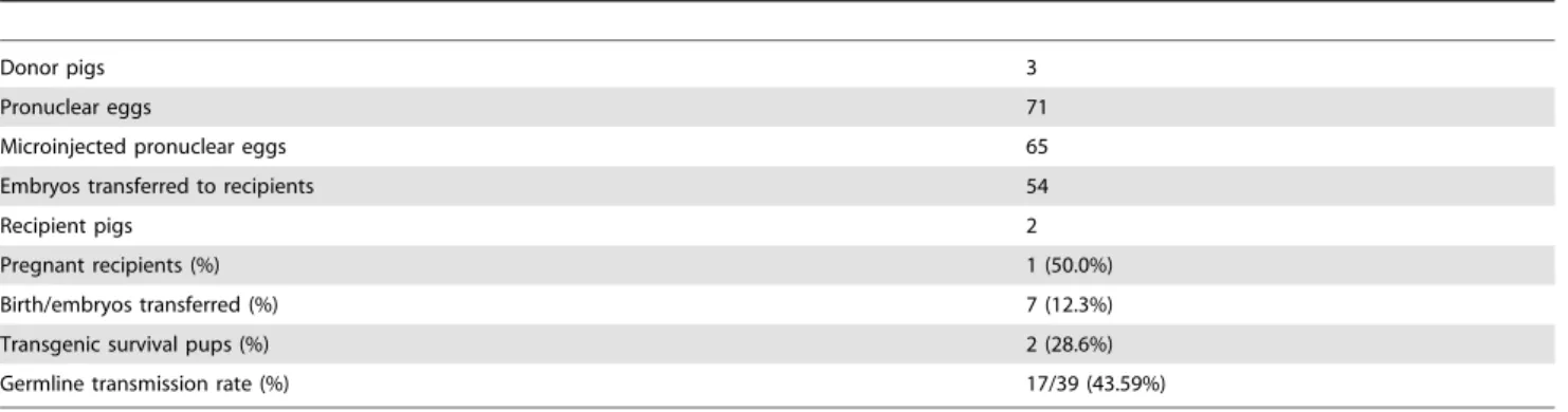
Table 1.Generation of red fluorescent protein (DsRed) transgenic pigs by pronuclear micro-injection.
Donor pigs 3
Pronuclear eggs 71
Microinjected pronuclear eggs 65
Embryos transferred to recipients 54
Recipient pigs 2
Pregnant recipients (%) 1 (50.0%)
Birth/embryos transferred (%) 7 (12.3%)
Transgenic survival pups (%) 2 (28.6%)
Germline transmission rate (%) 17/39 (43.59%)
doi:10.1371/journal.pone.0106864.t001
Figure 2. Transgenic founder pigs carrying the DsRed gene were detected by (a) PCR and (b) Southern blot analysis.The presence of transgenes in DsRed2 pigs were confirmed by using a DsRed primer, which produced a 780-bp PCR product. The genomic DNA of these 2 transgenic pigs (Nos. 1 and 3) were digested using XbaI and DraI. The digested DNA was subsequently hybridized using a 1.1-kb probe.
cell-sorting (FACS) buffer (PBS containing 2% fetal bovine serum), and then dissociated into single cells by gentle pipetting at a density of 16106cells/mL. The cells were stained with fluorescein isothiocyanate (FITC)-conjugated mouse anti-pig antibodies CD4a, CD31, CD44, and CD90 (eBioscience Inc., San Diego, CA, USA) at 4uC for 30 min. The isotype was used as the negative control. The cells were analyzed using a FACS scan flow cytometer (Beckman Coulter Inc., Fullerton, CA, USA).
Differentiation of pig amniotic fluid progentitor/stem cells
To evaluate the differentiation potency of the isolated pAFPCs, a series of in vitro differentiation experiments was conducted. To induce adipogenic differentiation, the pAFPCs were cultured in a StemPro Adipogenesis Supplement Medium (Gibco) for 3 wk; the differentiation medium was changed every 3 d. The pAFPCs were stained using 0.5% Oil-Red O (Sigma-Aldrich) to examine lipid droplet accumulation. For osteogenic differentiation, the pAFPCs were incubated in the StemPro Osteogenesis Supplement Medium (Gibco) for 1 wk, and the calcium deposition was confirmed by performing Alizarin red S (ARS; Sigma-Aldrich) staining. For chondrogenic differentiation, cell pellets were cultured in the StemPro Chondrogenesis Supplement Medium (Gibco) for 10 d. The production of proteoglycan was detected by performing toluidine blue (Sigma-Aldrich) staining.
Statistical analysis
SPSS 15.0 (SPSS, Chicago, IL) was used to conduct statistical analysis. Groups were compared by using Student’s t test for
continuous variables. APvalue less than 0.05 indicated statistical
significance.
Results
Sixty-five pronuclear embryos were injected with the DsRed gene, and 54 injected pronuclear embryos were transferred to 2 recipients. A total of 7 piglets were born, and 2 of the 7 harbored the DsRed gene. Our transgenic efficiency was comparable to that reported previously [10]. The germ-line transmission rate was approximately 43.59% (Table 1), which was concordant with Mendel’s laws.
Expression of red fluorescence
Genome typing revealed that Piglets 1 and 3 carried the Ds-Red gene, and this was confirmed through a PCR (Fig. 2a) and Southern blotting (Fig. 2b). Fluorescent images depicted the ubiquitous exterior expression of red fluorescence in the transgenic pigs (Fig. 3). All of the harvested organs (Fig. 4) and tissues (Fig. 5), namely the brain, eye, tongue, heart, lung, liver, spleen, pancreas, stomach, small intestine, large intestine, kidney, testis, uterus, ovary, muscle, and hoofs, strongly exhibited red fluorescence. Hematoxylin and eosin staining revealed no obvious histological difference between the control and DsRed pigs. A western blot analysis indicated higher DsRed protein expression in the pancreas, heart, muscle, and kidney than the other organs (Fig. 6).
Blood chemistry
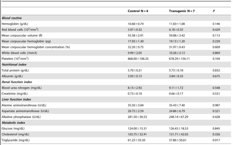
Hematological analysis revealed no significant difference regarding white blood cell counts, hemoglobin levels, or platelet counts. Blood biochemical analysis revealed that the DsRed pigs did not differ from the control pigs in nutrition (as indicated by serum total protein and albumin levels), renal function (as indicated by blood urea nitrogen and creatinine levels), liver function (as indicated by alanine aminotransferase, aspartate aminotransferase, and alkaline phosphatase levels), or metabolic status (as indicated by glucose, cholesterol, and triglyceride levels) (Table 2).
Computed tomography
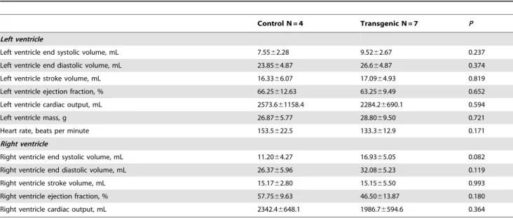
There were no significant differences between the control and transgenic offspring regarding cardiac structure and function (Table 3).
The postcontrast enhanced CT scans showed no structural difference in the brain, no focal lesions in the liver and kidneys, well-distended gall bladders, no intrahepatic bile duct dilatations, no differences in parenchymal perfusion and density, and similar renal cortex thicknesses between the 2 groups (Fig. 7).
Isolation, characterization, and differentiation of amniotic-fluid-derived progentitor/stem cells
The pAFPCs isolated from our transgenic pigs expressed red fluorescence protein (Fig. 8A). The pAFPCs expressed the markers CD44 and CD90, but not CD4a or CD31. Of the pAFPCs, CD44
Figure 3. Transgenic piglets and (white arrow) and nontransgenic controls (white triangle) were differentiated according to the presence of external red fluorescence.
cells accounted for 98.5%, and CD90 cells accounted for 7.4% (Fig. 8). pAFPCs have the ability of adipogenic, osteogenic, and chondrogenic differentiation.
Discussion
In this study, we successfully employed the pronuclear microinjection technique to produce pigs that ubiquitously
expressed red fluorescent protein (RFP). Their health was comprehensively confirmed through blood biochemistry and whole-body CT. In research applications, the use of this animal enables red-fluorescent-protein-tagged cells to be isolated, and can be detected using live imaging in chimera studies. In addition, tagged tissues can facilitate research in developmental biology and xenotransplantation. In this study, we proved the differentiation potential of the animals’ pAFPCs, which can serve as novel
Figure 4. Bright-field images of tissues from the transgenic pig offspring (left) depict a distinct red fluorescence in comparison with the nontransgenic control (right).
reagents for regenerative medicine with more effective cell-fate tracking.
The ability to monitor cell distribution, cell expression, and cell fate are fundamental to the development of cell therapy. Imaging of light emission from luminescent or fluorescent reporters enables real-time noninvasive in vivo analysis, and has become a mainstay of preclinical imaging [11,12]. Traditional direct in vitro labeling techniques, such as DiI for labeling membranes, or Hoechst 33342 for labeling nuclei, are subject to dilution by cell proliferation. The dyes released by dead cells can label neighboring host cells and yield false-positive results [13]. Furthermore, these intercalating dyes can be toxic to cells, and might affect the differentiation potential of stem cells in vivo. Our red fluorescent pig can provide an ample source of cells for grafting without these potential
confounding concerns, and can facilitate spatial, temporal, and quantitative analysis. Similar to our previous study on eGFP transgenic pigs [4], we observed various intensities of red fluorescence in different harvested organs and tissues. This phenomenon might be attributed to the differential assembly of transcription factors in each organ, which affected the spatial expression of our transgenic construct.
RFP was originally discovered in corals of the class Anthozoa [14]. This protein possesses 26%–30% of the conserved amino acid of aequorean GFP, and has a similar chromophoric structure. Unlike GFPs, the chromophore of DsRed contains an additional double bond that stretches its chromophore conjugation, resulting in its red color [15]. RFP offers the distinct advantage of low background autofluorescence [16]. However, the tight mutual
Figure 5. Fluorescence microscopy of the paraffin-embedded tissue sections of DsRed transgenic pigs revealed evident red fluorescence.
Figure 6. SDS-PAGE and western blot analysis of tissue samples from a DsRed transgenic pig. doi:10.1371/journal.pone.0106864.g006
Table 2.Blood routine and biochemistry between the transgenic offspring and the non-transgenic control pigs.
Control N = 4 Transgenic N = 7 P
Blood routine
Hemoglobin (g/dL) 10.6860.74 11.8361.08 0.146
Red blood cells (103/mm3) 5.97
60.32 6.1860.35 0.429
Mean corpuscular volume (fl) 55.5862.91 59.8663.42 0.113
Mean corpuscular hemoglobin (pg) 17.9361.30 19.1361.20 0.229
Mean corpuscular hemoglobin concentration (%) 32.2060.75 31.9760.43 0.609
White blood cells (/mm3) 9.9962.05 10.2662.13 0.869
Platelets (103/mm3) 868.00
6106.25 678.296156.11 0.104 Nutritional index
Total protein (g/dL) 5.7060.21 5.7360.18 0.832
Albumin (g/dL) 3.9360.15 3.8460.33 0.675
Renal function index
Blood urea nitrogen (mg/dL) 8.1562.92 9.1161.72 0.548
Creatinine (mg/dL) 0.7360.10 0.6660.17 0.531
Liver function index
Alanine aminotransferase (U/dL) 35.5063.84 35.4367.40 0.987
Aspartate aminotransferase (U/dL) 20.7562.59 24.8666.79 0.321
Alkaline phosphatase (U/dL) 281.50636.53 248.14667.29 0.428
Metabolic index
Glucose (mg/dL) 124.00615.31 126.43618.53 0.845
Cholesterol (mg/dL) 103.75632.41 131.71642.03 0.326
Triglyceride (mg/dL) 61.25635.50 57.86650.61 0.917
binding of the 4 subunits in the original RFP causes any protein fused with the DsRed becomes a tetramer [17]. Such a tetramer interferes with protein-protein interactions, and even causes aggregation or precipitation [18]. This tetramer can be toxic to
embryos [19] because it inhibits the expression of antiapoptotic proteins, such as the B-cell lymphoma extra-large protein, through translational regulation [20]. This is consequently the main hurdle in generating red fluorescence in transgenic animals. To solve the
Table 3.Cardiac structure and function between the transgenic offspring and the non-transgenic control pigs.
Control N = 4 Transgenic N = 7 P
Left ventricle
Left ventricle end systolic volume, mL 7.5562.28 9.5262.67 0.237
Left ventricle end diastolic volume, mL 23.8564.87 26.664.87 0.374
Left ventricle stroke volume, mL 16.3366.07 17.0964.93 0.819
Left ventricle ejection fraction, % 66.25612.63 63.2569.49 0.652
Left ventricle cardiac output, mL 2573.661158.4 2284.26690.1 0.594
Left ventricle mass, g 26.8765.77 28.8069.50 0.721
Heart rate, beats per minute 153.5622.5 133.3612.9 0.171
Right ventricle
Right ventricle end systolic volume, mL 11.2064.27 16.9365.05 0.082
Right ventricle end diastolic volume, mL 26.3765.96 32.0865.23 0.119
Right ventricle stroke volume, mL 15.1762.80 15.1565.50 0.993
Right ventricle ejection fraction, % 57.7569.63 46.50613.87 0.180
Right ventricle cardiac output, mL 2342.46648.1 1986.76594.6 0.364
doi:10.1371/journal.pone.0106864.t003
Figure 7. Whole-body CT of the normal and transgenic pigs.Neither structural differences nor abnormal lesions were observed in the DsRed pig.
tetrameric stoichiometry issue, Campbell et al. [21] successfully generated monomeric RFP, which improved the reliability of protein fusion.
We comprehensively characterized our transgenic pig and employed hematology analysis, blood biochemistry profiling, and CT imaging to ensure the health of the pig. To date, we have observed no noticeable reduction of red fluorescence for over 5 generations. Moreover, in routine daily observations, no distin-guishable behavioral differences between age-matched transgenic and control animals have been observed. In conjunction with various other fluorescent-tagged cells and organs, our red fluorescent pig forms the basis of a multicolored scheme for basic biomedical research on human diseases.
Conclusions
We established and characterized a pig harboring the DsRed-Monomer. As a large domestic animal available for medical research, this pig is an addition to the armamentarium for elucidating many cellular processes in vivo, and can provide reagents for cell therapy and transplantation studies.
Acknowledgments
The authors are grateful for the technical support provided by the Veterinary Medicine Digital Pathology Laboratory of National Taiwan University, Taiwan.
Figure 8.(a) Cytometric analyses of CD4a, CD31, CD44, and CD90 conjugated with FITC on DsRed pig amniotic fluid progenitor/stem cells (pAFPCs). pAFPCs were negative for the surface antigens CD4a and CD31, and positive for CD44 and CD90. (b) Mesoderm trilineage differentiation potential of DsRed pAFPCs. DsRed pAFPCs exhibited spindle-shaped morphology (A1) and expressed red fluorescence under fluorescent microscopy (A2). Lipid droplets were observed in the DsRed pAFPCs adipogenic differentiation culture by using Oil-Red O staining (B1). Calcium accumulation was observed in the osteogenic differentiation culture by using Alizarin red staining (C1). Collagen was detected in the chondrogenic differentiation culture by using toluidine blue staining (D1). Under fluorescent microscopy, red fluorescence was still evident after osteogenetic (B2), adipogenetic (C2), and chondrogenetic (D2) differentiation.
Author Contributions
Conceived and designed the experiments: WTC YPL SCW. Performed the experiments: CJC SYP MHW CCY YSL WTC YPL SCW. Analyzed the
data: CJC SYP MHW CCY YSL WTC YPL SCW. Contributed reagents/ materials/analysis tools: MHW WTC YPL SCW. Contributed to the writing of the manuscript: CJC SYP MHW CCY YSL WTC YPL SCW.
References
1. Schook LB, Beever JE, Rogers J, Humphray S, Archibald A, et al. (2005) Swine Genome Sequencing Consortium (SGSC): a strategic roadmap for sequencing the pig genome. Comp Funct Genomics 6: 251–255.
2. Walters EM, Wolf E, Whyte JJ, Mao J, Renner S, et al. (2012) Completion of the swine genome will simplify the production of swine as a large animal biomedical model. BMC Med Genomics 5: 55.
3. Sprangers B, Waer M, Billiau AD (2008) Xenotransplantation: where are we in 2008? Kidney Int 74: 14–21.
4. Hsiao FS, Lian WS, Lin SP, Lin CJ, Lin YS, et al. (2011) Toward an ideal animal model to trace donor cell fates after stem cell therapy: production of stably labeled multipotent mesenchymal stem cells from bone marrow of transgenic pigs harboring enhanced green fluorescence protein gene. J Anim Sci 89: 3460–3472.
5. Murakami T, Kobayashi E (2012) GFP-transgenic animals for in vivo imaging: rats, rabbits, and pigs. Methods Mol Biol 872: 177–189.
6. Sommer JR, Jackson LR, Simpson SG, Collins EB, Piedrahita JA, et al. (2012) Transgenic Stra8-EYFP pigs: a model for developing male germ cell technologies. Transgenic Res 21: 383–392.
7. Matsunari H, Onodera M, Tada N, Mochizuki H, Karasawa S, et al. (2008) Transgenic-cloned pigs systemically expressing red fluorescent protein, Kusa-bira-Orange. Cloning Stem Cells 10: 313–323.
8. Lu Y, Kang JD, Li S, Wang W, Jin JX, et al. (2013) Generation of transgenic Wuzhishan miniature pigs expressing monomeric red fluorescent protein by somatic cell nuclear transfer. Genesis 51: 575–586.
9. Okabe M, Ikawa M, Kominami K, Nakanishi T, Nishimune Y (1997) ‘Green mice’ as a source of ubiquitous green cells. FEBS Lett 407: 313–319. 10. Hirabayashi M, Takahashi R, Ito K, Kashiwazaki N, Hirao M, et al. (2001) A
comparative study on the integration of exogenous DNA into mouse, rat, rabbit, and pig genomes. Exp Anim 50: 125–131.
11. Hadjantonakis AK, Papaioannou VE (2004) Dynamic in vivo imaging and cell tracking using a histone fluorescent protein fusion in mice. BMC Biotechnol 4: 33.
12. Abe T, Fujimori T (2013) Reporter mouse lines for fluorescence imaging. Dev Growth Differ 55: 390–405.
13. Reinecke H, Minami E, Zhu WZ, Laflamme MA (2008) Cardiogenic differentiation and transdifferentiation of progenitor cells. Circ Res 103: 1058–1071.
14. Matz MV, Fradkov AF, Labas YA, Savitsky AP, Zaraisky AG, et al. (1999) Fluorescent proteins from nonbioluminescent Anthozoa species. Nat Biotechnol 17: 969–973.
15. Gross LA, Baird GS, Hoffman RC, Baldridge KK, Tsien RY (2000) The structure of the chromophore within DsRed, a red fluorescent protein from coral. Proc Natl Acad Sci U S A 97: 11990–11995.
16. Vintersten K, Monetti C, Gertsenstein M, Zhang P, Laszlo L, et al. (2004) Mouse in red: red fluorescent protein expression in mouse ES cells, embryos, and adult animals. Genesis 40: 241–246.
17. Baird GS, Zacharias DA, Tsien RY (2000) Biochemistry, mutagenesis, and oligomerization of DsRed, a red fluorescent protein from coral. Proc Natl Acad Sci U S A 97: 11984–11989.
18. Yanushevich YG, Staroverov DB, Savitsky AP, Fradkov AF, Gurskaya NG, et al. (2002) A strategy for the generation of non-aggregating mutants of Anthozoa fluorescent proteins. FEBS Lett 511: 11–14.
19. Hadjantonakis AK, Macmaster S, Nagy A (2002) Embryonic stem cells and mice expressing different GFP variants for multiple non-invasive reporter usage within a single animal. BMC Biotechnol 2: 11.
20. Zhou J, Lin J, Zhou C, Deng X, Xia B (2011) Cytotoxicity of red fluorescent protein DsRed is associated with the suppression of Bcl-xL translation. FEBS Lett 585: 821–827.