(1)doi: 10.3389/fmicb.2018.01441
Edited by:
Rebecca Thombre,
Pune University, India
Reviewed by:
Shahper Nazeer Khan,
Aligarh Muslim University, India
Rodolfo García-Contreras,
Universidad Nacional Autónoma de
México, Mexico
*Correspondence:
Pedro V. Baptista
pmvb@fct.unl.pt
Marta Martins
mmartins@tcd.ie
Alexandra R. Fernandes
ma.fernandes@fct.unl.pt
†Present Address:
Matthew P. McCusker,
Kerry Europe, Global Technology &
Innovation Centre, Naas, Ireland
Specialty section:
This article was submitted to
Antimicrobials, Resistance and
Chemotherapy,
a section of the journal
Frontiers in Microbiology
Received:31 March 2018
Accepted:11 June 2018
Published:02 July 2018
Citation:
Baptista PV, McCusker MP,
Carvalho A, Ferreira DA, Mohan NM,
Martins M and Fernandes AR (2018)
Nano-Strategies to Fight Multidrug
Resistant Bacteria—“A Battle of the
Titans”. Front. Microbiol. 9:1441.
doi: 10.3389/fmicb.2018.01441
Nano-Strategies to Fight Multidrug
Resistant Bacteria—“A Battle of the
Titans”
Pedro V. Baptista
1
*, Matthew P. McCusker
2†
, Andreia Carvalho
1
, Daniela A. Ferreira
3
,
Niamh M. Mohan
3,4
, Marta Martins
3
* and Alexandra R. Fernandes
1
*
1UCIBIO, Departamento de Ciências da Vida, Faculdade de Ciências e Tecnologia, Universidade Nova de Lisboa, Caparica,
Portugal,2School of Food Science and Environmental Health, College of Sciences and Health, Dublin Institute of Technology,
Dublin, Ireland,3Department of Microbiology, Moyne Institute of Preventive Medicine, Schools of Genetics and Microbiology,
Trinity College Dublin, University of Dublin, Dublin, Ireland,4Nuritas Limited, Dublin, Ireland
the use of nanoparticles still presents a challenge to therapy and much more research is
needed in order to overcome this. In this review, we will summarize the current research
on nanoparticles and other nanomaterials and how these are or can be applied in the
future to fight multidrug resistant bacteria.
Keywords: antimicrobial resistance, multidrug resistance, nanomaterials, nanoparticles, plant-based compounds,
novel antimicrobial agents, nanotheranostics
INTRODUCTION
Multidrug resistant (MDR) bacteria remain the greatest challenge
in public health care. The numbers of infections produced
by such resistant strains are increasing globally. This acquired
resistance of pathogens presents a key challenge for many
antimicrobial drugs. Recent advances in nanotechnology offer
new prospects to develop novel formulations based on distinct
types of nanoparticles (NPs) with different sizes and shapes and
flexible antimicrobial properties.
NPs may offer a promising solution as they can not only
combat bacteria themselves but can also act as carriers for
antibiotics and natural antimicrobial compounds (
Wang et al.,
2017a
). While various materials have been explored from
liposomal to polymer based nano-drug carriers, metallic vectors,
such as gold NPs, are attractive as core materials due to their
essentially inert and nontoxic nature (
Burygin et al., 2009
).
Arguably the most attractive aspect of NPs drug delivery systems
is their ability to introduce a wide range of therapeutics,
either bound to their large surface area or contained within
the structure, to the site of infection effectively and safely by
having a controlled rate of targeted delivery (
Pissuwan et al.,
2011; Gholipourmalekabadi et al., 2017
). By improving the
pharmacokinetic profile and therapeutic index of encapsulated
drugs compared to free drug equivalents, the dose required to
achieve clinical effects can be dramatically decreased (
Gao et al.,
2018
). This in turn, can reduce the toxicity and the adverse side
effects associated with high systemic drug concentrations and
frequent dosing (
Liu et al., 2009
).
This review covers the latest approaches in the development
of new nanobiotechnology approaches that may challenge the
medical practice to fight bacteria and particularly MDR bacteria.
NANOMATERIALS AGAINST BACTERIA
Nanomaterials have at least one dimension in the nanometer
scale range (1–100 nm) that convey particular physical and
chemical properties considerably different from those of bulk
materials (
Wang et al., 2017a
). Among the wide range of
nanomaterials, particular interest has been directed toward NPs.
NPs have a number of features, which make them favorable as
vectors for drugs to combat disease-causing pathogens. These
include their enhancement of drug solubility and stability (
Huh
and Kwon, 2011
); their ease of synthesis (
Gholipourmalekabadi
et al., 2017
); their biocompatibility with target agents; and their
modulated release, which can be controlled by stimuli, such
as light, pH and heat (
Wang Z. et al., 2017
). Their distinctive
functionality in drug delivery is achieved by their ultra-small
size and vast surface to volume ratios. This is a key competitive
advantage over conventional therapies in the treatment of
infections caused by intracellular pathogens and MDR strains.
Their functionalization with different (bio)molecules is another
important feature. These comprise NPs containing Ag, Au, Al,
Cu, Ce, Cd, Mg, Ni, Se, Pd, Ti, Zn, and super-paramagnetic Fe
(
Hemeg, 2017; Slavin et al., 2017
). AgNPs are considered the
most effective nanomaterial against bacteria but other metallic
NPs, such as CuONPs, TiONPs, AuNPs, and Fe3O2NPs, have
also demonstrated bactericidal effects (
Dakal et al., 2016; Hemeg,
2017; Slavin et al., 2017
).
While poor membrane transport limits the potency of many
antibiotics (
Andrade et al., 2013
), drug loaded NPs vehicles can
enter host cells
via
endocytosis, facilitating their intracellular
entry (
Wang Z. et al., 2017
). Membrane penetration can also be
achieved through interactions with surface lipids, for example,
using gold NPs in the co-administration of protein-based drugs
(
Huang et al., 2010
). The therapeutic appeal of NPs is enhanced
by their ability to confer physical protection against bacterial
resistance mechanisms (
Huh and Kwon, 2011
). Furthermore,
the potential to load multiple drug combinations into NPs
presents a highly complex antimicrobial mechanism of action,
to which, bacteria are unlikely to develop resistance (
Huh
and Kwon, 2011
). Although, this is usually believed to be the
case, there are some studies reporting development of bacterial
resistance against silver NPs (
Panáˇcek et al., 2018
). There is
also evidence that exposure of bacteria to this type of NPs
may increase its antibiotic tolerance (
Kaweeteerawat et al.,
2017
).
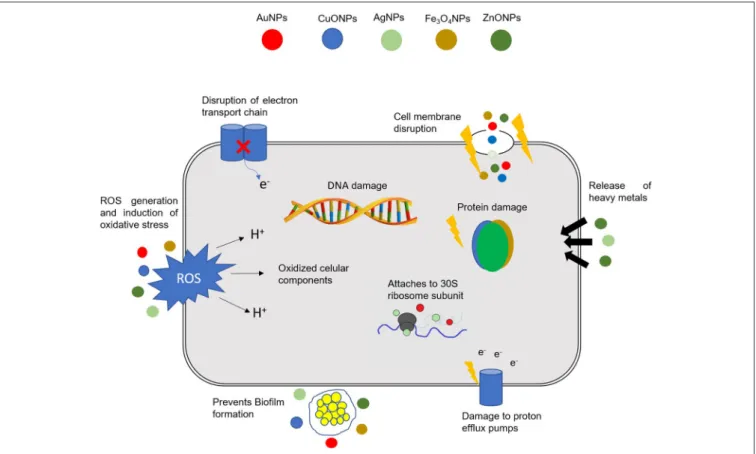
NPs can exert their antibacterial activity
via
a multitude of
mechanisms, such as: (1) direct interaction with the bacterial cell
wall; (2) inhibition of biofilm formation; (3) triggering of innate
as well as adaptive host immune responses; (4) generation of
reactive oxygen species (ROS); and (5) induction of intracellular
effects (
e.g
., interactions with DNA and/or proteins). Because
they do not present the same mechanisms of action of standard
antibiotics (
Figure 1
), they can be of extreme use against MDR
bacteria (
Singh K. et al., 2014; Aderibigbe, 2017; AlMatar et al.,
2017; Hemeg, 2017; Natan and Banin, 2017; Rai et al., 2017; Slavin
et al., 2017; Zaidi et al., 2017; Bassegoda et al., 2018; Katva et al.,
2018; Siddiqi et al., 2018
).
FIGURE 1 |Different mechanisms of action of NPs in bacterial cells. The combination in a single nanomaterial of a multitude of cellular effects may have a tremendous
impact in fighting MDR bacteria. DNA, deoxyribonucleic acid; ROS, Reactive oxygen species; AuNPs, gold NPs; CuONPs, Copper oxide NPs; AgNPs, silver NPs;
Fe3O4NPs, iron oxide NPs; ZnONPs, zinc oxide NPs.
2017; Wang et al., 2017a; Zaidi et al., 2017; Hadiya et al.,
2018
). Some of the advantages of using NPs as vectors are
due to their small and controllable size; their protective action
against enzymes that would otherwise destroy antimicrobial
compounds; their ability to actively deliver antibiotics; and
their capability to combine several therapeutic modalities onto
a single nanomaterial (
e.g.
, several antibiotics/compounds onto
the same NPs for combined action; combining silencing agents
and drugs, etc.) (
Turos et al., 2007; Huh and Kwon, 2011;
Mohammed Fayaz et al., 2011; Liu et al., 2013; Qi et al., 2013;
Li et al., 2014; Ranghar et al., 2014; Thomas et al., 2014;
Wang et al., 2014; Payne et al., 2016; Rai A. et al., 2016;
Singh J. et al., 2016; Yeom et al., 2016; Esmaeillou et al.,
2017; Zaidi et al., 2017; Zong et al., 2017; Hadiya et al.,
2018
).
NPs carriers can tackle bacterial threats “passively,” through
prolonged drug retention at the specific infection site, or
“actively,” through surface conjugation with active molecules
that bind a certain target (
Wang Z. et al., 2017
). The balance
between the surface modification interaction strength, the
compound release rate and the stability of the conjugate
should be carefully considered for the design of an effective
“active” delivery strategy (
Burygin et al., 2009; Pissuwan et al.,
2011
). In an attempt to overcome their therapeutic limitations,
various research groups have investigated the conjugation of
antibiotics to NPs (
Tiwari et al., 2011
). For example, Saha
et al. describe the direct conjugation of ampicillin, streptomycin
and kanamycin to gold NPs (
Saha et al., 2007
). The resulting
complexes were shown to have lower minimum inhibitory
concentration (MIC) than the free drug counterparts against
both Gram -negative and -positive bacteria. While the detailed
mechanism of these effects are not explained by the authors
in the above case, Fayaz et al. has attempted to uncover
how their vancomycin functionalized gold NPs demonstrated
activity against strains which are usually vancomycin resistant
based either on mutations (vancomycin resistant
Staphylococcus
aureus
), or membrane structure (
Escherichia coli
) (
Mohammed
Fayaz et al., 2011
). They propose that only when the antibiotic
was complexed with the NPs could this result in nonspecific,
multivalent interactions and anchoring of the carrier to the cell
wall synthesis proteins. Based on the presence of pits in the cells,
which was observed using transmission electron microscopy,
the authors concluded that the consequence of the non-specific
binding was compromised membrane integrity, and subsequent
cell death (
Mohammed Fayaz et al., 2011; Gao et al., 2018
).
ANTIBACTERIAL MECHANISM OF NPS
surface area in contact with bacteria through electrostatic
attraction, van der walls forces or hydrophobic interactions;
on the nanoparticle size and stability; together with the drug
concentration (
Chen et al., 2014; Gao et al., 2014; Li et al., 2015
).
The interaction of NPs with bacteria generally triggers oxidative
stress mechanisms, enzymatic inhibition, protein deactivation
and changes in gene expression. Still, the most common
antibacterial mechanisms are related to oxidative stress, metal ion
release, and non-oxidative mechanisms (
Wang et al., 2017a; Zaidi
et al., 2017
see
Figure 1
).
Oxidative stress induced by ROS is one of the most important
mechanisms assisting the antibacterial activity of NPs (
Dwivedi
et al., 2014; Rudramurthy et al., 2016
). ROS are natural
byproducts of cellular oxidative metabolism and have significant
important roles in the modulation of cell survival and death,
differentiation and cell signaling. In bacteria, ROS are formed
from aerobic respiration, and their production is balanced by
the cell antioxidant machinery but upon an additional ROS
insult, oxidation of biomolecules, and cell components result
in severe cellular damage (
Li et al., 2012b
). The excessive
production of ROS leads to a disturbed redox homeostasis
resulting in oxidative stress, affecting membrane lipids and
altering the structure of DNA and proteins (
Dwivedi et al., 2014
).
It has been shown that while O
−
2
and H2O2
can be neutralized
by endogenous antioxidants,
·OH and singlet oxygen (
1
[O2])
lead to acute microbial death (
Zaidi et al., 2017
). Different
NPs may generate distinctive ROS, such as superoxide (O
−
2
)
or hydroxyl radical (·OH), hydrogen peroxide (H
2
O
2
), and
1
[O2]) (
Wang et al., 2017a
). In this manner, the level of ROS
generated by NPs is dependent on the chemical nature of
the NPs themselves. Application of metallic NPs is currently
being considered to overcome bacterial infections since they
have shown antimicrobial efficacy due to their high
surface-to-volume ratio. An increase ratio is usually accompanied by
increased production of ROS, including free radicals.
Zhang
et al. (2013)
demonstrated that ROS generation and metal
ion release significantly enhanced the antibacterial activity
through uncoated AuNPs in aqueous suspension under UV
irradiation (365 nm). Umamaheswari (
Umamaheswari et al.,
2014
) demonstrated that the antibacterial activity of AuNPs
against
E. coli
,
Salmonella
Typhi,
Pseudomonas aeruginosa
and
Klebsiella pneumoniae
were due to oxidative stress caused by
increased intracellular ROS. A recent study (
Zhang et al., 2013
)
evaluated AuNPs and AuNPs -laser combined therapy against
C. pseudotuberculosis
and suggested that the mechanism of
action is related with ROS production, that causes an increase
of oxidative stress of microbial cells in the form of vacuole
formation as an indication of potent activity. This effect was
higher with AuNPs-laser, causing a rapid loss of bacterial cell
membrane integrity due to the fact that laser light enhances
at least one fold antimicrobial activity of AuNPs. Several other
studies have addressed the role of metal NPs to induce MDR
bacteria death
via
oxidative stress (
Table 1
) (
Foster et al., 2011;
Li et al., 2012b; Rai et al., 2012; Zhang et al., 2013; Reddy L.
S. et al., 2014; Singh R. et al., 2014; Pan et al., 2016; Courtney
et al., 2017; Ulloa-Ogaz et al., 2017; Zaidi et al., 2017
). Indeed,
titanium dioxide NPs were shown to adhere to the surface of
the bacterial cell and trigger the production of ROS, which in
turns lead to damage of the structure of cellular components
and consequent cell death (
Foster et al., 2011
). In another
important study using different metal NPs, AgNPs were shown
to generate superoxide radicals and hydroxyl radicals, whereas
Au, Ni, and Si NPs generated only singlet oxygen, which upon
entering the cell produced an antibacterial effect (
Zhang et al.,
2013
). More recently, Reddy and co-workers demonstrated that
ZnONPs alone can also act as an effective antibacterial agent
via
the generation of ROS (
Reddy L. S. et al., 2014
). Exposure
to UV irradiation may also potentiate the action of NPs.
Li
et al. (2012b)
reported the augmented antibacterial effects of
zinc oxide (ZnO) and titanium oxide (TiO) NPs triggered by
UV irradiation as the results of the increased production of
superoxide, hydroxyl and singlet oxygen radicals that potentiated
bacteria mortality by severe oxidative stress. Graphene oxide–
iron oxide NPs have also demonstrated maximum antibacterial
activity due to the generation of hydroxyl radicals and diffusion
into bacterial cells (
Pan et al., 2016
). More recently, Ulloa-Ogaz
and collaborators demonstrated that copper oxide NPs interact
with bacteria, generating an intracellular signaling cascade that
trigger oxidative stress and, thus, an antibacterial effect (
Ulloa-Ogaz et al., 2017
).
Metal oxides slowly release metal ions that are up taken
by the cell, reaching the intracellular compartment where they
can interact with functional groups of proteins and nucleic
acids, such as amino (–NH), mercapto (–SH), and carboxyl (–
COOH) groups (
Wang et al., 2017a
). This interaction alters
the cell structure, hampers enzymatic activity and interferes
with the normal physiological processes in the bacterial cell.
It has been shown that copper oxide (CuO) NPs cause a
significant alteration of the expression of key proteins and may
inhibit denitrification. Proteomic analysis showed that CuONPs
cause an alteration of proteins involved in nitrogen metabolism,
electron transfer and transport (
Su et al., 2015
). Also, the
interaction of gold–superparamagnetic iron oxide NPs with
bacterial proteins
via
disulfide bonds affects the metabolism and
redox system of bacterial cells (
Niemirowicz et al., 2014
). NPs
may also enter bacteria through absorption, releasing metal ions
to the surrounding medium and/or binding to the negatively
charged functional groups of the bacterial cell membrane. For
example, silver ions (from silver NPs) are adsorbed on the cell
membrane, leading to protein coagulation (
Jung et al., 2008
).
Jankauskaitl and collaborators described the bactericidal effect
of graphene oxide/Cu/Ag NPs against
E. coli
,
P. aeruginosa
,
K. pneumoniae
,
S. aureus,
and Methicillin-resistant
S. aureus
(MRSA) through a possible synergy between multiple toxic
mechanisms (
Jankauskaite et al., 2016
).
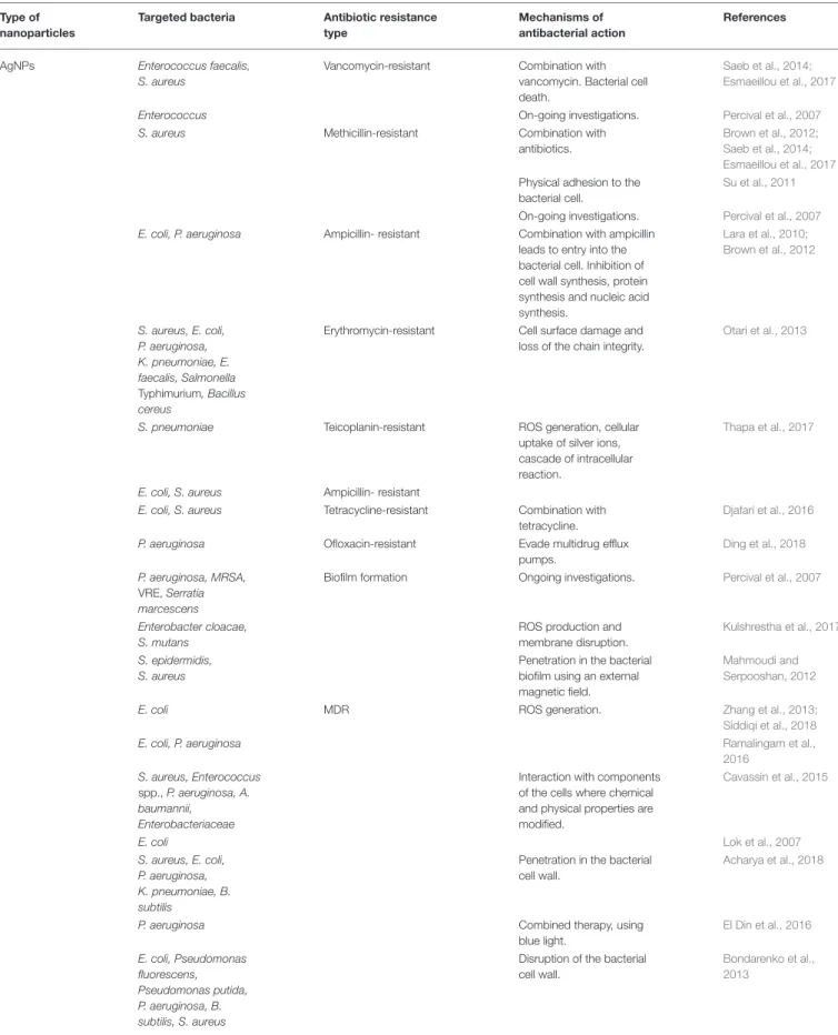
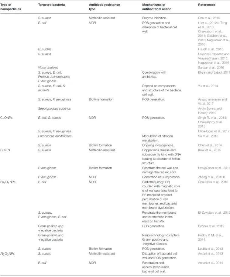
TABLE 1 |Nanoparticles against MDR pathogens and their mechanisms of action.
Type of
nanoparticles
Targeted bacteria Antibiotic resistance
type
Mechanisms of
antibacterial action
References
AgNPs Enterococcus faecalis,
S. aureus
Vancomycin-resistant Combination with
vancomycin. Bacterial cell
death.
Saeb et al., 2014;
Esmaeillou et al., 2017
Enterococcus On-going investigations. Percival et al., 2007
S. aureus Methicillin-resistant Combination with
antibiotics.
Brown et al., 2012;
Saeb et al., 2014;
Esmaeillou et al., 2017
Physical adhesion to the
bacterial cell.
Su et al., 2011
On-going investigations. Percival et al., 2007
E. coli, P. aeruginosa Ampicillin- resistant Combination with ampicillin
leads to entry into the
bacterial cell. Inhibition of
cell wall synthesis, protein
synthesis and nucleic acid
synthesis.
Lara et al., 2010;
Brown et al., 2012
S. aureus, E. coli,
P. aeruginosa,
K. pneumoniae, E.
faecalis, Salmonella
Typhimurium, Bacillus
cereus
Erythromycin-resistant Cell surface damage and
loss of the chain integrity.
Otari et al., 2013
S. pneumoniae Teicoplanin-resistant ROS generation, cellular
uptake of silver ions,
cascade of intracellular
reaction.
Thapa et al., 2017
E. coli, S. aureus Ampicillin- resistant
E. coli, S. aureus Tetracycline-resistant Combination with
tetracycline.
Djafari et al., 2016
P. aeruginosa Ofloxacin-resistant Evade multidrug efflux
pumps.
Ding et al., 2018
P. aeruginosa, MRSA,
VRE,Serratia
marcescens
Biofilm formation Ongoing investigations. Percival et al., 2007
Enterobacter cloacae,
S. mutans
ROS production and
membrane disruption.
Kulshrestha et al., 2017
S. epidermidis,
S. aureus
Penetration in the bacterial
biofilm using an external
magnetic field.
Mahmoudi and
Serpooshan, 2012
E. coli MDR ROS generation. Zhang et al., 2013;
Siddiqi et al., 2018
E. coli, P. aeruginosa Ramalingam et al.,
2016
S. aureus, Enterococcus
spp.,P. aeruginosa, A.
baumannii,
Enterobacteriaceae
Interaction with components
of the cells where chemical
and physical properties are
modified.
Cavassin et al., 2015
E. coli Lok et al., 2007
S. aureus, E. coli,
P. aeruginosa,
K. pneumoniae, B.
subtilis
Penetration in the bacterial
cell wall.
Acharya et al., 2018
P. aeruginosa Combined therapy, using
blue light.
El Din et al., 2016
E. coli, Pseudomonas
fluorescens,
Pseudomonas putida,
P. aeruginosa, B.
subtilis, S. aureus
Disruption of the bacterial
cell wall.
Bondarenko et al.,
2013
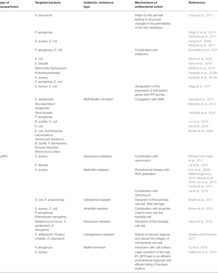
TABLE 1 |Continued
Type of
nanoparticles
Targeted bacteria Antibiotic resistance
type
Mechanisms of
antibacterial action
References
A. baumannii Attach to the cell wall
leading to structural
changes in the permeability
of the cell membrane.
Chang et al., 2017
P. aeruginosa Singh K. et al., 2014;
Salomoni et al., 2017
S. aureus, E. coli Jung et al., 2008;
Muniyan et al., 2017
P. aeruginosa, E. coli Combination with
antibiotics.
Esmaeillou et al., 2017
E. coli Karimi et al., 2016
E. faecalis Katva et al., 2018
SalmonellaTyphimurium McShan et al., 2015
Enterobacteriaceae Paná ˇcek et al., 2016b
S. aureus,
P. aeruginosa, E. coli
Paná ˇcek et al., 2016a
S. aureus, E. coli Upregulation of the
expression of antioxidant
genes and ATP pumps.
Nagy et al., 2011
S. epidermidis MDR/Biofilm formation Conjugation with AMP. Jaiswal et al., 2015
Mycobacterium
smegmatis
Mohanty et al., 2013
Vibrio fluvialis,
P. aeruginosa
Lambadi et al., 2015
B. subtilis, E. coli Liu et al., 2013
E. coli Pal et al., 2016
E. coli, Acinetobacter
calcoaceticus,
Aeromonas bestiarum,
B. Subtili, P. fluorescens,
Kocuria rhizophila,
Micrococcus luteus
Ruden et al., 2009
AuNPs S. aureus Vancomycin-resistant Combination with
vancomycin.
Mohammed Fayaz
et al., 2011
E. faecalis Lai et al., 2015
S. aureus Methicillin-resistant Photothermal therapy with
ROS generation.
Kuo et al., 2009;
Millenbaugh et al.,
2015; Mocan et al.,
2016; Hu et al., 2017;
Ocsoy et al., 2017
Combination with
vancomycin.
Lai et al., 2015
E. coli, K. pneumoniae Cefotaxime-resistant Disruption of the bacterial
cell wall, DNA damage.
Shaikh et al., 2017
S. aureus, E. coli,
P. aeruginosa,
Enterobacter aerogenes
Ampicillin-resistant Combination with ampicillin.
Lead to entry into the
bacterial cell.
Brown et al., 2012
Streptococcus bovis, S.
epidermidis, E.
Aerogenes
Kanamycin-resistant Disruption of the bacterial
cell wall.
Payne et al., 2016
K. 45ellular45, Proteus
mirabilis, A. baumannii
Carbapenems-resistant Disturb of osmotic balance
and disrupt the integrity of
cell bacterial cell wall.
Shaker and Shaaban,
2017
P. aeruginosa Biofilm formation Interaction with cell surface. Yu et al., 2016
S. aureus Laser excitation of the near
IR LSPR lead to an efficient
photothermal response with
efficient killing of bacteria
biofilms.
Pallavicini et al., 2014
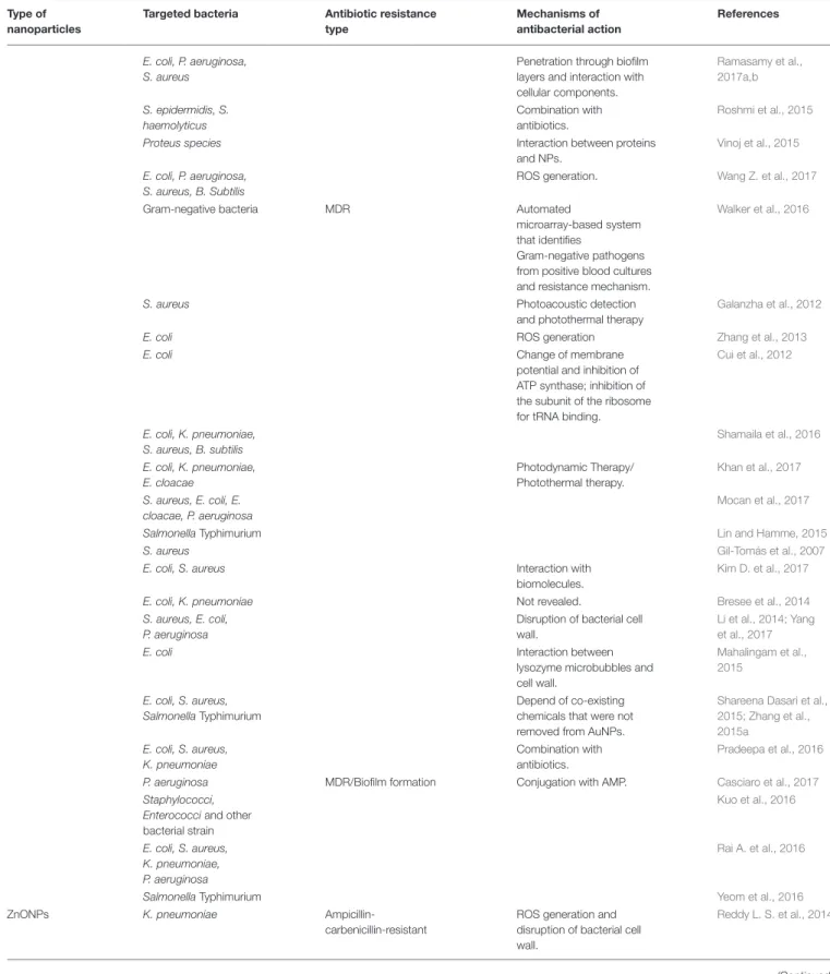
TABLE 1 |Continued
Type of
nanoparticles
Targeted bacteria Antibiotic resistance
type
Mechanisms of
antibacterial action
References
E. coli, P. aeruginosa,
S. aureus
Penetration through biofilm
layers and interaction with
cellular components.
Ramasamy et al.,
2017a,b
S. epidermidis, S.
haemolyticus
Combination with
antibiotics.
Roshmi et al., 2015
Proteus species Interaction between proteins
and NPs.
Vinoj et al., 2015
E. coli, P. aeruginosa,
S. aureus, B. Subtilis
ROS generation. Wang Z. et al., 2017
Gram-negative bacteria MDR Automated
microarray-based system
that identifies
Gram-negative pathogens
from positive blood cultures
and resistance mechanism.
Walker et al., 2016
S. aureus Photoacoustic detection
and photothermal therapy
Galanzha et al., 2012
E. coli ROS generation Zhang et al., 2013
E. coli Change of membrane
potential and inhibition of
ATP synthase; inhibition of
the subunit of the ribosome
for tRNA binding.
Cui et al., 2012
E. coli, K. pneumoniae,
S. aureus, B. subtilis
Shamaila et al., 2016
E. coli, K. pneumoniae,
E. cloacae
Photodynamic Therapy/
Photothermal therapy.
Khan et al., 2017
S. aureus, E. coli, E.
cloacae, P. aeruginosa
Mocan et al., 2017
SalmonellaTyphimurium Lin and Hamme, 2015
S. aureus Gil-Tomás et al., 2007
E. coli, S. aureus Interaction with
biomolecules.
Kim D. et al., 2017
E. coli, K. pneumoniae Not revealed. Bresee et al., 2014
S. aureus, E. coli,
P. aeruginosa
Disruption of bacterial cell
wall.
Li et al., 2014; Yang
et al., 2017
E. coli Interaction between
lysozyme microbubbles and
cell wall.
Mahalingam et al.,
2015
E. coli, S. aureus,
SalmonellaTyphimurium
Depend of co-existing
chemicals that were not
removed from AuNPs.
Shareena Dasari et al.,
2015; Zhang et al.,
2015a
E. coli, S. aureus,
K. pneumoniae
Combination with
antibiotics.
Pradeepa et al., 2016
P. aeruginosa MDR/Biofilm formation Conjugation with AMP. Casciaro et al., 2017
Staphylococci,
Enterococciand other
bacterial strain
Kuo et al., 2016
E. coli, S. aureus,
K. pneumoniae,
P. aeruginosa
Rai A. et al., 2016
SalmonellaTyphimurium Yeom et al., 2016
ZnONPs K. pneumoniae
Ampicillin-carbenicillin-resistant
ROS generation and
disruption of bacterial cell
wall.
Reddy L. S. et al., 2014
TABLE 1 |Continued
Type of
nanoparticles
Targeted bacteria Antibiotic resistance
type
Mechanisms of
antibacterial action
References
S. aureus Methicillin-resistant Enzyme inhibition. Cha et al., 2015
E. coli MDR ROS generation and
disruption of bacterial cell
wall.
Li et al., 2012b; Tong
et al., 2013;
Chakraborti et al.,
2014; Gelabert et al.,
2016; Nagvenkar et al.,
2016
B. subtilis Hsueh et al., 2015
S. aureus Lakshmi Prasanna and
Vijayaraghavan, 2015;
Nagvenkar et al., 2016
Vibrio cholerae Sarwar et al., 2016
S. aureus, E. coli,
Proteus, Acinetobacter,
P. aeruginosa
Combination with
antibiotics.
Ehsan and Sajjad, 2017
S. aureus, E. coli, S.
mutants
Depend on components
and structure of the bacteria
cell wall.
Yu et al., 2014
S. aureus, P. aeruginosa Biofilms formation ROS generation. Aswathanarayan and
Vittal, 2017
Streptococcus sobrinus Aydin Sevinç and
Hanley, 2010
CuONPs E. coli, S. aureus MDR ROS generation. Singh R. et al., 2014;
Chakraborty et al.,
2015
S. aureus, P. aeruginosa Ulloa-Ogaz et al., 2017
Paracoccus denitrificans Modulation of nitrogen
metabolism.
Su et al., 2015
S. aureus Biofilm formation Ongoing investigations. Chen et al., 2014
CuNPs S. aureus Methicillin-resistant Copper ions release and
subsequently bind with DNA
leading to disorder of helical
structure.
Kruk et al., 2015
P. aeruginosa Biofilm formation Penetrate the cell wall and
damage the nucleic acid.
LewisOscar et al., 2015
P. aeruginosa MDR Generation of Cu hydrosols. Zhang et al., 2015b
Fe3O4NPs E. coli MDR Radiofrequency (RF)
coupled with magnetic core
shell nanoparticles lead to
RF-mediated physical
perturbation of cell
membranes and bacterial
membrane dysfunction.
Chaurasia et al., 2016
S. aureus,
P. aeruginosa, E. coli
Penetrate the membrane
and interference in the
electron transfer.
El-Zowalaty et al., 2015
Gram-positive and
-negative bacteria
ROS generation. Behera et al., 2012
Gram-positive and
-negative bacteria
Nanotechnology to capture
Gram- positive and
-negative bacteria.
Reddy P. M. et al.,
2014
S. aureus Biofilm formation ROS generation. Leuba et al., 2013
Al2O3NPs S. aureus Methicillin-resistant Disruption of bacterial cell
wall and ROS generation.
Ansari et al., 2013
E. coli MDR Penetration and
accumulation inside
bacterial cell wall.
Ansari et al., 2014
TABLE 1 |Continued
Type of
nanoparticles
Targeted bacteria Antibiotic resistance
type
Mechanisms of
antibacterial action
References
TiO2NPs S. aureus Methicillin-resistant Release ions and react with
thiol group of proteins
present on bacteria surface.
Roy et al., 2010
E. coli MDR ROS generation and
disruption of bacterial cell
wall.
Li et al., 2012b
E. coliand
Gram-positive bacteria
Photocatalytic disinfection. Foster et al., 2011
E. coli Peroxidation and
decomposition of
membrane fatty acids.
Joost et al., 2015
Cu/Zn bimetal
NPs
S. aureus Methicillin-resistant Membrane disruption, DNA
damage, inhibition of protein
synthesis.
Ashfaq et al., 2016
Au/Ag
bimetallic NPs
Enterococcus Vancomycin-resistant Theranostic system for
SERS and aPDT.
Zhou et al., 2018
E. coli, S. aureus, E.
faecalis, P. aeruginosa
Biofilm formation Disruption of bacterial cell
wall and inactivate the
proteins and enzymes for
ATP production.
Ramasamy et al., 2016
B. subtilis E. coli,
K. pneumoniae,
S. aureus
MDR Combination with
antibiotics.
Baker et al., 2017
P. aeruginosa, E. coli,
S. aureus, Micrococcus
luteus
Fakhri et al., 2017
E. coli, S. aureus dos Santos et al., 2012
Au/Pt
bimetallic NPS
E. coli MDR Damage of the inner
membrane, increase
intracellular ATP level.
Zhao et al., 2014
Au/ Fe3O4NPs P. aeruginosa MDR Disruption of bacterial cell
wall.
Niemirowicz et al.,
2014
Cu/Ni
bimetallic NPs
S. aureus, E. coli,S.
mutans
MDR Adsorption of ions to the
bacteria cells.
Argueta-Figueroa et al.,
2014
MgF2NPs S. aureus Biofilm formation Attach and penetrate cell
surface leading to disruption
in membrane potential,
promotes the lipid
peroxidation and DNA
binding.
Lellouche et al., 2009;
Chen et al., 2014
Graphene
Oxide NPs
S. aureus Methicillin-resistant Combine antibiotics with
exposure to NIR.
Pan et al., 2016
E. coli, E. faecalis MDR UV irradiation lead to
generation of ROS.
Govindaraju et al.,
2016
E. coli, P. aeruginosa,
K. pneumoniae,
S. aureus
Multiple toxic mechanisms. Jankauskaite et al.,
2016
E. cloacae, S. mutans Biofilm formation ROS generation, release of
ions.
Kulshrestha et al., 2017
SeNPs S. aureus, E. coli MDR Theranostic nanoplatform
for selective imaging and
targeted therapy: Disruption
of the bacteria cell wall.
Huang et al., 2017
SiNPs S. aureus Methicillin-resistant Theranostics nanoprobe for
near-infrared fluorescence
imaging and photothermal
therapy: Disruption of the
bacteria cell wall.
peptidoglycans and abundant pores that allow the penetration
of foreign molecules, leading to covalent binding with proteins
and cellular components, interrupting the proper functioning
of the bacterial cell (
Sarwar et al., 2015
). In addition,
Gram-positive bacteria have a highly negative charge on the surface
of the cell wall. For example, LPS provides negatively charged
regions on the cell wall of Gram-negative bacteria that attracts
NPs; and, since teichoic acid is only expressed in Gram-positive
bacteria, the NPs are distributed along the phosphate chain. As
such, the antimicrobial effect is more foreshadowed in
Gram-positive than -negative bacteria (
Wang et al., 2017a
). Indeed,
Yu and colleagues synthesized a novel hydroxyapatite whisker
(HAPw)/zinc oxide (ZnO) NPs and evaluated the antimicrobial
effect against
S. aureus
,
E. coli,
and
Streptococcus mutans
. The
authors demonstrate that the antibacterial effect depends on
the components and structure of the bacterial cell wall. The
antibacterial action of these NPs could be improved for
Gram-positive bacteria and certain components could prevent the
adhesion of ZnO NPs to the bacterial cell barrier (
Yu et al.,
2014
). Ansari et al. reported that the accumulation on NPs in
the bacterial cell wall causes irregularly shaped pit, perforation
and disturbs metabolic processes (
Ansari et al., 2014
). In a study
carried out by Joost and co-workers, it was demonstrated that
a treatment with TiO2
NPs increased the bacterial cell volume,
resulting in membrane leakage (
Joost et al., 2015
).
BIOFILM FORMATION AND
QUORUM-SENSING
Biofilm formation plays an important role in bacterial resistance
protecting bacteria and allowing then to evade the action of
antibiotics (
Lebeaux et al., 2014; Khameneh et al., 2016
). The
most active fractions of bacteria are now recognized to occur as
biofilms, where cells are adhered to each other on surfaces within
a self-produced matrix of extracellular polymeric substance
(EPS). EPS provide a barrier allowing to inhibit the penetration
of antibiotics and further promote antibiotic resistance leading
to a serious health threat worldwide since biofilms are resistant
to antibiotics penetration and escape innate immune system
by phagocytes (
Hall-Stoodley et al., 2004; Bjarnsholt, 2013
).
Numerous experimental evidence show that NPs are capable
of disrupting the bacterial membranes and can hinder biofilm
formation thus reducing the survival of the microorganism
(
Peulen and Wilkinson, 2011; Leuba et al., 2013; Pelgrift and
Friedman, 2013; Slomberg et al., 2013; Chen et al., 2014; Miao
et al., 2016; Yu et al., 2016; Kulshrestha et al., 2017
). This way,
NPs provide an alternative strategy to target bacterial biofilms
with potential to use both antibiotic-free and antibiotic-coated
approaches (
Gu et al., 2003; Li et al., 2012a; Sathyanarayanan
et al., 2013
). Earlier reports demonstrated that NPs are able
to interfere with biofilm integrity by interacting with EPS and
with the bacterial communication - quorum sensing (QS). The
properties of NPs must be designed to be able to inhibit biofilm
formation namely through size and surface chemistry. The size
of NPs is important to it since they must be able to penetrate the
EPS matrix and surface chemistry will command the amount of
interactions with the EPS (
Lundqvist et al., 2008
). The majority
of the strategies to achieve inhibition of biofilm formation are to
target and interfere with QS molecules (
Singh et al., 2017
).
QS systems in bacterial populations act as major regulatory
mechanisms of pathogenesis, namely in the formation of
biofilm structures. These systems help bacteria to “communicate”
with each other, through the production and detection of
signal molecules (
Rutherford and Bassler, 2012; Papenfort and
Bassler, 2016
). Using this cell-to-cell communication, bacterial
populations are able to synchronize the expression of their
genes, acquiring competitive advantage to respond to changes
in the environment (
Rutherford and Bassler, 2012
). Therefore,
QS systems are known to promote the formation of antibiotic
tolerant biofilm communities. It is known that biofilm structures
are a recalcitrant mode of bacterial growth that increases
bacterial resistance to conventional antibiotics (
Reen et al., 2018
).
This way, bacterial biofilms pose a significant challenge to the
efficacy of conventional antibiotics being considered an essential
platform for antibiotic resistance (
Høiby et al., 2011
). Taking
this into account, it isn’t surprising that the targeting and
disruption of QS signaling systems and consequently, of the
biofilm production, set the pillar for future next-generation
anti-virulence therapies to be developed (
LaSarre and Federle, 2013;
Venkatesan et al., 2015; Jakobsen et al., 2017
).
Surface-functionalized NPs with
β
-cyclodextrin (
β
-CD) or
N-acylated homoserine lactonase proteins (AiiA) are able to
interfere with signaling molecules preventing these molecules
from reaching its cognate receptor, therefore inhibiting the
signal/receptor interaction. This process will “turn off” QS and
obstructing the bacterial communication (
Kato et al., 2006;
Ortíz-Castro et al., 2008
). Several papers reported inhibition of biofilm
formation namely by gold NPs (AuNPs). Acyl homoserine
lactones (AHL) are signaling molecules with a role in bacterial
QS and bind directly to transcription factors to regulate gene
expression Recently, Gopalakrishnan and colleges synthesized
(
Vinoj et al., 2015
) AuNPs coated AiiA purified from
Bacillus
licheniformis
. These AiiA AuNPs inhibited EPs production
and demonstrated potent antibiofilm activity against Proteus
species at 2–8
µ
M concentrations without being harmful for
the host cells at the 2
µ
M concentration.
Sathyanarayanan et al.
(2013)
demonstrated that using AuNPs there is a significant
reduction of
S. aureus
and
P. aeruginosa
biofilms applied in high
concentration (exceeding 50 mg/L). A recent study by
Yu et al.
(2016)
demonstrated that AuNPs were able to strongly attenuate
biofilm formation of
P. aeruginosa
. The inhibition observed in
this study was related with interruption of adhesin- mediated
interaction between the bacteria and the substrate surface due
to electrostatic attractions between the AuNPs and cell wall
surface of
P. aeruginosa
, instead of QS-related molecules. Positive
charge AuNPs inhibited significantly
S. aureus
and
P. aeruginosa
biofilm formation (while minimizing mammalian cytotoxicity)
(
Ramasamy et al., 2016
). The use of NPs demonstrates an
exclusive approach to penetrate infectious biofilms and target
bacterial communication, overcoming this major health issue
related with biofilm infections.
used by traditional antibiotics, combined therapeutic regimens
are promising strategies to tackle the surge of multidrug resistant
(MDR) bacteria bypassing their defense mechanisms (
Pelgrift
and Friedman, 2013; Singh K. et al., 2014; Hemeg, 2017; Zaidi
et al., 2017
). Additionally, NPs have been shown to activate
macrophages in a dose dependent manner (
Patel and Janjic, 2015
)
which promotes the host defenses (
Hemeg, 2017; Jagtap et al.,
2017
).
This multi-target action of NPs may overcome multidrug
resistance by circumventing several obstacles encountered by
traditional antibiotics (
Pelgrift and Friedman, 2013; Chen et al.,
2014; Singh K. et al., 2014; Hemeg, 2017; Jagtap et al., 2017;
Rai et al., 2017; Zaidi et al., 2017
).
Table 1
highlights several
types of NPs that have shown effective bactericidal activity
when administered isolated; combined with standard antibiotics;
and/or radiation or as vectors for biocidal delivery allowing
killing of MDR bacteria, and in some cases also inhibiting biofilm
production.
We will now focus on the different types of metal NPs
highlighting their most relevant mechanism/effects against MDR
bacteria and/or biofilms structures.
SILVER NANOPARTICLES (AGNPS)
Since the ancient times, silver has been recognized as having
antimicrobial effects (
Rai et al., 2009; Reidy et al., 2013
). Based
on all the evidence to date, AgNPs are probably one of the most
promising inorganic NPs that can be used for the treatment of
bacterial infections (
Natan and Banin, 2017
). These NPs may
be synthesized by traditional chemical reduction or
via
“green”
chemistry approaches using plant and/or microbial extracts
(
Iravani et al., 2014; Ribeiro et al., 2018
).
Several mechanisms have been proposed to understand
how AgNPs mediate cell death, including cell wall disruption
(
Lok et al., 2007; Bondarenko et al., 2013
), oxidation of
cellular components, inactivation of the respiratory chain
enzymes, production of ROS, and decomposition of the cellular
components (
Chen et al., 2014; Rizzello and Pompa, 2014;
Dakal et al., 2016
). The permeability of the membrane increases
after incorporation of AgNPs into the cell membrane. The
adsorption of the NPs leads to the depolarization of the cell
wall, altering the negative charge of the cell wall to become
more permeable. It was demonstrated disruption of the cell
wall with subsequent penetration of the NPs. The entry of
AgNPs induces ROS that will inhibit ATP production and
DNA replication (
Zhang et al., 2013; Dakal et al., 2016;
Durán et al., 2016; Ramalingam et al., 2016
). However, there
is evidence that AgNPs can release Ag
+
, known to exhibit
antimicrobial activity, when interacting with thiol-containing
proteins, which weaken their functions (
Durán et al., 2010
). The
precise method of the antibacterial mechanism of AgNPs is still
not completely understood (
Franci et al., 2015; Durán et al.,
2016
). All the existing data indicates that AgNPs exert several
bactericidal mechanisms in parallel, which may explain why
bacterial resistance to silver is rare (
Karimi et al., 2016
). Concerns
regarding the cytotoxicity and genotoxicity of AgNPs have
been raised (
Chopra, 2007
) but various authors have conducted
clinical trials based on AgNPs and no important clinical
alterations have been detected (
Munger et al., 2014a,b; Smock
et al., 2014
). Interestingly, AgNPs have been found to exhibit
higher antimicrobial activity than antibiotics like gentamicin or
vancomycin against
P. aeruginosa
and MRSA (
Saeb et al., 2014
).
Lara
et al
. showed the potential bactericidal effect of AgNPs
against MDR
P. aeruginosa
, ampicillin-resistant
E. coli
O157:H7
and erythromycin-resistant
Streptococcus pyogenes
(
Lara et al.,
2010
). Nagy
et al.,
reported that AgNPs were capable of inhibiting
the growth of
S. aureus
and
E. coli via
the up-regulation
of the expression of several antioxidant genes and ATPase
pumps (
Nagy et al., 2011
). Dolman
et al
. also showed that
the Ag-containing Hydrofiber
R
dressing and nanocrystalline
Ag-containing dressing are effective agents against antibiotic
sensitive Gram-negative and -positive bacteria as well as
antibiotic resistant bacteria, such as MRSA,
Vancomycin-resistant
Enterococci
(VRE) and
Serratia marcescens
, avoiding
the formation of biofilms on biomaterials (
Percival et al., 2007
).
Su and collaborators showed that AgNPs immobilized on the
surface of nanoscale silicate platelets (AgNP/NSPs) have strong
antibacterial activity against MRSA and silver-resistant
E. coli
via
generation of ROS (
Su et al., 2011
). Singh and collaborators
showed that AgNPs from
P. amarus
extract exhibited excellent
antibacterial potential against MDR strains of
P. aeruginosa
(
Singh K. et al., 2014
). Recently, two different shaped AgNPs
(spheres and rods) were used against Grampositive and
-negative bacteria, both showing promising antibacterial activity
against different strains (
Acharya et al., 2018
).
An emerging practice is to combine AgNPs with antibiotics
to enhance antimicrobial potency. Recently, Katya and
collaborators showed that the combination of gentamicin
and chloramphenicol with AgNPs has a better antibacterial
effect in MDR
E. faecalis
than both antibiotics alone (
Katva
et al., 2018
). McShan
et al
. described that AgNPs combined
with either one of two-different class of antibiotics (tetracycline
and neomycin) can exhibit a synergistic effect, showing an
enhanced antibacterial activity at concentrations below the
MIC of either the NPs or the antibiotic (
McShan et al., 2015
).
Other authors also reported similar results (
Thomas et al.,
2014; Panáˇcek et al., 2016a,b; Salomoni et al., 2017
). Djafari and
collaborators described the synthesis of water-soluble AgNPs
using the antibiotic tetracycline as co-reducing and stabilizing
agent (AgNPs@TC) and demonstrated their effectiveness against
tetracycline-resistant bacteria (
Djafari et al., 2016
).
membrane. Unfortunately, AMPs have poor enzymatic stability,
low permeability across biological barriers and may be rapidly
degraded in the human body by proteases, which greatly limits
their application (
Wang, 2014
). Immobilization of the peptides
onto NPs can increase their stability, enhancing the antimicrobial
properties of the NPs and therefore, has the potential to be used as
a new tool to tackle antibiotic resistant bacteria (
Brandelli, 2012;
Rai A. et al., 2016
). Indeed, the first author to demonstrate that
functionalized AgNPs with peptides increased their antibacterial
activity was Ruden and co-workers (
Ruden et al., 2009
). Based
on this strategy several researchers functionalized AgNPs with
AMPs (AgNP@AMP) with increases in the antimicrobial activity
compared with free AMPs (
Ruden et al., 2009; Liu et al.,
2013; Mohanty et al., 2013
). Polymyxin B is the most used
AMP and exhibits antibacterial activity
via
interaction with
the endotoxin LPS in the outer membrane of Gram-negative
bacteria (
Morrison and Jacobs, 1976; Lambadi et al., 2015
).
It was proved that AgNPs functionalized with polymyxin-B
removed almost completely endotoxins from solutions and
hindered the formation of biofilm onto surgical blades (
Jaiswal
et al., 2015; Lambadi et al., 2015
). Liu
et al.,
demonstrated
that the immobilization of peptides with AgNPs enhanced their
antimicrobial activity compared to an unbound peptide and also
minimized toxicity of AgNPs compared to using the AgNPs
alone (
Liu et al., 2013
). A recent study by Pal
et al
. describes
a system consisting of a cysteine containing AMP conjugated
with AgNPs, which demonstrated that the Ag-S bonds increased
stability and enhanced antimicrobial activity than conjugation
using electrostatic interactions (
Pal et al., 2016
).
Other methods have been used to improve the antibacterial
activity of AgNPs. One of these methods relies on the use
of visible blue light, which was previously shown to exhibit
strong antibacterial activity (
Dai T. et al., 2013; Maclean
et al., 2014
). El Din and collaborators demonstrated that
blue light combined with AgNPs exhibits therapeutic potential
to treat MDR infections and can represent an alternative
to conventional antibiotic therapy, since the antimicrobial
activity of the combination was greater than the components
alone. Moreover, this approach proved to be synergistic in
the treatment of an unresponsive antibiotic-resistant bacteria
responsible for a wound in a horse (
El Din et al., 2016
).
Spherical shaped thioglycolic acid-stabilized AgNPs
(TGA-AgNPs) conjugated with vancomycin were used as drug delivery
systems and demonstrated to possess increased antimicrobial
activity against MDR bacteria such as MRSA and VRE
(
Esmaeillou et al., 2017
).
GOLD NANOPARTICLES (AuNPs)
Metallic gold is considered inert and non-toxic, which may vary
when it shifts form metallic bulk to oxidation states (I and
II) (
Merchant, 1998
). Gold NPs (AuNPs) may be synthesized
by traditional chemical reduction of a gold salt or
via
“green”
chemistry approaches using plant and/or microbial extracts
(
Shah et al., 2014
). The most used and described method is
the chemical synthesis based on the reduction of chloroauric
acid by citrate (
Lee and Meisel, 1982; Fernandes and Baptista,
2017
). Some studies have addressed the potential of using AuNPs
as antibacterial agents, but some controversy still exists (
Cui
et al., 2012; Bresee et al., 2014; Shah et al., 2014; Shareena
Dasari et al., 2015; Zhang et al., 2015a; Shamaila et al.,
2016
).
According to Yu H and collaborators, AuNPs are usually not
bactericidal at low concentrations and weakly bactericidal at high
concentrations (
Shareena Dasari et al., 2015; Zhang et al., 2015a
).
This is possibly due to the effect of co-existing chemicals, such
as gold ions, surface coating agents, and chemicals involved in
the synthesis that were not completely removed (
Shareena Dasari
et al., 2015; Zhang et al., 2015a
). However, other authors describe
that the antibacterial mechanism of AuNPs is associated to (i) the
collapse in the membrane potential, hindering ATPase activity
causing a deterioration of the cell metabolism; (ii) hindering of
the binding subunit of the ribosome to tRNA (
Cui et al., 2012
);
and (iii) Shamaila and co-workers showed that AuNPs may
affect the bacterial respiratory chain by attacking nicotinamide
(
Shamaila et al., 2016
). Since AuNPs are non-toxic to the host
(
Conde et al., 2014; Li et al., 2014; Rajchakit and Sarojini, 2017
),
the possibility of fine tuning their conjugation chemistry to act
as carriers or delivery vehicles of antibiotics or other antibacterial
moieties may enhance their bactericidal effect and potentiate the
effect of antibiotics (
Zhao and Jiang, 2013; Conde et al., 2014;
Li et al., 2014; Uma Suganya et al., 2015; Zhang et al., 2015a;
Fernandes et al., 2017
).
Cationic and hydrophobic functionalized AuNPs were
shown to be effective against both Gramnegative and
-positive uropathogens, including MRSA. These AuNPs exhibited
low toxicity to mammalian cells (biocompatibility) and the
development of resistance to these NPs was very low (
Li
et al., 2014
). Vinoj
et al
. demonstrated that coating AuNPs
with N-acylated homoserine lactonase proteins (AiiA AuNPs)
resulted in a nanocomposite with activity against MDR species
compared with AiiA proteins alone (
Vinoj et al., 2015
). Other
approaches were also studied, as adsorbing AuNPs to
PVA-lysozyme micro bubbles potentiate the antibacterial activity due
to the interaction of AuNPs with cells membranes causing
bacterial lysis (
Mahalingam et al., 2015
). Galic acid capped
AuNPs have also been found to be active against Gram-negative
and -positive bacteria (
Kim D. et al., 2017
). Recently, Ramasamy
and collaborators described the direct one-pot synthesis of
cinnamaldehyde immobilized on gold nanoparticles (CGNPs)
with effective biofilm inhibition of more than 80% against
Gram-positive bacteria (methicillin-sensitive and -resistant strains of
S. aureus
, MSSA and MRSA, respectively) and Gram-negative
(
E. coli
and
P. aeruginosa
)
in vitro
and
in vivo
(
Ramasamy
et al., 2017a,b
). Also, the integration of AuNPs with ultrathin
graphitic carbon nitride was described as having high bactericidal
performance against both MDR Gram-negative and -positive
bacteria, and a high effectiveness in eliminating existing
MDR-biofilms and preventing the formation of new MDR-biofilms
in vitro
and collaborators develop a single-step synthesis of
kanamycin-capped AuNPs (Kan-AuNPs) with high antibacterial activity
against both Gram-positive and -negative bacteria, including
kanamycin resistant bacteria. The authors observed a significant
reduction in the MIC against all the bacterial strains tested
for Kan-AuNPs when compared to the free drug. This higher
efficacy was due to the disruption of the bacterial envelope,
resulting in leakage of the cytoplasmic content and consequent
cell death (
Payne et al., 2016
). Pradeepa and collaborators
synthesized AuNPs with bacterial exopolysaccharide (EPS) and
functionalized them with antibiotics (levofloxacin, cefotaxime,
ceftriaxone and ciprofloxacin). They observed that these AuNPs
exhibited excellent bactericidal activity against MDR
Gram-positive and -negative bacteria compared to free drugs.
E. coli
was the most susceptible MDR bacteria followed by
K. pneumoniae
and
S. aureus
(
Pradeepa et al., 2016
). Recently,
Yang and collaborators described the effect of small molecule
(6-aminopenicillanic acid, APA)-coated AuNPs to inhibit MDR
bacteria (
Yang et al., 2017
). They doped AuNPs into electrospun
fibers of poly(
ε
-caprolactone) (PCL)/gelatin to produce materials
that avoid wound infection by MDR bacteria and demonstrated
in vitro
and
in vivo
that APA-AuNPs reduced MDR bacterial
infections (
Yang et al., 2017
). Shaker and Shaaban evaluated
the surface functionalization of AuNPs with carbapenems
[imipenem (Ipm) and meropenem (Mem)] as a delivering
strategy against carbapenem resistant Gram-negative bacteria
isolated from an infected human. Both Ipm-AuNPs and
Mem-AuNPs, with 35 nm diameter showed a significant increase in
antibacterial activity against all the tested isolates (
Shaker and
Shaaban, 2017
). Also, Shaikh and collaborators described recently
the synthesis and characterization of cefotaxime conjugated
AuNPs to target drug-resistant CTX-M-producing bacteria. The
authors could invert resistance in cefotaxime resistant bacterial
strains (i.e.,
E. coli
and
K. pneumoniae
) by using
cefotaxime-AuNPs. Their results reinforce the efficacy of conjugating
an unresponsive antibiotic with AuNPs to restore its efficacy
against otherwise resistant bacterial pathogens (
Shaikh et al.,
2017
).
Combination of AuNPs with other approaches has also
been demonstrated. Indeed, one of the most extraordinary
properties of AuNPs is the capability to transform light into
heat under laser irradiation (
Mendes et al., 2017; Mocan
et al., 2017
). This property is extremely important because
it can be exploited to develop photothermal nanovectors to
destroy MDR bacteria at a molecular level (for a complete
review see
Mocan et al., 2017
). For example, Khan and
collaborators showed that the combination of Concanavalin-A
(ConA) directed dextran capped AuNPs (GNPDEX-ConA)
conjugated with methylene blue (MB) (MB@GNPDEX-ConA)
and photodynamic therapy (PDT) enhanced the efficacy and
selectivity of MB induced killing of MDR clinical isolates,
including
E. coli
,
K. pneumoniae
, and
E. cloacae
(
Khan
et al., 2017
). Gil-Tomas and collaborators described that the
functionalization of AuNPs covalently with toluidine blue O–
tiopronin forms an enhanced, exceptionally potent antimicrobial
agent when activated by white light or 632 nm laser light
(
Gil-Tomás et al., 2007
). Hu and collaborators prepared a
mixed charged zwitterion-modified AuNPs consisting of a weak
electrolytic 11-mercaptoundecanoic acid (HS-C10-COOH) and
a strong electrolytic (10-mercaptodecyl)trimethylammonium
bromide (HS-C10-N4) that exhibited
in vivo
and under
near-infrared (NIR) light irradiation an enhanced photothermal
ablation of MRSA biofilm with no damage to the healthy tissues
around the biofilm (
Hu et al., 2017
). Also, the antibacterial
activity of glucosamine-gold nanoparticle-graphene oxide
(GlcN-AuNP-GO) and UV-irradiated GlcN-AuNP-GO was
evaluated against
E. coli
and
E. faecalis
. Results show that UV
irradiation of GlcN-AuNP-GO results in higher antibacterial
activity than the standard drug kanamycin (
Govindaraju et al.,
2016
). Ocsoy
et al
. reported the development of DNA
aptamer-functionalized AuNPs (Apt@AuNPs) and gold nanorods
(Apt@AuNRs) for inactivation of MRSA with targeted PTT
(
Ocsoy et al., 2017
). The authors showed that although both
NPs could specifically bind to MRSA cells, Apt@AuNPs and
Apt@AuNRs increased resistant cell death for 5% and for more
than 95%, respectively through PTT. This difference in induction
of cell death was based on the relatively high longitudinal
absorption of NIR radiation and strong photothermal conversion
capability for the Apt@AuNRs compared to the Apt@AuNPs.
However, with the new developments of using AuNPs for
hyperthermia in the visible light (
Mendes et al., 2017
) might
additionally potentiate the Apt@AuNPs results observed for
these authors (
Ocsoy et al., 2017
). Recently, Mocan
et al
. also
described the synthesis of AuNPs by wet chemistry, their
functionalization with IgG molecules following laser irradiation.
Their results indicate that administration of IgG-AuNPs
following laser irradiation provided an extended and selective
bacterial death in a dose dependent manner (
Mocan et al.,
2016
).
In recent years, a new approach relying on the conjugation of
AuNPs with AMPs has shown interesting results (
Rajchakit and
Sarojini, 2017
). Indeed, Kuo and collaborators mixed
synthetic-peptides containing arginine, tryptophan and cysteine termini
[(DVFLG)2REEW4C and (DVFLG)2REEW2C], with aqueous
tetrachloroauric acid to generate peptide-immobilized AuNPs
[i.e., (DVFLG)2REEW4C-AuNPs and
(DVFLG)2REEW2C-AuNPs] that were effective antibacterial agents against
glycol) carboxylic acid (PEGCOOH) covalently bound to AMP
showed a significantly increase of the antibacterial and
anti-biofilm activity for resistant Gram-negative bacteria (
Casciaro
et al., 2017
). Yeom and co-workers demonstrated the most
advanced
in vivo
clinical application for AuNPs@AMP using
infected mice and resulting in the inhibition of
Salmonella
Typhimurium colonization in the organs of the animals (
Yeom
et al., 2016
). The reason behind the increased antimicrobial
activity of AuNPs@AMP over the free components is that
AuNPs can get a higher concentration of the antibiotic at the
site of action. These NPs can interact with LPS, proteins in
the membrane of the bacteria and in some cases, penetrate
the bacterial membrane through the porin channel. This
way they can interact with the inner membrane making the
AuNPs@AMP conjugate more efficient than the non-conjugated
form (
Katz and Willner, 2004; Wangoo et al., 2008; Chen J. et al.,
2009
).
BIMETALLIC NPS
Ag and Au may be used in a single NP to enhance the effects
of a drug and reduce the required dose. Alternatively, they
can be used alone since they possess antimicrobial properties
that are enhanced when combined in the form of bimetallic
NPs (
Arvizo et al., 2010; Singh R. et al., 2016
). The role of
Ag against MDR pathogens has been previously described.
However, AgNPs are difficult to functionalize with biomolecules
and drugs. Such limitation may be circumvented by means of
alloy/bimetallic NPs that excel their monometallic counterparts
providing improved electronic, optical and catalytic properties
(
Cho et al., 2005; Shah et al., 2012
). As reported above,
AuNPs constitute good vectors to the delivery of pharmacologic
compounds. Gold(Au)-silver(Ag) alloys are an optimal solution
since they combine the antimicrobial effect of silver with the
ease of functionalization and improved stability in complex
biological media provided by gold (
Doria et al., 2010; dos
Santos et al., 2012
). Fakhri and co-workers synthetized and
functionalized AgAuNPs with a tetracycline and concluded
that there exists a synergetic effect of the antibiotic with the
bimetallic nanoparticle, with greater bactericidal activity of
this form in detriment of its free forms. The mechanism of
action was established as being the generation of ROS (
Fakhri
et al., 2017
). Also recently, Baker and collaborators described
the synthesis and antimicrobial activity of bimetallic AgAuNPs
from the cell free supernatant of
Pseudomonas veronii
strain
AS41G inhabiting
Annona squamosa L
. The authors showed
their synergistic effect with standard antibiotics with 87.5,
18.5, 11.15, 10, 9.7, and 9.4% fold increased activity with
bacitracin, kanamycin, gentamicin, streptomycin, erythromycin
and chloramphenicol, respectively, against bacitracin resistant
strains of
Bacillus subtilis
,
E. coli,
and
K. pneumoniae
(
Baker
et al., 2017
). Zhao and collaborators have demonstrated the
antibacterial activity of AuPtNPs bimetallic NPs against sensitive
and drug-resistant bacteria
via
the dissipation of the bacterial
membrane potential and the elevation of adenosine triphosphate
(ATP) levels (
Zhao et al., 2014
).
Other types of bimetallic NPs have been studied and their
antibacterial activity explored, but in most cases as coating
agents and not as a delivery approach and antibacterial activity
(
Argueta-Figueroa et al., 2014
).
METAL OXIDES
Metal oxides NPs are among one of the most explored and
studied family of NPs and are known to effectively inhibit
the growth of a wide range of sensitive and resistant
Gram-positive and -negative bacteria, emerging as hopeful candidates
to challenge antimicrobial resistance (
Raghunath and Perumal,
2017; Reshma et al., 2017; Kadiyala et al., 2018
). Iron oxide
(Fe3O4), Zinc oxide (ZnO), and Copper oxide (CuO) possess
antimicrobial properties and can be applied in clinical care
(
Sinha et al., 2011
). Due to the intrinsic photocatalytic activity
of the metal oxides they generate ROS and become powerful
agents against bacteria (
Tong et al., 2013; Singh R. et al.,
2014
). These will be described in more detail on the following
sections.
IRON OXIDE (FE
3
O
4
)
The synthesis of iron oxide NPs may be achieved
via
different
routes (
Babes et al., 1999; Berry and Curtis, 2003
). The
antibacterial mechanism of these NPs is mainly attributed to
dissolved metal ions and the generation of ROS (
Wang et al.,
2017a
). It was shown that superparamagnetic iron oxide NPs
interact with microbial cells by penetrating the membrane and
interfering with the electron transfer (
Behera et al., 2012;
El-Zowalaty et al., 2015
). Additionally, it has been described that
iron oxide NPs can damage macromolecules, including DNA
and proteins, through the formation of ROS (
Leuba et al.,
2013
). Pan
et al
. developed a system of reduced graphene oxide
(rGO)-iron oxide nanoparticles (rGO-IONP) by the chemical
deposition of Fe
2+
/Fe
3+
ions on nanosheets of rGO in aqueous
ammonia. The
in vivo
results showed maximum antibacterial
activity due to the generation of hydroxyl radicals that can
cause physical and chemical damage, which inactivated MRSA
(
Pan et al., 2016
).
ZINC OXIDE (ZnO)
ZnO NPs are often used to restrict microorganism growth,
being effective against planktonic bacteria, and also inhibiting
the formation of biofilms (
Hsueh et al., 2015; Sarwar et al.,
2016
) (
Espitia et al., 2012
). These NPs can be synthesized
by various methods, from green chemistry to sonochemistry
(
Salem et al., 2015; Ali et al., 2016; Nagvenkar et al., 2016
).
The antibacterial mechanism of the NPs is partially attributed
to two principal factors, the dissolution of metal ion and the
generation of ROS (
Gelabert et al., 2016; Nagvenkar et al., 2016;
Sarwar et al., 2016
). ZnO releases Zn
2+
in liquid medium and