Article
J. Braz. Chem. Soc., Vol. 23, No. 11, 2011-2015, 2012. Printed in Brazil - ©2012 Sociedade Brasileira de Química 0103 - 5053 $6.00+0.00
A
*e-mail: [email protected]
A Sensitive Fluorescent Assay for Trypsin Activity in Biological Samples
using BSA-Au Nanoclusters
Xianxiang Wang,* Yanying Wang, Hanbing Rao and Zhi Shan
College of Life and Science, Sichuan Agricultural University, 625014 Ya’an, Sichuan, P. R. China
Neste artigo, foi desenvolvido um novo método fluorométrico, simples, sensível e seletivo, para a medição de tripsina em amostras biológicas. O método foi baseado na medição da supressão da intensidade da fluorescência de agregados de nanopartículas de ouro estabilizados com albumina sérica bovina (BSA), pela proteólise enzimática. A curva de calibração para tripsina foi atingida no intervalo de concentração de 1-60 nmol L-1 com coeficiente de correlação 0,995 e um limite de
detecção de 0,6 nmol L-1. O método foi também usado satisfatoriamente para avaliação da atividade
da tripsina e os resultados mostraram que os valores das constantes de Michaelis-Menten (Km)
e catalítica (Kcat), de tripsina para estes substratos de nanoagreagados BSA-Au, foram 1,6×10 -5
mol L-1 e 3,8 s-1 a 37 oC, respectivamente. Este biossensor enzimático é de interesse considerável
devido à simplicidade do procedimento, e o método estabelecido tem grande potencial na detecção de outras proteases em diagnósticos clínicos de várias doenças.
A novel, simple, sensitive, and selective fluorometric method was developed for measuring trypsin in biological samples in this article. The method was based upon measuring the quenching of the fluorescence intensity of the bovine serum albumin (BSA) stabilized Au nanoclusters by enzymatic proteolysis. The calibration plot for trypsin was achieved over the concentration range 1-60 nmol L-1 with a correlation coefficient of 0.995 and a limit of detection of 0.6 nmol L-1.
The method was also used satisfactorily for the assessment of the trypsin activity and the results showed that the Michaelis-Menten (Km) and catalytic (Kcat)constant values of trypsin for BSA-Au
nanoclusters substrate were 1.6×10⁻⁵ mol L-1 and 3.8 s-1 at 37 oC, respectively. This enzyme biosensor
is of considerable interest due its promise for simple procedure and the established method has great potential in detection of other proteases in clinical diagnostics of various diseases.
Keywords:BSA-Au nanoclusters, trypsin, fluorescence, enzyme activity
Introduction
Trypsin is a catalyst for the hydrolysis of amide bonds in peptides and proteins, which plays an important role in numerous important physiological processes such as protein turnover, digestion, cell differentiation and
growth, immunological defense, cancer, and apoptosis.1
Consequently, the monitoring of trypsin activity plays a central role in biochemical, pharmaceutical, and in the clinical application. The most common methods for
detection degradative enzyme activity utilize fluorogenic2
or chromogenic substrates3 or fluorescence resonant
energy transfer-based substrates4 or surface acoustic
wave-impedance sensor.5 Unfortunately, these methods
require considerable sample preparation and sophisticated
instrumentation, thereby making the assays time-consuming, costly and error-prone. Furthermore, these methods do not meet the performance parameters necessary for rapid detection of low levels of enzyme activity directly in biological samples, making them unsuitable for point-of-case diagnostic applications.
To overcome these difficulties, it is important to develop a more efficient and simple method for monitoring the trypsin activity in biological samples. Among the reported methods for detecting enzyme activity, fluorometric analysis offers apparent advantage over other methods, such
as electrophoretic method,6,7 by virtue of their sensitivity
necessary. Bovine serum albumin (BSA) is used generally as a model protein substrate to study the proteases activity, but these methods exclude fluorometric analysis because itself has no fluorophore. Recently, natural and efficient biosynthesis of the unique fluorescent nanomaterials have
gained increasing interest for many scientists.8-10 Among
fluorescent nanomaterials, BSA-templated Au nanoclusters are of particular interest due to their facile synthesis, red
emission, high quantum yield and good biocompatibility.10
The prepared BSA-Au nanoclusters formed in the BSA solution could have been stabilized by a combination of
Au-S bonding with the protein (via the 35 Cys residues in
BSA), and the steric protection due to the bulkiness of the protein. If the surrounding BSA biomolecules is destroyed by other reagents, the fluorescence intensity of the Au nanoclusters will be decreased, for example, the cases of
Cu2+ and glutathione (GSH)-Au nanoclusters,11 or hydrogen
peroxide and BSA-Au nanoclusters.12 Since trypsin is an
efficient hydrolase for BSA digestion, the structure of the BSA-Au nanoclusters will be destroyed in the present of trypsin. Therefore, this enzymatic proteolysis reaction will decrease the fluorescence of BSA-Au nanoclusters. Inspired by this observation, we herein used originally the fluorescent BSA-Au nanoclusters as a probe to measure the trypsin activity in biological samples. Furthermore, this assay is simple, fast and sensitive.
Experimental
Chemicals and apparatus
Bovine serum albumin (BSA) and trypsin from
bovine pancreas (T1426, activity about 10,000 units per
mg proteins) were bought from Sigma-Aldrich (China).
HAuCl4.3H2O and other salts were purchased from Kelong
Reagent Co., Chengdu, China. All other chemicals, such as sodium hydroxide, were of analytical grade and used without further purification. All solutions were prepared with water purified by a Milli-Q purification system (Millipore, USA).
Fluorescence measurements were conducted with a Thermo Electron Varioskan (Thermo Fisher Scientific Inc.) spectral multimode reader using 384-well fluorescence plates, the excitation and emission wavelengths is 370 nm and 621 nm, respectively. UV-Vis absorption spectra were recorded by using a SHIMADZU UV-Vis 2450 spectrophotometer (Japan). The molecular weights of BSA and BSA-Au nanoclusters were analyzed with matrix-assisted laser desorption/ionization-time-of-flight (MALDI-TOF) mass spectrometry on a Bruker Daltonics Autoflex II TOF/TOF system (Germany). PHS-3E pH
meter (Shanghai Feile Co., Ltd., China) was used during the preparation of buffers.
Synthesis of red fluorescent BSA-Au nanoclusters
Red fluorescent BSA-stabilized Au nanoclusters was synthesized in aqueous solution following a previous
reported method.10 In a typical experiment, all glassware
used in the experiments were cleaned in a bath of freshly
prepared aqua regia (HCl:HNO3, 3:1, v/v), and rinsed
thoroughly in water prior to use. HAuCl4 solution (15 mL,
10 mmol L-1, 37 °C) was added to BSA solution (15 mL,
50 mg mL-1, 37 °C) under magnetic stirring. Then, NaOH
(1 mol L-1, 1.5 mL) solution was introduced and the mixture
was allowed to incubate at 37 °C under vigorous magnetic stirring for 24 h. The colour of the solution changed from light yellow to deep brown. The solution was then dialyzed in double distilled water for 48 h to remove unreacted
HAuCl4 or NaOH. The final solution was stored at 4 °C in
refrigerator when not in use.
Procedures for trypsin activity assay
Trypsin activity assays were conducted at 30 oC
with 0.1 mmol L-1 BSA-Au nanoclusters substrate in
10 mmol L-1 phosphate buffered saline (abbreviated PBS),
pH 7.2, and 100 mmol L-1 NaCl. A 50 µL aliquot of BSA-Au
nanoclusters was added and incubated for 10 min, and then the fluorescence intensity was recorded by Thermo Electron Varioskan spectral multimode reader using 384-well fluorescence plates. Next, an aliquot of trypsin solution was added, and the fluorescence intensity was recorded automatically at set intervals. Then, the fluorescence intensities were converted to substrate concentration as a function of time by using equation 1, which is derived from the Stern-Volmer equation.
[Qt] = [Q0] (1 – It /I0) (1)
In equation 1, [Qt] is the BSA-Au nanoclusters
concentration at time t, [Q0] is the initial BSA-Au
nanoclusters concentration, It is the fluorescence intensity
at time t after trypsin addition and I0 is the initial
fluorescence intensity of the BSA-Au nanoclusters. From the slope of this plot in the equation 1, the activity of the enzyme was calculated in the usual way. The
Michaelis-Menten (Km) and catalytic (Kcat) constant values were
determined by Lineweaver-Burk regression. Kcat is defined
as Vmax/[enzyme].
hospital. The plasma were used directly and diluted 100 times with ultrapure water before analysis. 100 µL of
1 µmol L-1 BSA-Au nanoclusters and 900 µL diluted plasma
solution were mixed thoroughly, and left for another 10 min to measure the fluorescence. Then, an aliquot of trypsin were added in the mixed solution and left for another 5 min to detect the fluorescence intensity.
Results and Discussion
Characterization of BSA-Au nanoclusters
The aqueous solution of as-prepared BSA-Au nanoclusters was deep brown in color and exhibited bright-red fluorescence under ultraviolet light. The spectral characteristics of the BSA-Au nanoclusters are consistent
with the literature.10 The absorbance and emission spectra
of the BSA-Au nanoclusters (Figure 1) show that the emission spectrum of the BSA-Au nanoclusters displayed about 620 nm upon excitation at 370 nm. Referred to
previously studies, BSA-Au nanoclusters were formed in
situ by reduction the entrapped Au ions with the activated
BSA molecules, and the Au nanoclusters formed in the BSA solution could have been stabilized by a combination of Au-S bonding with the protein (via the 35 Cys residues in BSA). MALDI-TOF mass spectrometry (Figure 2) confirmed that the BSA molecular weight was about 66 kDa, and the prepared BSA-Au nanoclusters showed a peak shift of about 5 kDa, which was attributed to the 25 gold atoms in the nanoclusters, and the structure is highly stability, and corresponded to the most common magic cluster size.
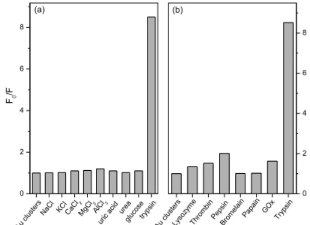
Effect of foreign species and other proteases
The validity and selectivity of the developed fluorescence method was tested by studying the influence of a series of potentially interfering species co-existed in biological
samples, e.g., NaCl, KCl, CaCl2, MgCl2, and AlCl3, uric
acid, urea and glucose. The concentration of these foreign
species was 100-fold than the trypsin (1 mmol L-1) in
PBS buffer of pH 7.4 at 10 min contact time at 37 oC
with 1 mmol L-1 BSA-Au nanoclusters. Figure 3a reveals
that no significant change on the fluorescence intensity of BSA-Au nanoclusters in the presence of the tested interfering species except trypsin. The interaction between BSA-Au nanoclusters and other proteases was also studied. Figure 3b showed that no obvious decrease in fluorescence intensity by adding other proteases, such as lysozyme, thrombin, pepsin, bromelain, papain and glucose oxidase (GOx). These results demonstrated that the enzymolysis of the BSA-Au nanoclusters by trypsin specifically caused the decrease in fluorescence intensity, and the BSA-Au nanoclusters could be used as a fluorescent probe for the sensitive detection of trypsin with minor interference from other proteases.
Figure 1. Absorption and fluorescence spectra of BSA-Au nanoclusters. All solutions were prepared using PBS buffer solution at pH 7.4.
Figure 2. MALDI-TOF mass spectra of BSA and BSA-Au nanoclusters.
Figure 3. (a) Effect of foreign species and (b) other proteases toward 1 mmol L-1 BSA-Au nanoclusters by monitoring the relative fluorescence
intensity F0/F (where F0 and F is the fluorescence intensity in the absence and in the presence of foreign species, respectively). The foreign species and other proteases concentrations were as follows: 100 mmol L-1
(NaCl, KCl, CaCl 2, MgCl2, AlCl3, uric acid, urea and glucose, lysozyme,
Trypsin activity assays
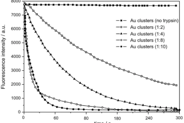
The BSA-stabilized Au nanoclusters can be hydrolyzed by trypsin. Figure 4 shows that the fluorescence intensity of prepared BSA-Au nanoclusters remains stable due to high resistance to photobleaching, which decreased only about 2% within 5 min. However, with the addition of trypsin, a concentration dependent decrease in fluorescence is observed, and more enzymes were favorable to enzymatic proteolysis.
From the time-course experiment (Figure 5a), we found that the rate of hydrolysis increased linearly with time of incubation for more than 1 min when the trypsin
concentration was 0.001 mmol L-1 of reaction solution and
BSA-Au nanoclusters concentration was 0.025 mmol L-1.
Simultaneously, linearity was observed between relative
fluorescence intensity (F0/F) and the trypsin concentration
over the range of 1 to 60 nmol L-1 (Figure 5b), with a
correlation coefficient of 0.995. The limit of detection
(LOD) is defined by the equation LOD = (3σ/k), where σ
is the standard deviation of blank measurements (n = 11) and k is the slope of calibration graph. Here LOD was
0.6 nmol L-1. So, this novel approach has potential to fast
and quantitatively detect trypsin in biological samples. Kinetic constants were derived from a double-reciprocal plot by the Lineweaver-Burk method and are based on the average of triplicate determinations. Figure 6 showed that
the Km and Kcat values for the hydrolysis of as-prepared
BSA-Au nanoclusters were 1.6×10⁻⁵ mol L-1 and 3.8 s-1 at
37 oC. The K
m value was much smaller than other reported
fluorogenic substrates,13,14 indicating that trypsin has high
affinity to the BSA-Au nanoclusters as compared with other substrates.
Analytical applications
In order to demonstrate the potential application of the proposed method for trypsin assays in clinical diagnosis, the biological samples were prepared by adding fixed amount of trypsin into human blood plasma taken from four healthy volunteers. The results are shown in Table 1. The accuracy of the method was investigated by recovery studies. The
Table 1. Analytical results (mean ± s; n = 3) for the determination of trypsin in healthy human blood plasma
Sample Trypsin added / (nmol L-1)
Trypsin found / (nmol L-1)a
Recovery / %
plasma 1 0 -
-plasma 2 5 4.9 ± 0.02 98
plasma 3 10 9.7 ± 0.04 97
plasma 4 15 14.8 ± 0.04 99
aMean of three determinations.
Figure 4. Fluorescence decay of BSA-Au clusters with variable amounts of trypsin.
Figure 5. (a) Hydrolysis of BSA-Au nanoclusters by trypsin. Trypsin concentration was 0.001 mmol L-1 of reaction solution and BSA-Au
nanoclusters concentration was varied with 0.005, 0.0125 and 0.025 mmol L-1. (b) F
0/F upon the interaction of BSA-Au nanoclusters
with different amount of trypsin. The inner graph shows the linear plot that reveals detectable concentration range of 1-60 nmol L-1 and the linear
coefficient is 0.995, where F0 and F are the relative fluorescence intensities
of BSA-Au nanoclusters in the absence and in the presence of trypsin with incubation at 37 oC for 2 min.
recoveries of spiked trypsin in the diluted human blood plasma ranged from 97 to 99%, thus demonstrating the potential applicability of the BSA-Au nanoclusters as fluorescence probe for the quantitative detection of trypsin in biological samples.
Conclusions
In summary, a sensitive, selective and simple fluorescent method for detection of trypsin activity using BSA-Au nanoclusters was developed. The mechanism is based on enzymolysis of the BSA-Au nanoclusters templates. A linear calibration plot for trypsin is in the range of 1 to
60 nmol L-1 with a limit of detection of 0.6 nmol L-1, and the
Km and Kcat values are 1.6×10⁻⁵ mol L-1 and 3.8 s-1 at 37 oC,
respectively. Furthermore, the present fluorescent method is also promising for the detection of other proteases by using target-specific peptide stabilized fluorescent metal nanoclusters.
Acknowledgments
This work was financially supported by the Science and Technology Department of Sichuan Province (N. 2011JY0134) and the Education Department of Sichuan Provincial (N.10ZC086).
References
1. Turk, B.; Nat. Rev. Drug Discovery 2006, 5, 785. 2. Farmer, W. H.; Yuan, Z.; Anal. Biochem. 1991, 197, 347. 3. Baustert, J. H.; Wolfbeis,O. S.; Moser, R.; Koller, E.; Anal.
Biochem. 1988, 171, 393.
4. Matayoshi, E.; Wang, G.; Krafft, G.; Erickson, J.; Science 1990, 247, 954.
5. Cai,Q.Y.; Wang, R. H.; Wu, L. Y.; Nie, L. H.; Yao, S. Z.; Microchem. J. 1997, 55, 367.
6. Lefkowitz, R. B.; Marciniak, J. Y.; Hu, C. M.; Schmid-Schonbein, G. W.; Heller, M. J.; Electrophoresis 2010, 31, 403. 7. Shimura, K.; Matsumoto, H.; Kasai, K.; Electrophoresis 1998,
19, 2296.
8. Eisenstein, M.; Nat. Methods 2005, 2, 6.
9. Cui, R.; Liu, H. -H.; Xie, H. Y.; Zhang, Z. L.; Yang, Y. R.; Pang, D. W.; Xie, Z. X.; Chen, B. B.; Hu, B.; Shen, P.; Adv. Funct. Mater. 2009, 19, 2359.
10. Xie, J. P.; Zheng, Y. G.; Ying, J. Y.; J. Am. Chem. Soc. 2009, 131, 888.
11. Chen,W. B.; Tu, X. J.; Guo, X. Q.; Chem. Commun. 2009, 1736. 12. Jin, L.; Shang, L.; Guo, S.; Fang, Y.; Wen, D.; Wang, L.; Yin, J.;
Dong, S. J.; Biosens. Bioelectron. 2011, 26, 1965.
13. Ahn,T.; Kim, J. S.; Choi, H. I.; Yun, C. H.; Anal. Biochem. 2002, 306, 247.
14. Finehout, E. J.; Cantor, J. R.; Lee, K. H.; Proteomics 2005, 5, 2319.
Submitted: January 4, 2012