Chronic subdural hematoma: epidemiological and prognostic
Chronic subdural hematoma: epidemiological and prognostic
Chronic subdural hematoma: epidemiological and prognostic
Chronic subdural hematoma: epidemiological and prognostic
Chronic subdural hematoma: epidemiological and prognostic
analysis of 176 cases
analysis of 176 cases
analysis of 176 cases
analysis of 176 cases
analysis of 176 cases
Hematoma subdural crônico: análise epidemiológica e prognóstica de
Hematoma subdural crônico: análise epidemiológica e prognóstica de
Hematoma subdural crônico: análise epidemiológica e prognóstica de
Hematoma subdural crônico: análise epidemiológica e prognóstica de
Hematoma subdural crônico: análise epidemiológica e prognóstica de
176 casos
176 casos
176 casos
176 casos
176 casos
JAMIL FARHAT NETO1; JOÃO LUIZ VITORINO ARAUJO1; VINÍCIUS RICIERI FERRAZ1; LUCIANO HADDAD1; JOSÉ CARLOS ESTEVES VEIGA TCBC-SP1
A B S T R A C T A B S T R A C T A B S T R A C T A B S T R A C T A B S T R A C T
Objective Objective Objective Objective
Objective: To characterize patients with chronic subdural hematoma undergoing surgery and to identify prognostic indicators. Methods
Methods Methods Methods
Methods: We conducted a retrospective analysis of patients diagnosed with chronic subdural hematoma (CSDH) undergoing surgical treatment. We analyzed: age, period from trauma to diagnostic imaging, pre and postoperative Glasgow coma scale, type of surgery, associated comorbidities, use of postoperative drainage and outpatient treatment. ResultsResultsResultsResultsResults: The sample consisted of 176 patients, 126 male and 50 female patients (ratio 2.5 : 1), ages ranged from six months to 97 years, with an average of 59.3 years. CSDH was caused by trauma in 52% of patients, with the time from trauma to imaging averaging 25.05 days; 37.7% were hypertensive patients and 20% had a neurological disease. Eighty-five (48.3%) patients were elderly and altered consciousness was present in 63% of cases. Of the 91 (51.7%) non-elderly patients, 44% presented with headache, altered consciousness occurred in 40% and motor abnormalities in 27.5%. The CSDH was located on the right in 41%, left in 43% and bilaterally in 16% of patients. ConclusionConclusionConclusionConclusion: the change of consciousness was the most common clinical alteration in the elderly and headache inConclusion non-elderly. The most associated comorbidity was the arterial hypertension and the most frequent cause, head trauma. The trepanation with two oriffices associated with a closed drainage system was the most used operating, with high efficacy and low complication rate.
Key words: Key words: Key words: Key words:
Key words: Hematoma, Subdural, Chronic. Intracranial Hemorrhages. Neurosurgery. Epidemiology. Prognosis.
1. Disciplina de Neurocirurgia da Faculdade de Ciências Médicas da Santa Casa de São Paulo, SP, Brasil.
INTRODUCTION
INTRODUCTION
INTRODUCTION
INTRODUCTION
INTRODUCTION
C
hronic Subdural Hematoma (CSDH) is one of the most frequent types of intracranial hemorrhage, with favorable prognosis when treated properly. However, as it tends to occur in older patients, its evolution may suffer interference from postoperative complications 1. It istherefore important to accurately assess complications, recurrences and other factors related to better treatment 2.
Currently, there is a steady increase in the incidence of CSDH in inhabitants of developed countries due to the increase in life expectancy of this population, with incidence values reaching up to 0.0074% in the group of patients over 70 years of age 1.
Surgical treatment of CSDH is widely accepted as the most effective method 3. Although techniques are
diverse and vary among services, the following can be used: one or two trepanations with the use of drainage catheters; small craniotomy and endoscopic removal; subdural shunt as an alternative for pediatric patients; wide craniotomy with removal of the hematoma and resection of the membrane; and others 1.
This study aims to characterize patients with chronic subdural hematoma undergoing surgery and to identify related prognostic factors.
METHODS
METHODS
METHODS
METHODS
METHODS
We analyzed charts of patient diagnosed with CSDH who were treated consecutively at the Division of Neurosurgery, Department of Surgery of Santa Casa de São Paulo, from November 2001 to September 2008. The analysis included patients aged zero to 97 years. We analized: age, time from trauma to diagnostic imaging, Glasgow coma scale pre and post surgery, type of surgery, associated comorbidities, use of postoperative drainage and outpatient follow-up. To assess symptoms, we classified the patients by age, over 65 years and below 65 years, in accordance with the definition of elderly population of the World Health Organization (WHO) 4.
Glasgow coma scale. Data were analyzed for statistical significance using the Student’s t test, p <0.05 being considered significant.
RESULTS
RESULTS
RESULTS
RESULTS
RESULTS
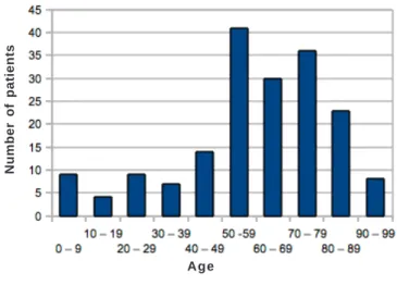
Our sample consisted of 176 patients, 126 male and 50 female (ratio 2.5: 1), ages ranged from six months to 97 years, with an average of 59.3 years (Figure 1) .
CSDH was caused by trauma in 92 (52%) patients, the time from trauma to diagnosis confirmed by imaging averaging 25 days. Twelve patients (6.5%) were in neurological operations postoperative period, five of them underwent ventricular shunt (VPS) and three, surgical treatment of acute subdural hematoma. Regarding associated diseases, 37.7% of patients were hypertensive, 20% had some neurological disease, 7.5% being cerebrovascular and 12.5%, malformations and tumors. Nineteen percent of patients were alcoholics (Table 1). Ongoing medications were inhibitors of angiotensin-converting enzyme (ACE) in 19%, 10% antiplatelet therapy, 9.6% diuretics and 9% oral hypoglycemic agents (Table 2).
We found 85 (48.3%) patients over 65 years, 25.6% of whom had altered consciousness and 17% changes in motor function. Among the 91 (51.7%) patients younger than 65 years, 22.7%had headache, 21% altered consciousness, and 14.2%, motor abnormalities (Table 3). As for the prognosis, in our sample there was no statistical difference among the elderly and non-elderly, with RR ranging from 0.49 to 1.25.
The Glasgow Coma Scale on admission ranged from 4 to 15 with an average of 13, and after surgery it
ranged from 7 to 15 with an average of 14. Anisocoria was present in 7.5% of patients on admission. Motor deficit was distributed among the four limbs, varying from 1 to 5 with an average of 4 in the four limbs, corresponding to a subnormal force that allows walking and performing simple tasks.
ICU admission was required in 13.7% of patients and tracheostomy was performed in 7%. Clinical complications were present in 10% of cases, infection being the most frequent, especially airway infection, meningitis and skin infection. Surgical rapprochement was necessary in 9% of cases, with an average of 106 days interval, there being no statistical difference as for prognosis of patients surgically re-approached in our sample, with RR ranging from 0.82 to 8.61.
Diagnostic imaging was done by Computerized Tomography (CT) in 92% and by MRI at 8%. The hemato-ma was located on the right in 41% of patients, on the left in 43%, and in 16% it was bilateral. Midline deviation was described in 42% of patients. There was no statistical difference in the prognosis of patients with midline deviation (RR 0.23 to 2.85) and bilateral CT findings (RR 0.08 to 3.55).
Table 1 Table 1 Table 1 Table 1
Table 1 - Associated Diseases.
D i s e a s e s D i s e a s e sD i s e a s e s D i s e a s e s
D i s e a s e s %%%%%
Trauma 52
Systemic hypertension 37.7
Neurological diseases (non vascular) 20
Alcoholism 19
Diabetes Mellitus 17
Cardiovascular Diseases 14.4
Smoking 10.3
Cerebrovascular diseases 7.5
Malignant neoplasms 6.2
Nephrological Diseases 4.1
Epilepsy 3.4
Dyscrasias 2.7
Pulmonary Diseases 2.7
Psychiatric Diseases 1.4
HIV + 0.7
Source: Discipline of neurosurgery, Surgery Department, Santa Casa de São Paulo (2001-2008).
Table 2 Table 2 Table 2
Table 2 Table 2 - Medication in use.
M e d i c i n e s M e d i c i n e sM e d i c i n e s
M e d i c i n e sM e d i c i n e s %%%%%
No medication 55
Angiotensin-converting enzyme (ACE) inhiibitors 19
Diuretics 9.6
Antiplatelet agents 9.6
Hypoglycemic agents 9
Anticonvulsants 7.5
Beta-blockers 5.5
Cardiotonics 4.1
Statins 3.4
Nonsteroidal anti-inflammatory drugs (NSAIDs) 1.4
Source: Discipline of neurosurgery, Surgery Department, Santa Casa de São Paulo (2001-2008).
Figure 1 Figure 1 Figure 1
Figure 1 Figure 1 - Age Classification.
Age
The treatment of choice was trepanation in 94% and craniotomy in 6%. When the CSDH was unilateral we held two trepanation orifices in 86% and on in 14%; when it was bilateral, on its turn, we performed four orifices in 70%were, two in 25% and three in 5% of operations. The closed drainage system was used in 85% of patients, with a mean time of 2.57 days, ranging from one to seven days. The average Glasgow coma scale was four at follow-up. Only 62% of patients returned after three months, 32% in six months and 21% in one year.
Hospital stay ranged from two to 177 days, with an average of 12.1 days. Sequelae were present in 6.5% of patients, 10% of patients died, 7.8% for clinical cau-ses.
DISCUSSION
DISCUSSION
DISCUSSION
DISCUSSION
DISCUSSION
Chronic subdural hematoma is characterized by a well defined and encapsulated collection between the dura mater and arachnoid membranes containing a mixture of fluid and coagulated blood in various stages5,6. It is one
of the most common forms of intracranial hemorrhage, being considered a benign lesion, though chronically progressive, but in most cases, the evolution without the imposition of surgical treatment can be fatal due both to brain compression exerted by hematoma and to the associated diseases. On the other hand, early diagnosis and surgical drainage allow complete recovery in most cases7.
Most patients are in the third decade or older, with the highest incidence between the fifth and sixth decades8, as was found in our series, average 59.3 years,
which was lower than that found in cases of review studies1,9,10. In all series men were more commonly affected
than women, corroborating our findings.
The pathophysiology of CSDH is not fully understood. There are two major theories proposed to explain its growth: the osmotic theory and the theory of
recurrent bleeding in encapsulated hematoma. The osmotic theory is based on the assumption that the liquefaction of hematoma increases the protein content and osmotic pressure, attracting fluid from blood vessels into the neighboring cavity by an osmotic pressure gradient across a semipermeable membrane (hematoma capsule)11.
However, Weir questioned this theory by demonstrating that the osmolarity of the hematoma fluid is identical to the blood and cerebrospinal fluid ones12. The recurrent
bleeding theory is the most accepted. The hematoma capsule possesses abnormal and dilated blood vessels, being a source of bleeding. Ito et al.13, after the administration of
red blood cells labeled with Cr, six to 24 hours before the hematoma drainage, showed that the hematoma content was 0.2 to 28% new blood. They also demonstrated that hematoma relation with the use of antiplatelet agents and anticoagulants, reinforcing this theory. Since most patients have cortical atrophy and decreased blood buffering effect, there is a contribution to the gradual expansion of CSDH11.
The most common cause of hematoma in all series is traumatic, being associated with chronic alcoholism, implantation of vascular shunts,coagulation disorders, epilepsy, and trauma8.
Our series is in line with the literature 2, since over
50% of patients had history of head trauma, and, for patients in neurosurgical postoperative period, the use of bypass systems was the most frequent previous operation 2.
The imaging exam of choice for the diagnosis remains the TC, mainly because it is faster and less costly compared to MRI, and also can be used in patients with metallic implants and cardiac pacemakers14,15. The most
common type of CSDH is hypodense (70.5%), followed by hematoma with various densities (19.6%), isodense (7.5%), and less commonly, hyperdense (2.4%) 1. MRI is useful to
mark the various stages of hematoma and provides detailed information on size, age and complexity of the hematoma, but is only indicated for the diagnosis of isodense or bilate-ral hematoma.
Table 3 Table 3 Table 3 Table 3
-Table 3 - Symptoms.
Symptoms Symptoms Symptoms Symptoms
Symptoms A g eA g eA g eA g eA g e
< 6 5 < 6 5< 6 5
< 6 5< 6 5 > 6 5> 6 5> 6 5> 6 5> 6 5 %
%%
%% NNNNN %%%%% NNNNN
Headache 22.7% 40 10.2% 18
Altered consciousness 21% 37 25.6% 45
Motor disorder 14.2% 25 17% 30
Sensitivity disorder 0.5% 1 0 0
Seizure 5.1% 9 3.4% 6
Vomiting 5.1% 9 1.1% 2
Incidental 1.1% 2 1.7% 3
Fever 1.7% 3 0 0
Aphasia 1.1% 2 2.8% 5
Urinary incontinence 0 0 1.1% 2
Functional results were satisfactory at discharge, that is, with good recovery for patients with Glasgow score greater than 13 and outpatients with higher scores than 3, ie with independent living. Our patients showed good recovery in 82% of cases, a result similar to that found in the literature 2,16,17, which shows that, if treated properly,
CSDH has a good prognosis.
Spontaneous resolution or clinical treatment of CSDH is well documented, but in some series hospitalization ranged from three weeks to 42 days and some patients are eventually surgically treated18,19. Several treatment options
are proposed: bed rest, corticosteroids, mannitol and other hypertonic solutions have been used. However, the clinical treatment is not the choice for most patients, since surgical techniques are minimally invasive and recovery is made in less than a week for most patients. In our series, only 24% of patients were in hospital for more than a week. The type of drainage to be employed has been much discussed over the years, but there is a current trend of using trepanation for drainage of the hematoma, given the increased mortality when performing craniotomy1.
Neuroendoscopic techniques are being used to multiloculated or septate hematomas, but are still rarely recommended.
Recurrence of CSDH after the first drainage is not uncommon and studies show values of 7 to 18%2,16,17;
in our series it was 9%. Risk factors for recurrence of the hematoma are usually divided into three categories: factors associated with the patient, such as age, sex, alcohol consumption, tendency to bleeding, cortical atrophy or intracranial hypotension; factors associated with the pathogenesis of CSDH, as the structure of neomembrane or hematoma characteristics; and factors associated with surgery, how long it took until the beginning of the
operation, additional irrigation procedures or closed drainage system. Studies20,21 have shown that age, coagulation
disorders, alcoholism and bilateral CSDH are risk factors for increased hematoma recurrence. Nonetheless, Oishi et al. 22 did not observe risk factors associated with patient
characteristics, but only with the pathogenesis and operation. As for pathogenesis,they found time of operation as a factor, since they observed a in higher reoperation rate in patients operated early due to a rapidly expanding hematoma 22,23.
The CSDH the cure rate after drainage by trepanation is high, but the neurological deterioration occasionally complicates the course of the postoperative period, requiring postoperative surveillance24,25. Acute
subdural hematoma, intracranial hypertensive hemorrhage, hypertensive pneumoencephalus and other cerebrovascular diseases can occur as postoperative complications2,26-28. In
our series there were no cases of surgical postoperative complications, only clinical ones, such as infections and heart disease, in 9% of patients.
CSDH is a common disease in the neurosurgeon practice, but still associated with significant morbidity and mortality27,28. In our patients, the change of consciousness
was the most frequent clinical alteration in elderly patients, and headache in the non-elderly. The disease most associated with CSDH was arterial hypertension and the most frequent cause was traumatic.
In conclusion, the change of consciousness was the most common clinical alteration in the elderly and headache in non-elderly. The most commonly associated comorbidity was the SAH and the most frequent cause, head trauma. The trepanation with two orifices associated with a closed drainage system was the most used operation, with high efficacy and low complication rate.
R E S U M O R E S U M O R E S U M O R E S U M O R E S U M O
Objetivo: Objetivo: Objetivo: Objetivo:
Objetivo: caracterizar os pacientes com hematoma subdural crônico submetidos à intervenção cirúrgica e identificar os indicadores prognósticos. Métodos:Métodos:Métodos:Métodos:Métodos: análise retrospectiva de pacientes diagnosticados com hematoma subdural crônico (HSDC) submetidos a tratamento cirúrgico. Foram analisados: idade, período do trauma ao diagnóstico por imagem, escala de coma de Glasgow pré e pós-operatório, tipo de intervenção cirúrgica, comorbidades associadas, utilização de drenagem pós-operatória e acompanhamento ambulatorial. Resultados:Resultados:Resultados:Resultados:Resultados: a amostra consistiu em 176 pacientes, 126 do sexo masculino e 50 pacientes do sexo feminino (propor-ção de 2,5:1), a idade variou de seis meses a 97 anos, com uma média de 59,3 anos. O HSDC foi causado por trauma em 52% dos pacientes, com o intervalo do trauma ao diagnóstico por imagem, em média, de 25,05 dias. Eram hipertensos 37,7% dos pacientes e 20% possuíam alguma doença neurológica. Oitenta e cinco (48,3%) pacientes eram idosos e a alteração da consciência esteve presente em 63% dos casos. Não eram idosos 91 (51,7%)p pacientes, 44% aprresentaram cefaleia, alteração da consciência ocorreu em 40% dos pacientes e as alterações motoras, em 27,5%. O HSDC localizou-se à direita em 41%, à esquerda em 43% e, bilateral em 16% dos pacientes. Conclusão:Conclusão:Conclusão:Conclusão:Conclusão: a alteração de consciência foi a alteração clínica mais comum nos idosos e a cefaleia em não idosos. A comorbidade mais associada foi a HAS e a causa mais frequente, o traumatismo craniano. A trepanação com dois orifícios associada ao sistema de drenagem fechado foi a operação mais utilizada, com alta efetividade e baixo índice de complica-ções.
Descritores: Descritores: Descritores: Descritores:
REFERENCES
REFERENCES
REFERENCES
REFERENCES
REFERENCES
1. Gelabert-Gonzáles M, Iglesias-Pais M, García-Allut A, Martínez-Rumbo R. Chronic subdural haematoma: surgical treatment and outcome in 1000 cases. Clin Neurol Neurosurg. 2005;107(3):223-9.
2. Mori K, Maeda M. Surgical treatment of chronic subdural hema-toma in 500 consectuive cases: clinical characteristics, surgical outcome, complications, and recurrence rate. Neurol Med Chir. 2001;41(8):371-81.
3. Yamamoto H, Hirashima Y, Hamada H, Hayashi N, Origasa H, Endo S. Independent predictors of recurrence of chronic subdural hematoma: results of multivariate analysis performed using a logistic regression model. J Neurosurg. 2003;98(6):1217-21.
4. World Health Organization (2002). Active Ageing: A Police Framework. A contribution of the World Health Organization to the Second United Nations World Assembly on Ageing. Madrid, Spain, April, 2002. Disponível em: http://whqlibdoc.who.int/hq/2002/ WHO_NMH_NPH_02.8.pdf.
5. Ernestus RI, Beldzinski P, Lanfermann H, Klug N. Chronic subdural hematoma: surgical treatment and outcome in 104 patients. Surg Neurol. 1997;48(3):220-5.
6. Yasuda CL, Morita ME, Nishimori FY, Yasuda AM, Alves HL. Hema-toma subdural crônico: estudo de 161 pacientes operados e a relação com alterações no coagulograma. Arq Neuro-Psiquiatr. 2003;61(4):1011-4.
7. Pittella JEH, Duarte JE. Prevalência e padrão de distribuição das doenças cerebrovasculares em 242 idosos, procedentes de um Hospital Geral, necropsiados em Belo Horizonte, Minas Gerais, no período de 1976 a 1997. Arq Neuro-Psiquiatr. 2002;60(1):47-55. 8. Misra M, Salazar JL, Bloom DM. Subddural-peritoneal shunt: treatment for bilateral chronic subdural hematoma. Surg Neurol. 1996;46(4):378-83.
9. Rhode V, Graf G, Hassler W. Complications of burr-hole craniotomy and closed-system drainage for chronic subdural hematomas: a retrospective analysis of 376 patients. Neurosurg Rev. 2002;25(1-2):89-94.
10. Krupp WF, Jans PJ. Treatment of chronic subdurl haematoma with burr-hole craniostomy and closed drainage. Br J Neurosurg. 1995;9(5):619-27.
11. Adhiyaman V, Asghar M, Ganeshram KN, Bhowmick BK. Chronic subdural haematoma in the elderly. Postgrad Med J. 2002;78(916):71-5.
12. Weir B. The osmolality of subdural hematoma fluid. J Neurosurg. 1971;34(4):528-33.
13. Ito H, Yamamoto S, Saito K, Ikeda K, Hisada K. Quantitative estimation of hemorrhage in chronic subdural hematoma using the 51Cr erythrocyte labeling method. J Neurosurg. 1987;66(6):862-4.
14. Gelabert-González M, Fernández-Villa JM, López-García E, García-Allut A. Chronic subdural hematoma in patients over 80 years of age. Neurocirurgia. 2001;12(4):325-30.
15. Tsutsumi K, Maeda K, Iijima A, Usui M, Okada Y, Kirino T. The relationship of preoperative magnetic resonance imaging findings and closed system drainage in the recurrence of chronic subdural hematoma. J Neurosurg. 1997;87(6):870-5.
16. Ernestus RI, Beldzinski P, Lanfermann H, Klug N. Chronic subdural hematoma: surgical treatment and outcome in 104 patients. Surg Neurol. 1997;48(3):220-5.
17. Mellergård P, Wisten O. Operations and re-operations for chronic subdural haematomas during a 25-year period in a well defined population. Acta Neurochir. 1996;138(6):708-13.
18. Kurti X, Xhumari A, Petrela M. Bilateral chronica subdural haematomas: surgical or non-surgical treatment. Acta Neurochir. 1982;62(1-2):87-90.
19. Voelker JL. Nonoperative treatment of chronic subdural hemato-ma. Neurosurg Clin N Am. 2000;11(3):507-13.
20. Asano Y, Hasuo M, Takahashi I, Shimosawa S. Recurrent cases of chronic subdural hematoma—its clinical review and serial CT findings. No To Shinkei. 1992;44(9):827-31.
21. Fukuhara T, Gotoh M, Asari S, Ohmoto T, Akioka T. The relationship between brain surface elastance and brain reexpansion after evacuation of chronic subdural hematoma. Surg Neurol. 1996;45(6):570-4.
22. Oishi M, Toyama M, Tamatani S, Kitazawa T, Saito M. Clinical factors of recurrent chronic subdural hematoma. Neurol Med Chir. 2001;41(8):382-6.
23. Araújo JFM, Iafigliola MG, Balbo RJ. Hematoma subdural crônico: análise de 35 casos. Arq Neuro-Psiquiatr. 1996;54(1):71-4. 24. De Jesús O, Pacheco H, Negron B. Chronic and subacute
hemato-ma in the adult population. The Puerto Rico experience. P R Health Sci. J. 1998;17(3):227-33.
25. Tagle P, Zamorano L, Villar S. Hematoma, subdural crónico: consideraciones terapéuticas y pronóstico. Rev chil neuro-psiquiatr. 1984;22(2):155-8.
26. Kwon TH, Park YK, Lim DJ, Cho TH, Chung YG, Chung HS, et al. Chronic subdural hematoma: evaluation of the clinical significance of postoperative drainage volume. J Neurosurg. 2000;93(5):796-9.
27.Tseng JH, Tseng MY, Liu AJ, Lin WH, Hu HY, Hsiao SH. Risk factors for chronic subdural hematoma after a minor head injury in the elderly: a population-based study. Biomed Res Int. 2014;2014:218646.
28. Jung YG, Jung NY, Kim E. Independent predictors for recurrence of chronic subdural hematoma. J Korean Neurosurg Soc. 2015;57(4):266-70.
Received: 18/02/2015
Accepted for publication: 10/05/2015 Conflict of interest: none.
Source of funding: none.
Mailing address: Mailing address: Mailing address: Mailing address: Mailing address: João Luiz Vitorino Araujo