INTRODUCTION
Address to: Dr. Gesner Pereira Lopes. Depto de Clínica Médica/UFTM. Av. Getúlio Guaritá 130, Bairro Abadia, 38025-440 Uberaba, MG, Brasil.
Phone: 55 34 3318-5119; Fax: 55 34 3318-5279
e-mail: r-g31@uol.com.br
Received 27 August 2013
Accepted 29 November 2013
Length and caliber of the rectosigmoid colon among
patients with Chagas disease and controls from
areas at different altitudes
Gesner Pereira Lopes
[1],
Márcia Maria Ferreira-Silva
[2],
Angel Anibal Ramos
[3],
Helio Moraes-Souza
[2],
Aluízio Prata
†
and Dalmo Correia
[4][1]. Setor de Radiologia e Diagnóstico por Imagem, Departamento de Clínica Médica, Faculdade de Medicina, Universidade Federal do Triângulo Mineiro, Uberaba, MG. [2] Disciplina de Hematologia e Hemoterapia, Hemocentro Regional de Uberaba, Universidade Federal do Triângulo Mineiro, Uberaba, MG. [3]. Departamento de Radiologia, Hospital Manuel Nuñes Butron, Puno, Peru.[4]. Disciplina de Doenças Infecciosas e Parasitárias, Departamento de Clínica Médica, Faculdade de Medicina, Universidade Federal do Triângulo Mineiro, Uberaba, MG. †In memoriam.
ABSTRACT
Introduction: In this study, we investigated radiological changes in the sigmoid colon in chagasic patients by comparing their colon lengthsand caliber with those of non-chagasic living in the same region and non-chagasic living at high altitudes. Methods:
A total of 317 individuals were evaluated using clinical, serological and radiological methods and divided into three groups: 1) one hundred and nine non-chagasic individuals fromUberaba, Brazil; 2) sixty-one non-chagasic from Puno, Peru; 3) one hundred forty-seven chagasics examined in Uberaba, being 62 without megacolon (3A), 72 with megacolon (3B) and 13 with doubtful diagnosis of megacolon (3C). Results: In group 2, the sigmoid colon had a signifi cantly larger caliber (p=0.001) and the
rectosigmoid colon was longer (p<0.001) than group 1. In subgroup 3A, the sigmoid colon (p<0.001) and rectum (p<0.001) had a signifi cantly larger caliber and the rectosigmoid was longer (p<0.001) than that of the non-chagasic individuals. In subgroup 3B, the rectosigmoid was longer in all patients, and the caliber of the sigmoid was signifi cantly larger than that of subjects in subgroups 3A and 3C (p<0.001). Conclusions: Morphometric analysis confi rms that Chagas disease may increase the caliber
and length of the rectosigmoid. Our results suggest that altitude, ethnicity and diet may have infl uenced the size and length of the rectosigmoid of andean patients.
Keywords:Chagas disease. Radiology. Megacolon. Sigmoid. Morphometry. Altitude.
Human Chagas disease (HCd), which affects between 2 and 3 million Brazilians, is an important societal problem1, with 75-90 million Latin Americans at risk of becoming infected2.
The association between megacolon and Trypanosoma cruzi
was reported by Chagas and Villela3. The etiopathogenesis of megacolon in HCd is a lesion caused by T. cruzi in the intramural nerve plexus of the autonomic nervous system in the large intestine. Decreased numbers of neurons (hypoganglionosis) is necessary and suffi cient for development of megacolon4,5. Intramural denervation leads to dyskinesia of the affected segment and typically culminates in dilatation and elongation of the terminal colon, which is responsible for deposition.
Moreover, secretion, absorption, and motility are altered, leading to chagasic megacolon5.
Altitude is thought to infl uence colon caliber and length. A study by Frisancho6 showed that dolichomegacolon of those living in the Andes mountains has several specifi c clinical, radiological, and anatomopathological characteristics as well as complications that distinguish it from other types of megacolon, particularly chagasic megacolon7,8.
In the Andes, megacolon is common among inhabitants of the South American regions located 3,000m above sea level; this condition is thought to be unrelated to HCd. Increased colon caliber and length are attributed to racial factors, cocaine use, and vitamin B defi ciency among patients in Sucre, Bolivia9. These morphological changes were also seen in patients in Peruvian hospitals located at altitudes >3,000m above sea level in the Andes10. Several possible causes of megacolon in Andean patients have been proposed, including anatomical elements, longer sigmoid colon, and aggravating factors including dietary habits, low atmospheric pressure, and racial factors.
METHODS
radiological investigation to analyze the rectosigmoid colon and compare differences between controls and patients with HCd. The rectosigmoid colon was chosen for the analysis because it is easily accessible and generally affected by HCd. Therefore, the aim of this study was to examine the radiological alterations within the sigmoid colon in patients with HCd and compare the measurements with those in control individuals in Brazil and control individuals living at high altitudes.
The study was conducted in region with altitude of 762 feet above sea level in Brazil, and at the mountainous region of Peru at an altitude of 3,850 feet above sea level.
Patients
A total of 317 individuals were examined and divided into 3 groups as follows: Group 1: one hundred nine individuals with negative serological results for HCd from Triângulo Mineiro
and Alto Paranaíba, State of Minas Gerais, Brazil (35 men with a median age of 45 years and 74 women with a median age of 42 years); Group 2: sixty-one individuals with negative serological results for HCd from the region of Puno, Peru (40 men with a median age of 37 years and 21 women with a median age of 36 years); and Group 3: one hundred forty-seven patients with HCd from Triângulo Mineiro, Brazil (median age, 58 years; 62 without megacolon [subgroup 3A], 72 with megacolon [subgroup 3B], and 13 with an unconfi rmed diagnosis of megacolon [subgroup 3C]).
Patient selection
The Brazilian individuals were selected from among patients with chronic HCd who were seeking outpatient services. The patients underwent a clinical examination in which all data were collected in a standard form prepared for this study. They were then subjected to a series of complementary examinations such as repeated serology for HCd, blood cultures for T. cruzi,and tests to characterize the clinical form of HCd.
Control individuals (Group 1), were from several clinical departments from Clinics Hospital, and did not have complaints related to HCd. Subjects from high-altitude regions (Group 2) were selected from those living within Puno, Peru. These individuals were chosen from employees and patients at the Puno’s Hospital; their family members were also selected. Individuals in both groups with diarrhea, constipation for >3 days, or evidence of infl ammatory or tumor-like intestinal lesions and those who were <20 or >60 years of age were excluded from the study.
A clinical evaluation was previously conducted in patients and controls to rule out secondary causes of megacolon dysfunction, such as congenital or toxic megacolon, intestinal parasitic disease, cancer, or metabolic causes (diabetes mellitus and hypothyroidism).
Serological exams for human Chagas disease
Blood samples were collected from all patients, and serological reactions such as indirect hemagglutination (IHA), indirect immunofl uorescence reaction (IFR), and enzyme-linked
immunosorbent assay (ELISA) were conducted to diagnose HCd. Only samples that were positive for at least 2 of the 3 performed tests were considered seropositive.
Blood samples of the patients from Puno were acquired via fi nger prick, collected on Whatman® qualitative fi lter paper, Grade 1 (Sigma-Aldrich, USA), and tested using the same serological tests.
Radiological exams
Colopathy was diagnosed by measuring the caliber and length of the rectosigmoid colon.
Thoracic teleradiography was used to evaluate the heart. Conventional radiography of the abdomen with the patient in the supine position was used to evaluate the distribution of intestinal gas before administration of opaque enema. These tests were performed using medium-sized devices at 500mA and 125kVp without an image intensifi er.
In Group 1, the colon test was preceded by intestinal preparation with administration of a laxative the day before and an aqueous enema 4h before the test. In all other groups, contrast colonoscopy (opaque enema) was conducted without intestinal preparation, according to the simplifi ed technique reported by Ximenes et al.13. In all patients, 300mL of barium sulfate was diluted in 900mL of water for a total volume of 1,200mL. Radiography was performed for each patient in the supine, prone, and side positions for a total of 3 radiographs each. The technique reported by Ximenes et al.13 was also used in Puno.
To diagnose megacolon, we determined whether the colon was enlarged by dilatation or elongation. We identifi ed different sigmoid parameters and classifi ed them as I, II, III, or undetermined based on the Farrar guidelines to determine the morphology of the sigmoid colon in control individuals in Uberaba11.
To measure the length of the rectosigmoid colon, a curvimeter was used and passed through the central axis of the colon using a backward motion from the anus up to the left iliac crest, which was assumed to be the beginning of the sigmoid colon. The caliber and length of the sigmoid colon and rectum were measured using a ruler based on the results of the side radiograph of the rectum. These measurements were always conducted at the level of the third sacral vertebra or where the sigmoid colon had a larger diameter. The caliber of the rectum was measured below the S3 level to the sigmoid colon. All measurements were conducted twice at different time points by the same investigator so that reproducibility of this method could be ensured.
Statistical analysis
RESULTS
TABLE 1- Comparisons between non-chagasic individuals examined in Uberaba, State of Minas Gerais, Brazil, (group 1) and in Puno, Peru,
(group 2) and between non-chagasic individuals of Uberaba (group 1) and chagasics without megacolon (subgroup 3A).
Median Group 1 Group 2 P value Subgroup 3A P value
(n = 109) (n = 61) Groups (1x2) (n = 62) Groups (1x3A)
Caliber of the sigmoid 3.1cm 5cm 0.001 3.4cm 0.013
<3.5cm 73 (67%) 2 (3.3%) 0.001 32 (50%) 0.271
>4.5cm 1 (0.9%) 39 (63.9%) >0.001 5 (8%) <0.001
Caliber of the rectum 4.5cm 4cm 0.008 5.45cm <0.001
<3.5cm 12 (11%) 2 (3.3%) 0.142 3 (4.8%) 0.001
>7.4cm 1 (0.9%) 1 (1.6%) 0.749 2 (3.2%) 0.276
Length of the rectosigmoid 57 cm 65 cm <0.001 65 cm <0.001
<50cm 18 (16.5%) 1 (1.6%) 0.007 7 (11.3%) 0.481
>74cm 1 (0.9%) 12 (19.7%) <0.001 10 (16.1%) <0.001
To compare categorical variables among groups, the χ2 test with Yates’s correction was used as needed or Fisher’s exact test was employed. Probabilities < 5% (p < 0.05) were considered statistically signifi cant.
Ethical considerations
This study was approved by the Institucional ethics committee of our Federal University. The study participants were informed of the tests to be performed and provided written consent.
A total of 317 patients with HCd and controls were divided into 3 groups and examined from 2000 to 2005. Analysis of subject age showed that individuals with Chagas tended to be older than controls (p < 0.005). There was no age difference between the HCd subgroups.
In control individuals (Group 1), the caliber of the sigmoid colon was 2-6cm with a median of 3.1cm. There was a signifi cant gender-related difference (p = 0.029) in caliber (median, 3.3cm in males vs. 3cm in females). The caliber of the rectum was 2.5-8.5 cm with a median of 4.5cm. Individual gender, age, and height did not affect caliber. The length of the rectosigmoid colon was 32-75cm. The median length was 57cm and was signifi cantly infl uenced by patient gender, age, and height
(Table 1).
Based on the tests performed on 109 control individuals (Group 1) and using a 99% percentile for comparison with other groups, we considered the following as the upper normal limits: sigmoid caliber, 4.5cm; rectum caliber, 7.4cm; and rectosigmoid length, 74cm.
In control individuals (Group 2), the caliber of the sigmoid colon was 3.2-8.0cm with a median of 5cm. The caliber of the rectum was 2.5-8cm with a median of 4cm. In 15 (24.6%)
individuals, the rectum had a higher caliber than that of the sigmoid colon. The length of the rectosigmoid colon was 45-84cm with a median of 65cm (Table 1). None of these measurements were infl uenced by gender, age, or height.
In the radiological exam of the sigmoid colon and rectum of 147 patients with HCd (Group 3), we diagnosed megacolon in 72 (49%) patientes. Thirteen (8.8%) had a unconfirmed diagnosis, whereas 62 (42.2%) did not have megacolon.
In patients with HCd but without megacolon (subgroup 3A), the caliber of the sigmoid colon was2.5-5.0cm with a median of 3.4cm. The caliber of the rectum was 3.0-7.6cm (median, 5.4cm).The length of therectosigmoidcolon insubgroup 3A was 31-86 cm (median, 65cm) (Table 1).
We observed that the caliber of the sigmoid colon was signifi cantly greater in subjects from Puno than in those from Uberaba (5.0cm and 3.1cm, respectively; p=0.001). Moreover, their rectosigmoid colons were larger (65cm in Puno vs. 57cm in Uberaba; p<0.001) (Table 1 and Figure 1).
The measurements of only 3 (2.7%) patients in this group exceeded the limits established for Group 1, whereas 41 (66.1%) individuals in Group 2 had measurements that exceeded the normal limits (Table 1).
Calibers of the sigmoid colon (3-4cm) and rectum (5-45cm) and the length of the sigmoid colon (65cm) of the patients with HCd but without megacolon (subgroup 3A) were signifi cantly higher than those of the control individuals from Group 1, who had measurements of 3.1, 4.5, and 57cm, respectively (Table 1 and Figure 2).
All 72 patients diagnosed with megacolon (subgroup 3B) had increased rectosigmoid colon length. The calibers of the rectums were measured in 54 patients with megacolon and were 3.0-13.5cm (median, 6.1cm).The calibers of the rectums were 3.5-10cm (median, 6.3cm). No patients had a caliber <3.5cm, and the caliber did not exceed 7.4cm in 39 (72.2%) patients
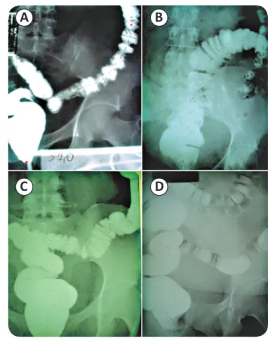
A
B
C
D
A
B
C
D
FIGURE 1 - Different morphologies of the rectosigmoid colon: increased sigmoid caliber and length (A), increased rectum caliber only (B), increased rectum caliber with normal sigmoid caliber (B) and (D), and elongated sigmoid (C).
FIGURE 2 - Normal (A) and altered morphometry (B). Normal rectosigmoid colon of a patient from Uberaba (C) and a patient from Puno (D).
TABLE 2 - Comparisons between chagasics without diagnosis of megacolon (subgroup 3A) and those diagnosed with megacolon (subgroup
3B) and between chagasics with megacolon (subgroup 3B) and those with doubtful diagnosis of megacolon (subgroup 3C).
Median Subgroup 3A Subgroup 3B P value Subgroup C
(n = 62) (n = 72) Groups (3Ax3B) (n = 13) Groups (3Bx3C) P value
Caliber of the sigmoid 3.4cm 6.10cm <0.001 4.1cm <0.001
<3.5cm 32 (50%) 1 (1.8%) <0.001 2 (16.6%) 0.083
>4.5cm 5 (8%) 50 (92.6%) 0.001 5 (41.6%) <0.001
Caliber of the rectum 5.45cm 6.3cm <0.001 6.75cm 0.987
<3.5cm 3 (4.8%) 0 0.293 0
->7.4cm 2 (3.2%) 15 (27.8%) <0.001 3 (25%) 1
All patients from subgroup 3B showed a large increase in sigmoid colon length, making this measurement impossible. When there were large alterations, the calibers of both the rectum and sigmoid colon increased.
All patients with an unconfi rmed diagnosis of megacolon (subgroup 3C) had a rectosigmoid length that exceeded the
normal limit (74cm). None was <80cm, and the median was 88.5cm. We observed that 41.6% of the calibers of the sigmoid colon and 25% of the calibers of the rectum exceeded the maximum normal limits established in this study (Table 2). The calibers of the sigmoid colon and rectum were signifi cantly higher in subgroup 3B than in subgroup 3A (p < 0.001) (Table 2).
patients. The lengths of the rectosigmoid colon were higher in subgroup 3B patients (with megacolon) than in subgroup 3C patients. Additionally, the sigmoid calibers were signifi cantly higher in subgroup 3B patients; however, there was no difference in rectum calibers (Table 2). Of the 13 patients in subgroup 3C, only 1 (7.7%) had a simultaneous increase in sigmoid and rectum calibers, while this simultaneous increase was observed in 12 (22.2%) patients in subgroup 3B. In subgroup 3C, there were 5 (41.6%) patients with calibers higher than the normal limits, while this was observed in only 3 (7.4%) patients of subgroup 3B. A caliber increase in either the sigmoid colon or the rectum was observed in 7 (58.3%) patients of subgroup 3C and in 39 (74.1%) patients of subgroup 3B.
DISCUSSION
The chronic phase of HCd includes a period of subpatent parasitemia after the fi rst 60 days of acute infection14. The factors infl uencing the clinical variability of the phases of HCd have not been elucidated, but genetic variability of both the host and the parasite may be important15.
The most frequently observed alterations of the digestive form of HCd include esophagopathy and colopathy, which cause swallowing diffi culties and constipation, respectively16,17. Colopathy manifests as prolonged constipation, which may have serious complications such as fecaloma and volvulus.
This study aimed to contribute to the diagnosis of colopathy, particularly small dilatations, by proposing the standard for measuring rectosigmoid dimensions. Thus, we established normal limits for sigmoid and rectum calibers and rectosigmoid lengths.
Compared to our results, studies have reported different median sigmoid and rectum values. Rezende et al.18 found that the diameter of a normal sigmoid colon should be <6cm. Hernandez et al.19 studied approximately 60 individuals and identifi ed a mean sigmoid caliber of 4.38, rectum caliber of 6.2cm, and rectosigmoid length of 51.2cm; all of these values are lower than those found in this study. However, the maximum values in control individuals are similar. This similarity suggests that the differences in limits established by earlier previous studies may be due to the measurement methodology.
Occurrence of megacolon in Puno and other regions within the Andes represents a special situation that requires further study, despite studies that have already been conducted by researchers from Bolivia, Argentina, Chile, and Peru. These studies describe changes in the colon in those living in the Andes and the existence of another type of endemic non-chagasic megacolon. They draw attention to the frequency of volvulus20,21 and changes in intestinal transit7 in certain countries of the Andes, which motivated us to include this population in our study.
The unique significant difference in rectosigmoid measurements between patients from Uberaba and those from Puno was that the patients from Puno had rectums with smaller calibers.
The initial hypothesis that dolichomegacolon in the Andes is due to altitude appears to have no basis because there are no reports of megacolon occurrence in other populations living at the same or even higher altitudes such as in the Alps or the Himalayas10. However, we unexpectedly observed a higher frequency of intestinal movement in patients from Puno, with a mean interval of 0.73 days in contrast to the interval of 2 days observed in controls from Uberaba. This difference may be related to the smaller caliber of the rectum observed in Puno despite the higher sigmoid caliber. However, other factors such as diet, cocaine use, and race must be considered and investigated further. Patients with HCd have greater calibers of the sigmoid colon and rectum and grater rectosigmoid lengths than those of control individuals. Sigmoid colon caliber was signifi cantly greater among individuals with HCd and evidence of megacolon than among individuals without megacolon and those with unconfi rmed diagnosis. A similar study by Castro12 of 291 patients showed similar fi ndings regarding sigmoid measurements; however, the authors observed that the mean diameters of the rectums in patients with HCd did not differ signifi cantly from those of control individuals.
We used morphometry to confi rm the presence of megacolon in both cases classifi ed as unconfi rmed and others previously considered normal. In these cases, calibers of the sigmoid colon and rectum and rectosigmoid colon lengths were signifi cantly higher in control individuals (8%, 3.2%, and 16.1%, respectively), that were higher than normal. In 16 (25.8%) patients, morphometry showed incipient organ enlargement. Such fi ndings show that colopathy occurs more frequently than what can be evaluated using conventional radiology.
In addition to being relevant for diagnosis, the use of morphometry revealed the importance of rectosigmoid elongation in pathophysiology of the megacolon. It showed that even in cases of advanced megacolon, sigmoid and rectum calibers may remain normal, which confi rms the results of earlier studies showing that elongation may precede dilatation22. In contrast to the esophagus, elongation of the megacolon occurs earlier and more frequently, the fecal content is retained, and an intestinal transit delay occurs22.
Among the 3 radiological measurements obtained, only the rectosigmoid length had increased in all radiographs with a megacolon diagnosis. Even among the 13 patients for whom diagnosis was unconfi rmed after radiography, morphometry enabled detection of alterations, thus allowing the diagnosis of megacolon. The 10 patients with a colon >74cm (subgroup 3A) were considered to have incipient megacolon. However, without using morphometry, it is extremely diffi cult to identify such patients.
In summary, our results suggest that factors such as diet (corn, potatoes, and starchy foods), cocaine use, ethnicity, and altitude may have infl uenced rectosigmoid calibers and lengths of Andean patients. Morphometry revealed that HCd may increase rectosigmoid caliber and length. Moreover, measurement results are dependent on the observer.
ACKNOWLEDGMENTS
REFERENCES
The authors declare that there is no confl ict of interest.
CONFLICT OF INTEREST
The authors acknowledge to all staff include on perform this study. We also thank those who collaborated with us, especially Angel Anibal Ramos, Victor Alberto Laguna-Torres, Valdo José Dias da Silva, Mario León Silva-Vergara, and Alejandro Luquetti Ostermeyer. We extend our deepest thanks to Prof. Dr. Aluízio Prata for his boundless energy and wisdom. It was an honor to have him as a mentor.
1. Ministério da Saúde. Secretaria de vigilância Sanitária. Aspectos epide-miológicos da doença de Chagas [Internet]. Brasília: Ministério da Saúde; 2011. [Cited 2012 February 12]. Available at: http://portal.saude.gov.br/
portal/saude/profi ssional/visualizar_texto.cfm?idtxt=31454/.
2. Coura JR, Dias JCP. Epidemiology, control and surveillance of HCd 100 years after its discovery. Mem Int Oswaldo Cruz 2009; 104:31-40.
3. Chagas C, Villela E. Forma cardíaca da Trypanosomiase Americana. Mem Inst Oswaldo Cruz 1922; 14:457-544.
4. Köberle F. Patogênese dos megas. Rev Goiana Med 1956; 2:101-110.
5. Köberle F. Megaesophagus. Gastroenterology 1958; 34:460-466.
6. Frisancho PD. Conceitos atuais sobre el Dolicomegacolon Andino. Peru. Rev Gastroenterology 1993; 1:2-15.
7. Frisancho PD, Jorge Banda D, Peña G, Salas P, Oscar Frisancho V. Tempo de transito intestinal em nativos del Altiplano. Peru. Rev Gastroenterology 1985; 13:45-51.
8. Frisancho D, Frisancho O, Chacón P. Mesocolitis Retrátil: Fisiopatologia y complicaciones. Rev Gastroenterology 1998; 18:113-118.
9. Molina B. Morfologia del asa sigmóide en nuestro medio y su relación con la frecuencia del vólvulo. [Masther Thesis]. [Bolivia]: Universidad del Sucre; 1941. 87p.
10. Franco FE. El colon ilio-pélvico en los peruanos. [Thesis]. [Peru]: Universidad Nacional Mayor de San Marcos; 1965. 65p.
11. Farrar CW. Patterns of sigmoid colon and their implications for barium enema radiography. Med Radio Photog 1979; 55:3-28.
12. Castro C, Hernandez EB, Rezende J, Prata A. Estudo radiológico do megacólon em área endêmica de doença de Chagas. Rev Soc Bras Med Trop 2010; 43:562-566.
13. Ximenes CA, Rezende JM, Moreira H, Glória M. Técnica simplifi cada
para o diagnóstico radiológico do megacólon chagásico. Rev Soc Bras Med Trop1984; 17:23.
14. Kirchoff LV. American trypanosomiasis (Chagas’ disease). A tropical disease now in the United States. New Engl J Med 1993; 329:639-644.
15. Macêdo AM, Machado CR, Oliveira RP, Pena SD. Trypanosoma cruzi: genetic structure of populations and relevance of genetic variability to the pathogenesis of HCd.Mem Inst Oswaldo Cruz 2004; 99:1-12.
16. Matsuda MN, Miller SM, Évora PRB. The chronic gastrointestinal manifestations of HCd. Clinics 2009; 64:1219-1224.
17. Santos CM, Cassiani RA, Dantas RO. Avaliação clínica na deglutição na doença de Chagas. Rev Soc Bras Fonoaudiol 2011; 16:215-220.
18. Rezende JM, Ximenes CA, Moreira C, Vaz MM, Luquetti AO, Milano MC. Alongamento do colo distal em pacientes com a forma digestiva da doença de Chagas. In: III Reunião Anual sobre Pesquisa Aplicada em Doença de Chagas; 1985.
19. Hernandez EB, Rezende JM, Macêdo V, Castro CN. Estudo radiológico do cólon em indivíduo área endêmica de doença de chagas através da técnica
simplifi cada de Ximenes. Rev Soc Bras Med Trop 2002; 35 (supl III):21.
20. Frisancho PD, Frisancho O. Dolicomegasigmoides volvulado. Peru, Rev Gastroentrology 1983; 3:73-81.
21. Ballaga VI, López LAO. Vólvulo Del sigmoid em La enfermedad da Chagas. Medisan 2011; 15:1014-1017.