297 297 297 297 297 Mem Inst Oswaldo Cruz, Rio de Janeiro, Vol. 92(3): 297-316, May/Jun. 1997
This research received financial support from Faperj, CNPq, PAPES and CABBIO.
+Corresponding author. Fax: +55-21-270.9997 Received 24 July 1996
Accepted 21 October 1996
The Changing Face of the Epidemiology of Tuberculosis due
to Molecular Strain Typing - A Review
Philip N Suffys
+, Marcelo E Ivens de Araujo, Wim M Degrave
Laboratório de Biologia Molecular e Diagnóstico de Doenças Infecciosas, Departamento de Bioquímica e Biologia Molecular, Instituto Oswaldo Cruz, Av. Brasil 4365, 21045-900 Rio de Janeiro, RJ, Brasil
About one third of the world population is infected with tubercle bacilli, causing eight million new cases of tuberculosis (TB) and three million deaths each year. After years of lack of interest in the disease, World Health Organization recently declared TB a global emergency and it is clear that there is need for more efficient national TB programs and newly defined research priorities. A more complete epidemiology of tuberculosis will lead to a better identification of index cases and to a more efficient treatment of the disease. Recently, new molecular tools became available for the identification of strains of Mycobacterium tuberculosis (M. tuberculosis), allowing a better recognition of transmission routes of defined strains. Both a standardized restriction-fragment-length-polymorphism-based methodology for epidemiological studies on a large scale and deoxyribonucleic acids (DNA) amplification-based methods that allow rapid detection of outbreaks with multidrug-resistant (MDR) strains, often charac-terized by high mortality rates, have been developed. This review comments on the existing methods of DNA-based recognition of M. tuberculosis strains and their peculiarities. It also summarizes literature data on the application of molecular fingerprinting for detection of outbreaks of M. tuberculosis, for identification of index cases, for study of interaction between TB and infection with the human immuno-deficiency virus, for analysis of the behavior of MDR strains, for a better understanding of risk factors for transmission of TB within communities and for population-based studies of TB transmission within and between countries.
Key words: tuberculosis - molecular epidemiology - fingerprinting - restriction fragment length polymorphism
Upon opening the World Wide Web (Internet) version of the World Health Organization (WHO) Report on the Tuberculosis (TB) Epidemic, three major headlines stand out: TB will kill 30 million people this decade; someone is infected with TB every second and TB drugs may become useless. This dramatic introduction is followed by: The TB epidemic is growing larger and more dangerous each year. If the TB epidemic continues to be ne-glected, future generations will remember this de-cade as the time when humanity allowed deadly bacilli that travel through the air to become drug-resistant and incurable throughout the world. In 1993, WHO declared TB as a global emergency since it is globally the leading cause of death asso-ciated with infectious diseases. Tuberculosis is especially predominant in developing countries where it causes 26% of all avoidable deaths and it is expected that during the next ten years, the num-ber of TB cases will still increase substantially.
This is partly due to the interaction between TB and the human immunodeficiency virus (HIV) epidemic; by the end of the century, TB is likely to be the leading cause of death among HIV-positive persons. Furthermore, due to poorly managed TB control programs, multidrug-resistant (MDR) TB is increasing all over the world. Some of MDR-TB is incurable and several reports mention cases of HIV-positive patients who deceased as a conse-quence of infection with MDR-TB within a couple of weeks.
con-298 298 298 298
298 Molecular Epidemiology of TB • PN Suffys et al.
trol program should therefore be identification and treatment of acid-fast bacillus smear positive cases and this can be achieved better by understanding the factors that foster transmission of Mycobacte-rium tuberculosis. Transmission of TB is influenced by a large number of risk factors and transmission dynamics are therefore different in distinct geo-graphic regions. Furthermore, due to the increased mobility of the world population, globalization of TB has been intensified and caused an even more complex pattern of TB spreading and epidemiol-ogy. Because of political, social and ethical rea-sons, it is difficult to prevent the migration of in-fected individuals and an optimal control strategy has therefore to be developed, taking into consider-ation the social and biologic factors that foster TB transmission.
Traditional epidemiology continues to be the corner-stone for the understanding of TB spread-ing but the dynamics of the latter will only be bet-ter understood upon study of the spreading of in-dividual strains of M. tuberculosis within a certain population. Until recently, detection of person to person transfer of M. tuberculosis clones was a difficult, if not impossible task; only subgroups of strains could be identified through phage typing and occurrence of some special drug-resistance profiles. Only upon the recent development of molecular methods based on recognition of se-quences of deoxyribonucleic acids (DNA) and their rearrangements, it has been possible to fingerprint single strains of M. tuberculosis. This has resulted in a better detection of outbreaks of particular MDR strains of M. tuberculosis and one can now prove the spillover of these strains from high-risk groups to the general population. Also, TB-spreading in particular geographic regions can now be evalu-ated and the construction of international networks on DNA fingerprinting of M. tuberculosis strains should allow epidemiology of TB on a global scale, hopefully improving the combat of the “Captain of all of these Men of Death”.
SOME GENERAL FEATURES OF TB
TB is a bacterial disease caused mostly by in-fection with M. tuberculosis, an organism belong-ing to the M. tuberculosis-complex which includes
M. tuberculosis, M. bovis, M. bovis-BCG, M. microti and M. africanum. M. tuberculosis is a facultative intracellular parasite, generally invad-ing macrophages, and although the disease affects mostly the lung, virtually every tissue or organ can be infected. Mycobacteria can be detected in clini-cal samples after acid-fast staining and microscopic examination. Tuberculosis is acquired through airborne transmission of droplet nuclei and risk of infection increases with their concentration and
with time of exposure (Bass et al. 1990). Once an individual has been infected, he remains infected for a long time, possibly progressing to active dis-ease, sometimes years after the initial infection, in about 10% of the infected cases when no immuno-suppressive disorders are present. Conditions such as physical and emotional stress or immunosupression upon HIV-infection increase the chance to develop active TB. Upon infection with M. tuberculosis, most individuals develop some degree of delayed-type hypersensitivity to tuberculin, providing as such a measure for infec-tion status. However, this measure varies with age and ethnic and geographic origin of the popula-tion under study, parameters that also seems to determine the efficiency of protection of BCG vac-cines against TB (Fine 1989). Age also influences the risk for developing progressive disease after primary infection, being highest among young chil-dren, while TB in adults generally appears many years after infection. Development of progressive disease can be due to reactivating a latent form of the disease or to reinfection; the attribution of each of the two mechanisms depends on the risk of in-fection within the community and the immune sta-tus of the individual (Sutherland 1976).
299 299 299 299 299 Mem Inst Oswaldo Cruz, Rio de Janeiro, Vol. 92(3), May/Jun. 1997
TABLE I
Estimated global tuberculosis incidence and mortality in 1990a
Region Tuberculosis incidence Tuberculosis mortality
Cases Rateb Deaths Rate
Southeastern Asia 3 106 000 237 1 087 000 84
Western Pacificc 1 839 000 136 644 000 48
Africa 992 000 191 393 000 76
Eastern Mediterranean 641 000 165 249 000 64
Americasd 569 000 127 114 000 25
Eastern Europe 194 000 47 29 000 7
Industrialized countriese 196 000 23 14 000 2
All regions 7 537 000 143 2 530 000 48
a: as published by Raviglione et al. 1995; b: incidence and mortality rate per 100 000 population; c: all countries of the region except Australia, Japan and New Zealand; d: all countries of the region except Canada and United States;
e: Western Europe, Australia, Canada, Japan, New Zealand and United States.
CURRENT SITUATION OF TB
Worldwide - Tuberculosis is the world’s ma-jor cause of death associated with a single infec-tious disease in adults. It has been estimated that approximately 1.7 billion people were infected with
M. tuberculosis until 1990, with the great majority of infected persons residing in developing coun-tries (for data on TB infection and disease see Murray et al. 1990, Kochi 1991, Dolin et al. 1994). The estimated number of TB cases and deaths in different geographic areas in 1990 is presented in Table I. That year, about 7.5 million cases of TB occurred all over the world, about 95% in third world countries. Due to the improvement of the standard of living in industrialized countries and to the introduction of chemotherapy, there has been a gradual decline in the number of TB cases dur-ing the twentiest century. The rate of this decline varies in different parts of the world and strongly depends on the economic situation in a country. In some countries, the rate of decline has reversed (Table II). Surprisingly, some of the countries known to have very high standards of living such
as Denmark and Switzerland now present an in-crease in the number of TB cases (Bloom 1992).
In developing countries - The death rate in Europe and USA has decreased from 300 (around 1850) to 1-2 per 100 000, while that of developing countries has declined much less: from 100 to 50 per 100 000 (Kochi 1994). Due to the less effec-tive application of chemotherapy and the large in-crease of the population, the annual number of TB deaths in developing countries has reached 2.7 million and there appears to be no tendency for a significant decrease of the disease. Lack of reli-able statistics in many countries hampers a clear picture, but current estimates show that the largest number of cases occur in Asia while the highest incidence is seen in sub-Saharan Africa (Grzybowski 1991). Many factors contribute to the size of the TB problem but the most important ones are: (i) the stage of the epidemic reached within each country; (ii) the efficacy of the tuber-culosis program and (iii) the prevalence of HIV infection. In many industrialized countries, the TB epidemic reached its heights at the end of the
eigh-TABLE II
The epidemiological pattern of tuberculosisa
Annual risk of infection
Areas of the world Health
resource Current Annual decline availability
level (%) trend (%)
I Industrialized 0.1-0.01 >10 Excellent
II Middle-income in Latin America, 0.5-1.5 5-10 Good
West Asia and North America
III Middle-income in East and Southeast Asia 1.0-2.5 <5 Good
IV Sub-Saharan and Indian subcontinent 1.0-2.5 0-3 Poor
300 300 300 300
300 Molecular Epidemiology of TB • PN Suffys et al.
teenth century while in many developing countries, this occurred much later. The stage of an epidemic is reflected by an age-specific incidence of mor-bidity and mortality and in many developing coun-tries, TB is mainly a disease of young adults. Che-motherapy, if properly used, can reduce enor-mously TB in the community but because of the fragile structure of treatment programs in many countries, although saving many lives, TB cases are not completely cured and patients remain in-fectious for a much longer time. Another impor-tant consequence of poor treatment is development of drug resistance: in many developing countries, primary drug resistance appears to be 25% while secondary resistance sometimes reaches 75% (Grzybowski 1991). HIV infection is quite com-mon in developing countries; it is widespread in Sub-Saharan Africa and rapidly increasing in Asia. HIV infection has a disastrous effect on TB taking it back to the heights of the epidemic (Schultzer et al. 1992).
In Brazil - The implementation of a national program against TB in Brazil is complex due to its large geographical area, the size of its population, its federal structure, its extreme variability in popu-lation density and economic development, high levels of internal migration, a large number of the population living in marginal conditions, and the reorganization and deterioration of the health sec-tor. The situation is worst in the north and north-east of the country, regions that are characterized by a high poverty. Passive case finding is realized in about 4 000 Health Centers, coordinated by 23 State Reference Centers, 7 Regional Reference Centers and 1 National Reference Center, screen-ing about 400 000 individuals yearly. Accordscreen-ing to the Ministry of Health, in 1992, 86 000 new cases of TB were reported, representing about 75% of the real total number of new cases, being 80% with a pulmonary form and with about 62% of these confirmed by microscopy. Curing rate is around 80% but every year more than 5 000 patients die. The national incidence rate is 66/100 000 but this number is much higher in Brazil’s largest cities: São Paulo, the country’s leader in number of cases, and Rio de Janeiro, with an incidence of 165/100 000 (SUS, Informe Epidemiológico, novembro 1992, Boletim de Pneumologia Sanitária, Número Especial, 1993). In these cities, the number of re-ported cases, the mortality rate and the number of patients abandoning treatment are increasing and some recent studies demonstrated the increase in primary resistance both in HIV-negative and -posi-tive TB patients (Pinto et al. 1995, Fadinho et al. 1995) and transmission of MDR-TB to contacts (Kritski et al. 1995). In the large cities, there is a
strong association between TB and HIV-seroposi-tivity and TB is the second most important oppor-tunistic disease among individuals infected with HIV. In 1988, 1.5% of TB patients in Rio de Janeiro were HIV-positive; this number has in-creased to 11% (Kritski et al. 1993).
STRAIN TYPING BY NON-DNA BASED METHODS
301 301 301 301 301 Mem Inst Oswaldo Cruz, Rio de Janeiro, Vol. 92(3), May/Jun. 1997
MOLECULAR METHODS FOR
FINGERPRINT-ING OF M. TUBERCULOSIS
The first reports on differentiation of strains of the M. tuberculosis-complex and strains of M. tu-berculosis using nucleic acid-based technology were based on strain-specific differences and fre-quencies of certain DNA sequences in the chro-mosomal DNA, as demonstrated upon digestion of genomic DNA with specific restriction enzymes and analysis of the generated patterns after separa-tion of the DNA fragments on agarose gel (Collins & Lisle 1984, Patel et al. 1986). The advantage of this kind of analysis is that it is technically simple: no hybridization step with defined probes is needed. However, interpretation of the results is a somehow intricate task because the large number of fragments generate a complex pattern and only a small number of different restriction fragment length polymorphism (RFLP) types was observed. Upon using less-cutting restriction enzymes that generate fragments with relative high molecular weight and separation of these fragments under special conditions in pulsed field gel electrophore-sis (PFGE), one can simplify RFLP analyelectrophore-sis. Be-cause of the necessity to digest intact genomic DNA
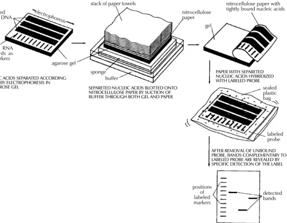
and because of the lysis-resistant cell wall of my-cobacteria, culture conditions and DNA extraction procedures are quite demanding. Strains of M. tu-berculosis and other mycobacteria have been dif-ferentiated with PFGE (Varnerot et al. 1992, Zhang et al. 1992) but the main limitation of the tech-nique is that the small polymorphism characteris-tic for different strains will not always produce different patterns. DNA polymorphism can also be demonstrated through hybridization of digested nucleic acids with genomic DNA or cloned frag-ments (Fig. 1). Several strategies for differentia-tion of strains of M. tuberculosis through hybrid-ization pattern analysis were described. One of these, using a total DNA probe and four-base re-striction enzymes, could differentiate between a small number of strains of M. tuberculosis ana-lyzed (Ross et al. 1991) but the use of the com-plete genome as a probe usually results in consid-erable background and affects the interpretation of the results. An alternative is the use of repetitive DNA sequences as probes. Some organisms have been typed by ribotyping, a technique using part of the coding sequence for rRNA as a probe; how-ever mycobacteria are characterized by the
pres-Fig. 1: detection of specific DNA molecules by gel-transfer hybridization.
unlabeled RNA or DNA
labeled RNA standards as size markers
agarose gel
sponge buffer
electrophores is
stack of paper towels
nitrocellulose paper
gel
nitrocellulose paper with tightly bound nucleic acids
sealed plastic bag
labeled probe
positions of labeled markers
detected bands
NUCLEIC ACIDS SEPARATED ACCORDING TO SIZE BY ELECTROPHORESIS IN
AN AGAROSE GEL SEPARETED NUCLEIC ACIDS BLOTTED ONTO NITROCELLULOSE PAPER BY SUCTION OF BUFFER THROUGH BOTH GEL AND PAPER
PAPER WITH SEPARETED NUCLEIC ACIDS HYBRIDIZED WITH LABELED PROBE
302 302 302 302
302 Molecular Epidemiology of TB • PN Suffys et al.
ence of only one or two copies of rDNA (Bercovier et al. 1986), which limits the polymorphism be-tween strains belonging to this genus. At the end of the eighties, some groups used cloned repeti-tive DNA from M. tuberculosis as a probe (Eisenach et al. 1986, 1988) and one of them could actually differentiate all individual strains of M. tuberculosis analyzed (Zainuddin & Dale 1989). Very recent, the use of an oligonucleotide probe (GTG)5, that recognizes polymorphic loci was shown to differentiate between several strains of
M. tuberculosis and could have some use when other, more specific probes are not informative (Wiid et al. 1994).
CHARACTERIZATION OF REPETITIVE
SEQUENCES WITHIN THE GENOME OF M.
TUBERCULOSIS AND THEIR USE FOR
DETECTION OF STRAIN POLYMORPHISM
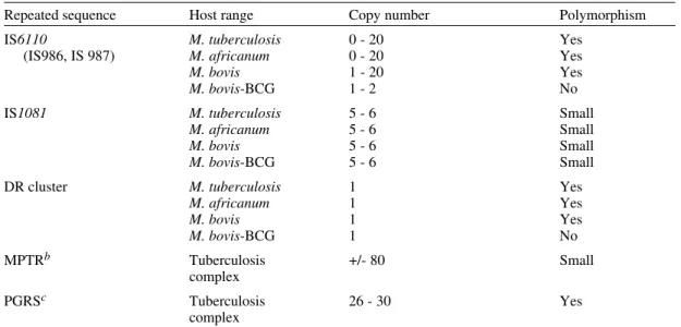
Repetitive elements and insertion sequences are frequently used as target sequences for differen-tiation between bacterial strains. Several of these elements have been characterized in the TB-com-plex and some of them are considered as mobile genetic elements (Poulet & Cole 1995a, b, Dale 1995). Table III summarizes the repetitive se-quences encountered in members of the TB-com-plex and in some additional mycobacterial species. For use of repetitive sequences in epidemiologic studies, the position of the sequence in the bacte-rial genome must be sufficiently stable but RFLP between different strains must be present.
The repeated DNA sequence with potential for epidemiological applications that was first charac-terized is an insertion sequence belonging to the enterobacterial IS3 family (McAdam et al. 1990). This sequence was detected by Southern analysis (Eisenach et al. 1988), shows homology with a plas-mid isolated from M. fortuitum (Zainuddin & Dale 1989) and, depending on the organism in which it was characterized is called IS6110 or IS986 (M. tuberculosis) or IS987 (M. bovis BCG; Eisenach et al. 1990, Hermans et al. 1990b, Thierry et al. 1990a, b). It is a 1 361 base pair long sequence that was detected exclusively in members of the TB-complex and differences of only a few nucle-otides have been detected between the sequenced copies. The sequence is flanked by two 28-base pair repeats and has two open reading frames (ORF) that show homology with genes coding for putative transposases of other elements of the IS3 family, which are typical features of mobile ele-ments. Although transposition of IS6110 has not yet been experimentally demonstrated in M. tuber-culosis, mobility of IS986 has been observed in
M. smegmatis (Fomukong & Dale 1993). The number of copies of IS6110 present in the genome is species- and strain-dependent (Table III); most strains of M. tuberculosis carry between 8 to 15 copies in different positions of the genome, al-though several single-copy strains of M. tubercu-losis have been reported. It appears that the posi-tion of IS6110 in some BCG strains and in single-copy M. tuberculosis strains is quite conserved and
TABLE III
Repetitive DNA sequences in Mycobacterium tuberculosisa
Repeated sequence Host range Copy number Polymorphism
IS6110 M. tuberculosis 0 - 20 Yes
(IS986, IS 987) M. africanum 0 - 20 Yes
M. bovis 1 - 20 Yes
M. bovis-BCG 1 - 2 No
IS1081 M. tuberculosis 5 - 6 Small
M. africanum 5 - 6 Small
M. bovis 5 - 6 Small
M. bovis-BCG 5 - 6 Small
DR cluster M. tuberculosis 1 Yes
M. africanum 1 Yes
M. bovis 1 Yes
M. bovis-BCG 1 No
MPTRb Tuberculosis +/- 80 Small
complex
PGRSc Tuberculosis 26 - 30 Yes
complex
303 303 303 303 303 Mem Inst Oswaldo Cruz, Rio de Janeiro, Vol. 92(3), May/Jun. 1997
located within a so-called “hot spot integration re-gion”; multi-copy strains nearly always carry an IS6110 within this site and few strains carry none or two copies of IS6110 in this region (Hermans et al. 1991). This “hot spot” is also characterized by the presence of virtually identical direct repeats (DR) separated by spacer DNA which, on their turn, serve as epidemiologic markers.
Collins and Stephens (1991) identified a 1 324 bp insertion sequence called IS1081, typical for the TB-complex and with homology to IS256 from
Staphylococcus aureus. Similar to IS6110, IS1081
is flanked by 15 bp inverted repeats and has an ORF with homology to a putative transposase. However, its mobility has never been demonstrated and its low transposition activity results in a very limited degree of polymorphism (Van Soolingen et al. 1992, 1993). Also, the copy-number of IS1081 is lower than that of IS6110, limiting its use in epidemiological studies to differentiation of
M. bovis-BCG from the other members of the TB-complex (Van Soolingen et al. 1992).
Several short repeated sequences have been identified in the genome of members of the TB-complex. The hot spot of integration of IS6110
contains a variable number DR of 36 bp, separated by unique spacer sequences of 35 to 41 bp. Both the number of DR (ranging from 10-50 copies) and the presence of determined spacer sequences var-ies from strain to strain, allowing the development of several fingerprinting methods (Van Soolingen et al. 1995). Strain typing on the basis of varia-tions in the DR cluster has been performed through Southern analysis (Van Soolingen et al. 1993, Sahadevan et al. 1995) and two different PCR-based methods PCR-based on variations within the DR cluster have been developed.
Another repetitive element called “major poly-morphic tandem repeat” (MPTR) was character-ized as a nonperfect repeat of 10 bp separated by 5 bp spacers. Although a copy number of around 80 was initially estimated (Hermans et al. 1992), re-cent data indicate the presence of less copies (Poulet & Cole 1994). The presence of MPTR, in contrast to the aforementioned repetitive elements, is not restricted to organisms belonging to the TB-com-plex (Table III) and it was postulated that these elements could be involved in genetic rearrange-ments (Hermans et al. 1990a). The MPTR dem-onstrated limited RFLP but a PCR-based assay, detecting the variability in the distance between IS6110 and MPTR seems to be more promising. Other mycobacteria demonstrate larger MPTR-based RFLP and the use of this sequence as an epidemiologic marker for some pathogenic species has been suggested (Hermans et al. 1992).
The most abundant repetitive element in the TB-complex is a polymorphic GC-rich repetitive sequence (PGRS) that appears to have some simi-larity with the MPTR on the basis of its sequence and host range. Several copies of this element have been cloned and sequenced independently (De Wit et al. 1990, Ross et al. 1992, Doran et al. 1993, Poulet & Cole 1994) and PGRS consists of many tandem repeats of a 9 bp GC-rich consensus se-quence. The repetitive sequence is present in at least 26 sites of the M. tuberculosis chromosome (Poulet & Cole 1995a) and was detected in some other mycobacterial species not belonging to the TB-complex (Table III). Contrary to the MPTR, PGRS is quite polymorphic and has been used in some epidemiologic studies of both human and bovine TB (Ross et al. 1992, Doran et al. 1993, Cousins et al. 1993, Dwyer et al. 1993, Van Soolingen et al. 1993). Taking advantage of the PGRS polymorphism, some PCR-based typing methods use this repetitive element as one of the target sequences.
Recently, a GC-rich repetitive sequence that could eventually be used as a target for RFLP-based fingerprinting was characterized in M. tuberculo-sis and some other mycobacteria. However, pre-liminary hybridization results on a small number of M. tuberculosis isolates indicate that the posi-tion of the sequence within the genome of M. tu-berculosis is probably too stable for its use as a marker (Verma et al. 1995).
USE OF THE INSERTION SEQUENCE IS6110
AS A TOOL FOR TYPING OF M.
TUBERCULOSIS: DEVELOPMENT OF A
STANDARDIZED PROTOCOL
IS6110: occurrence, characteristics and some limitations - Even before IS6110 was sequenced and completely characterized, its value for epide-miological studies of TB had already been sug-gested since unrelated strains of M. tuberculosis
showed different RFLP patterns after hybridiza-tion with cloned fragments containing the inser-tion sequence (Eisenach et al. 1988, Zainuddin & Dale 1989), an observation that was later confirmed by many other groups. Initial studies on stability, polymorphism and copy number of IS6110 dem-onstrated the important epidemiological value of this system. Hermans et al. (1990b) showed that
suffi-304 304 304 304
304 Molecular Epidemiology of TB • PN Suffys et al.
ciently mobile to differentiate between epidemio-logically unrelated strains; yet strains from an out-break or from household contacts show identical fingerprints. Also, isolation of several isolates from the same individual over a period of one or several years yielded the same DNA pattern (Van Soolingen et al. 1991, Otal et al. 1991, Cave et al. 1994), and the RFLP pattern of strains isolated from patients before and after development of resistance
in vivo (Godfrey-Faussett et al. 1993) also remained the same. Several studies showed the presence of 5 to 15 copies per genome using strains from Afri-can and European origin but the number of copies could be related with their geographic origin, as suggested by the higher copy number found in iso-lates from Hong Kong (Das et al. 1993). Van Soolingen et al. (1993) reported that many strains from the Far East contain a single IS6110 element as was also the case for about 30% of the strains derived from India; in another study, 40% of the strains from Madras were reported to have either no or a single copy of IS6110 (Das et al. 1995). Within a total of 41 strains from patients of Viet-namese origin, five strains had a single IS6110 copy while four strains did not contain the sequence (Yuen et al. 1993). Several single-copy strains were also characterized from Malaysia, Tanzania and Oman (Fomukong et al. 1994). Interestingly, many of the low copy number strains isolated in Tanzania (Yang et al. 1995b) and Denmark (Yang et al. 1995a), were obtained from patients origi-nating from Asia. As in most of the cases where only one IS6110 copy is present, the insertion se-quence is localized on a 1.5-kb PvuII fragment, and in this case, many of the epidemiologically unrelated M. tuberculosis strains give the same RFLP pattern. It was also observed that some un-related M. tuberculosis strains carrying two IS6110
elements at identically sized PvuII fragments could be distinguished by other typing methods, demon-strating that additional typing techniques have to be considered when dealing with low IS6110 copy-number strains (Van Soolingen et al. 1993).
A standardized protocol for RFLP typing of M. tuberculosis strains - In the last couple of years, several molecular methods for typing M. tubercu-losis strains have been developed. Although us-ing the same target sequence in their hybridization system, some laboratories used different restric-tion enzymes or different parts of a specific se-quence as a probe to generate RFLP patterns, mak-ing it impossible to compare fmak-ingerprints from dif-ferent laboratories. Even when adopting the same methodology, variations in experimental conditions and in laboratory practices generated RFLP-pat-terns that are difficult to reproduce. Therefore, a
standardized method for RFLP typing of M. tu-berculosis strains has been developed (Van Emb-den et al. 1993a) consisting basically of extraction and purification of genomic DNA of M. tubercu-losis from a well-grown culture; digestion of nucleic acids with PvuII; separation of DNA frag-ments under well-defined conditions on agarose gel, transfer of the DNA to a nylon membrane and hybridization with a fragment of the IS6110 se-quence generated by PCR (Fig. 1). Importantly, a specific amount of internal molecular weight marker is mixed with each digested DNA sample and after hybridization with the IS6110 probe, a second hybridization with internal marker is per-formed. Besides these internal markers, an exter-nal marker consisting of PvuII-digested DNA from a reference M. tuberculosis strain (Mt14323) is included in each gel. This method has several ad-vantages since many parameters influencing the quantity and position of the bands of the RFLP pattern can be compensated for; moreover, IS6110
has an internal PvuII site and the probe hybridizes with one side only, generating bands with compa-rable intensity, rendering analysis more simple.
305 305 305 305 305 Mem Inst Oswaldo Cruz, Rio de Janeiro, Vol. 92(3), May/Jun. 1997
present, similarity coefficients become more reli-able (Godfrey-Faussett et al. 1993). Recently, soft-ware packages such as GelCompar (Applied Maths) and BioImage (Millipore Systems) have been constructed for the storage and analysis of a large amount of normalized DNA patterns for vi-sualization of strain relation and comparison of a certain DNA pattern with a database.
APPLICATIONS OF MOLECULAR STRAIN TYPING AS AN ADDITIONAL TOOL FOR
EPIDEMIOLOGY OF TUBERCULOSIS: A REVIEW OF THE LITERATURE
Outbreaks and detection of index cases - By means of traditional identification methods such as tuberculin skin test conversion of contacts with a suspected index case and their follow up, spread of new infections with TB in institutional settings was well recognized before the use of fingerprint-ing (Kfingerprint-ing & Geis 1977, Stead et al. 1985, Nardell et al. 1986). This methodology has specific limi-tations and the use of molecular strain identifica-tion furnishes more convincing evidence for whether a single strain is being transmitted in a certain setting. Although identity of fingerprints from two strains does not prove that one individual has passed the strain to the other, combination of traditional and molecular investigation can lead to the index case. The first evaluation of IS6110 -RFLP for epidemiological purposes was published by Hermans et al. (1990b), comparing 15 strains from various regional hospitals and laboratories. Nine strains were found to have the same finger-print and all originated from patients that had been treated by the same physician. Pioneer work on the use of fingerprinting for tracing outbreaks de-tected an epidemic among homeless people in Amsterdam, identified two persons contaminated during bronchoscopy on the same day in the same clinic, demonstrated that a woman contaminated her husband and showed transmission within
cer-tain families in Czechoslovakia (Van Soolingen et al. 1991), identified sources of infection in a pub and a discotheque (Van Embden et al. 1993b) and demonstrated infection by a neighbor (Godfrey-Faussett et al. 1992a). The importance of trans-mission of TB within shelters for the homeless was also demonstrated in a study with TB patients from Australia (Dwyer et al. 1993) and by means of con-tact investigation by IS6110-RFLP, it was shown that TB was actively transmitted in a neighborhood bar in Minneapolis, where an index case infected 41 of his contacts (Kline et al. 1995). These stud-ies clearly demonstrate the value of RFLP analy-sis in situations where traditional contact tracing alone would not be able to detect clusters of TB; it also shows that single, highly infectious cases can have a severe influence on TB transmission within a community and, as a consequence, on TB pro-grams. Tuberculosis has been known to be en-demic in correctional facilities for many years (Stead 1978) and active transmission of TB in a New York jail has been confirmed by phage typ-ing and RFLP-analysis (Pelletier et al. 1993). Most outbreaks of TB have been observed in hospitals and patient care settings and many of these involve recent transmission of TB to patients with immunodepression, as demonstrated in a renal transplant unit (Jereb et al. 1993), in housing fa-cilities or care centers for HIV-infected persons (Daley et al. 1992, Kent et al. 1994) and in hospi-tal facilities (Edlin et al. 1992, Coronado et al. 1993, Beck-Sagué et al. 1992, Wenger et al. 1995). In 1990 through 1992, the Centers for Disease Con-trol (CDC) investigated seven outbreaks of MDR-TB involving over 200 persons with most cases occurring among persons with HIV infection (CDC unpublished data) and one of these outbreaks in-volved a M. tuberculosis strain resistant to seven drugs causing extremely high mortality rates. Not only HIV-infected patients but also health care workers such as nurses, house keepers and
306 306 306 306
306 Molecular Epidemiology of TB • PN Suffys et al.
tory workers (HIV-infected or not) developed MDR-TB during an outbreak (Jereb et al. 1995). Using an alternative typing technique by PFGE, it was shown that even upon short exposure to an index case, high rates of tuberculosis infection and disease in health care workers can occur (Griffith et al. 1995).
Community transmission - By the use of fin-gerprinting, one can nowadays better identify risk factors for transmission of TB within a certain com-munity. A study using IS6110-RFLP recently showed that extensive transmission of TB is tak-ing place in Europe in the same social setttak-ings as those reported in the USA (Genewein et al. 1993); more than one quarter of the patients in Berne (Switzerland) were linked to other patients and mainly associated with outbreaks within families or with drug addicts and homeless persons. Im-portantly, transmission was not limited to these high risk groups as there was a “spill over” to the gen-eral population. Other studies on transmission of TB in large urban settings through cluster analysis (Alland et al. 1994, Small et al. 1994, Friedman et al. 1995a, Sepkowitz et al. 1995) demonstrated that besides drug resistance, young age, race, social sta-tus and being health care worker are risk factors.
Influence of HIV on TB transmission - Infec-tion with HIV and development of AIDS has a se-rious impact on the global epidemiology of TB, due to the increase of the risk for developing TB. Impaired immunity allows latent M. tuberculosis
to proliferate and evolve to active disease; also, whether or not previously infected with TB, HIV-infected persons have less control on new infec-tions. Although reactivation of TB was thought to be the major cause of disease among AIDS patients, few data were available to determine the relative contribution of reactivation and recent (re-)infec-tion. During the last couple of years, molecular typing methods have demonstrated the important contribution of recent transmission of M. tubercu-losis for rapid development of TB in HIV-serop-ositive individuals. First, as stated earlier, several outbreaks with drug susceptible or MDR-TB strains have been observed among HIV-infected patients. Also, typing of representative M. tuberculosis
strains in community based studies has revealed that persons with HIV have a considerable higher risk of developing a recently transmitted TB (Alland et al. 1994, Tabet et al. 1994, Shafer et al. 1995, Sepkowitz et al. 1995). Comparing strains from HIV-seropositive patients with those from AIDS patients, increased probability of belonging to a recent transmission was found to be associ-ated with advanced immunodepression (Small et al. 1994). It has been suggested that strains
infect-ing AIDS patients could have some peculiar char-acteristics but a recent study done with TB patients from Tanzania, comparing fingerprints of M. tu-berculosis strains isolated from HIV-seropositive and -seronegative cases showed no evidence for particular clones playing a dominant role in a HIV-positive population (Yang et al. 1995b).
Laboratory cross-contamination - Laboratory cross-contamination is suspected when cultures yield positive for M. tuberculosis in the absence of clinical signs, and this can be the result of speci-men mix-up, of contamination of specispeci-mens or re-agents with environmental mycobacteria or be-cause of transfer from bacteria from one sample to another. Until recently, cross-contamination in the laboratory was demonstrated on the basis of anti-microbial susceptibility patterns, by some special biochemical characteristics and by phage typing, all of them typing methods with little differentiat-ing power (Jones Jr 1988, Smith & Vance 1991). Thanks to molecular typing, it has been possible to identify cross-contamination and the strain-typ-ing results were used in decisions regardstrain-typ-ing pa-tient care and for identifying problematic labora-tory procedures (Small et al. 1993a, b).
Laboratory acquired infection - Laboratory workers have a higher risk for developing TB than the general population and at least two clear cases of laboratory contamination have been supported by the use of RFLP-IS6110. In the first study, it was shown that a laboratory worker got infected with the reference strain “Erdman” with which he had been working (Mazurek et al. 1991). A sec-ond study revealed infection of a technician with a clinical isolate of M. tuberculosis during prepara-tion of inocula for identificaprepara-tion after scratching his hand on the edge of a safety cabinet (Peerbooms et al. 1995). These kind of studies allow a better knowledge on laboratory safety conditions and a better intervention in case of an accident.
307 307 307 307 307 Mem Inst Oswaldo Cruz, Rio de Janeiro, Vol. 92(3), May/Jun. 1997
RFLP-patterns was isolated, after exclusion of ac-quired resistance (Theisen et al. 1995). These kind of studies suggest that repeated resistance testing in patients with delayed response to therapy is of value.
Influence of drug resistance on fingerprint pat-terns - During the last couple of years, many re-ports on outbreaks of MDR-TB (caused by strains resistant to at least rifampicin and isoniazide) have been reported in the United States. Two important questions arise when dealing with strain typing and drug resistance: (i) does a change in drug resis-tance induce a change in the isolate’s fingerprint? and (ii) are strains with specific drug resistance patterns correlated with particular fingerprint pat-terns? To answer the first question, one has to con-sider the stability of IS6110-generated DNA pat-terns in time and it has been shown in various stud-ies that RFLP-patterns do not change rapidly in vivo which means that no major DNA rearrange-ments occur during a 2- to 3-year period (Otal et al. 1991). No changes in fingerprint were found in a large number of sequential isolates from the same patients before and after acquired antibiotic resistance (Godfrey-Faussett et al. 1993), suggest-ing the occurrence of mutations caussuggest-ing resistance instead of a selection of a resistant sub-population. Reinfection is the most probable mechanism un-derlying the fingerprint pattern change during de-velopment of drug resistance, as suggested by study of TB-patients in Hong Kong (Yuen et al. 1995) and of HIV-infected patients (Small et al. 1993b). In relation to the second question, many studies on fingerprinting of a large number of M. tuberculo-sis strains from specific populations looked for a correlation between strain clustering and drug re-sistance profile, so far without any evidence that a certain resistance is associated with a particular fingerprint type (e.g. Thierry et al. 1993). If any clustering is found among drug-resistant and MDR strains, it is because of the higher chance of these strains having been transmitted recently (Shafer et al. 1995). However, it is still possible that drug resistant strains have some peculiar behavior that influences their chance for clustering.
Population/geographic studies on transmission within and between countries -In an early report on the existence of region-specific strain relation-ships, it was suggested that this phenomenon is more frequent in high prevalence African coun-tries than in councoun-tries where prevalence is low such as The Netherlands (Van Soolingen et al. 1991). Godfrey-Faussett et al. (1992a, b), demonstrated separate clustering between strains isolated from English TB patients from the Midlands and from the Indian subcontinent (1992a) and between strains from patients from Malawi and Kenya
(1992b), confirming sufficient heterogeneity of strains from high incidence countries to allow trans-mission studies with molecular typing. Since then, a limited number of studies on RFLP analysis of strains from specific geographic regions from sev-eral countries have been published, including coun-tries with high, medium and low prevalence in TB. Takahashi et al. (1993) analyzed strains isolated in 18 Japanese sanatoria and observed a certain similarity in their banding patterns, possibly asso-ciated with a relative high prevalence of TB in Ja-pan. Although his samples seemed to be chosen randomly, strains were included from several small outbreaks which could bias towards a higher de-gree of strain similarity. In an effort to identify the importance of single cases for the spread of TB, no clustering was found among strains iso-lated from 31 patients in Austria, a low-incidence country, unless those derived from a married couple (Vogetseder et al. 1994). A large scale study was performed on 201 strains from Tunesia, a country in which TB is endemic, where apparently unre-lated strains were collected from four district hos-pitals in the northern part of the country (Chevrel-Dellagi et al. 1993). This study indicates that in some regions, microepidemics still occur while population heterogeneity in the other regions could bias to strain difference. Hermans et al. (1995) observed that 62% of the Tunisian isolates be-longed to three genetically related groupings and they explained these regional differences by the fact that transmission rates of TB in Tunesia differ greatly from province to province. The influence of geographic isolation on DNA polymorphism of
308 308 308 308
308 Molecular Epidemiology of TB • PN Suffys et al.
from Tanzania showed that a certain cluster could only be found in patients from the Kilimanjaro re-gion, while another was found in patients belong-ing to the Masai tribe (Gillespie et al. 1995). Iso-lates from the People’s Republic of China and Mongolia belonged to a distinct group of strains which share more than two-thirds of their bands and have a very particular spoligotyping pattern, the so-called “Beijing family” (Van Soolingen et al. 1995). The authors suggest that strains have diverged from a common ancestor and that “Beijing family” related strains could have spread more aggressively in the South-Asian region. Finally, considerable differences in strain clustering be-tween a high- (Ethiopia), a medium- (Tunesia) and a low-incidence (The Netherlands) country was demonstrated (Hermans et al. 1995). In this study, both clustering of identical and of similar patterns was considered and it was shown that transmis-sion rate is not necessarily directly correlated with homogeneity of the strain population; other fac-tors such as migration, regional differences within a country and BCG-vaccination could all have an influence on population dynamics of M. tubercu-losis. It was once more confirmed that in high in-cidence countries, M. tuberculosis strains have a tendency to belong to a limited number of fami-lies. Also, some patterns specific for Ethiopia or Tunesia were encountered in The Netherlands, al-lowing tracing of TB across national borders or continents.
EPIDEMIOLOGICAL STUDIES USING ADDITIONAL MOLECULAR MARKERS AND
AMPLIFICATION BASED METHODS
Molecular markers different from IS6110
-Because a significant number of strains show a particularly low copy number or even lack IS6110,
and unrelated low-copy number strains often gen-erate the same fingerprint (see above), Van Soolingen et al. (1993) analyzed the differentiat-ing power of other genetic markers on a large num-ber of strains, including strains that showed the same one or two band RFLP pattern after IS6110 -RFLP. They showed that DR and PGRS-based fin-gerprinting can differentiate low-IS6110 copy number strains but that both IS6110 and PGRS are the best markers known for differentiating M. tu-berculosis strains; IS6110 still has more discrimi-native power when a large number of copies are present. The same conclusion was drawn from a study comparing both typing methods on strains derived from patients before and after developing drug resistance (Yuen et al. 1995) and in a study showing transmission of TB among the homeless in Australia (Dwyer et al. 1993). The DR region is also sufficiently polymorphic to allow
differentia-tion of low IS6110 copy number strains while both IS1081 and MPTR are too stable to allow signifi-cant differentiation. The larger discriminative power of IS6110 as compared to DR has also been shown for strains from Denmark and again, IS1081
was found little discriminatory (Yang et al. 1994).
Amplification based methods - One of the ma-jor limitations of RFLP-based typing systems is the requirement of a well grown culture for DNA extraction. The time-lag between isolation of M. tuberculosis from a clinical sample and the growth of a mycobacterial culture is often too long, some-times resulting in the use of non optimized treat-ment protocols and, as a consequence, spread of eventual MDR bacilli. Therefore, quicker typing techniques have been developed and most of these are based on enzymatic amplification of a deter-mined DNA sequence of the M. tuberculosis ge-nome, including often IS6110. One of the biggest advantages of PCR-based typing methods, al-though not always elaborated in the studies pre-senting these systems, is the possibility of typing
M. tuberculosis strains directly in clinical samples, increasing substantially the speed of identification of the organism. Moreover, this kind of analysis can be fairly easily automated and could be used when only non-viable isolates are available or when isolates can not be restricted. Most of the systems described however lack reproducibility or have less discriminating power than IS6110-RFLP (Table IV). One of these systems is called “amplityping” and is based on the use of oligonucleotide primers, hybridizing with the ends of IS6110 and generat-ing a PCR reaction directed away from the inser-tion sequence, generating amplificainser-tion products through primer annealing with neighboring IS6110
309 309 309 309 309 Mem Inst Oswaldo Cruz, Rio de Janeiro, Vol. 92(3), May/Jun. 1997
drawback is the limited number and size of the generated PCR products, decreasing the informa-tion on strain relatedness in comparison with the standardized method (Plikaytis et al. 1993). Am-plification of the spacer region between the genes coding for 16S and 23S rRNA and posterior di-gestion of the amplicon with restriction enzymes was also performed for differentiation of M. tu-berculosis strains (Abed et al. 1995a). A better discrimination using this amplification system was obtained upon random amplified polymorphic DNA analysis (RAPD) of the amplified product (Abed et al. 1995b). Although RAPD performed with genomic DNA generates patterns containing too many bands for simple analysis, RAPD per-formed with the amplified 16S-23S spacer region generates patterns that can be analyzed easily and seem to have a high discriminatory power. Unfor-tunately, both the reproducibility and the final dis-criminative power of the RAPD-based method was found limited (Frothingam 1995, Glennon & Smith 1995). A recent study tested the use of a large number of arbitrary primers on total M. tuberculo-sis genomic DNA in the hope of finding a repro-ducible RAPD-based typing method and, interest-ingly, the most useful primers were homologous to insertion sequences, decreasing the “random-ness” of the PCR-system (Linton et al. 1994). Clus-tering obtained with this kind of fingerprinting was comparable to that of IS6110-RFLP, except for low copy number strains (Linton et al. 1995). How-ever, even when using these primers, and after con-siderable efforts to standardize the PCR conditions, reproducible profiles were hard to obtain. Another method described some years ago by Haas et al. (1993) is based on PCR using a primer comple-mentary to IS6110 and a second primer
comple-mentary to a linker ligated to the genomic DNA digested with a restriction enzyme. In contrast to most PCR-based typing methods, mixed-linker polymerase chain reaction (ML-PCR) typing some-times generates more bands than traditional fin-gerprinting and can be applied directly on smear-positive clinical specimens. The reproducibility and applicability of the method using it directly in clinical samples for screening of TB-patients still has to be proven but the PCR-based RFLP method was used on TB-cultures derived from renal trans-plant patients to demonstrate an outbreak of TB in a transplant unit (Jereb et al. 1993) and to demon-strate nosocomial transmission of M. tuberculosis
in an outbreak among health care workers (Zaza et al. 1995). Another elegant method for strain typ-ing of M. tuberculosis strains is based on detection of DNA polymorphism in the DR cluster and is called “direct variable repeat polymer chain reac-tion” (DVR-PCR, Groenen et al. 1993). The method is based on outward amplification of IS6110 and the DR, generating as such a strain-specific banding pattern upon hybridization with a DR-probe. One of the main advantages of the method is that the degree of DNA polymorphism is not reflected by size variation in banding pat-terns but rather by the presence or absence of cer-tain bands belonging to a multimer family, simpli-fying comparison and analysis. The method has a good differentiating power when a limited num-ber of strains is tested but the stability of the DR region is higher than that of IS6110 and several strains differentiated by RFLP analysis of Alu I-di-gested chromosomal DNA hybridized with DR were found identical by DRV-PCR. Another method that seems to have a comparable differen-tiating power as traditional fingerprinting is TABLE IV
Amplification-based typing methods for Mycobacterium tuberculosis
Method Target sequence Polymorphisma No. of bands Limitationsa
Amplityping IS6110 Similar High Low reproducibility
IS6110-ampliprinting IS6110/MPTRb Similar Low Small PCRc products
RAPDd analysis 16S-23S spacer High Low Low reproducibility
RAPD analysis insertion sequences Similar Low Low reproducibility
Mixed-linker PCR IS6110/linker High High None
Direct variable repeat PCR IS6110/DRe Similar High Complex PCR
Double repetitive element PCR IS6110/PGRSf Similar Low None
Spoligotyping DR/spacers Lower High None
310 310 310 310
310 Molecular Epidemiology of TB • PN Suffys et al.
“double-repetitive-element PCR” (DRE-PCR), based on outward amplification of IS6110 and PGRS, generating a banding polymorphism be-cause of the differences in distance between these elements (Friedman et al. 1995b). The positive predictive value of this method as compared to the traditional system was found to be 96% and the DNA patterns obtained through DRE-PCR seem to be sufficiently stable to use the method for epi-demiology. Finally, an amplification-based tech-nique called spoligotyping is based on amplifica-tion of the DR-region and subsequent differential hybridization of the amplified products with mem-brane bound oligonucleotides, complementary to the variable spacer regions localized between the DRs (Van Soolingen et al. 1995). The presence of these spacer sequences is variable in different M. tuberculosis strains and is visualized by a spot on a fixed site of the membrane, which simplifies to a large extend visual comparison and analysis of a large number of M. tuberculosis strains, allowing data to be stored and compared with a simple soft-ware. The differentiating power of spoligotyping is less than traditional fingerprinting when high IS6110 copy number strains are analyzed; in the case of strains with few IS6110 elements, discrimi-nation is better achieved with spoligotyping. This method can distinguish easily between M. tuber-culosis and M. bovis, can be used with culture posi-tive clinical samples and has been used for dem-onstration of the clonal nature of the so-called “Beijing family” from the People's Republic of China and Mongolia (Van Soolingen et al. 1995).
MOLECULAR TYPING OF BRAZILIAN
ISOLATES OF M. TUBERCULOSIS
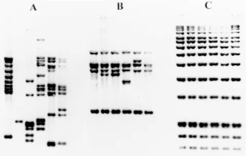
We are currently typing strains of M. tubercu-losis using the standardized IS6110-RFLP method. For this, DNA is extracted (Santos et al. 1992) from a Lowenstein-Jensen culture of tubercle bacilli, followed by analysis of nucleic acids by RFLP as recommended by a standardized protocol (Van Embden et al. 1993a). IS6110-specific banding patterns are generated and normalized by a com-puter program (GelCompar, Applied Maths, Kortrijk, Belgium) through superposition of IS6110
banding patterns with the internal markers. Fin-gerprints are then analyzed and stored in a data-base. An example of DNA fingerprints of M. tu-berculosis strains isolated from patients from Rio de Janeiro is shown in Fig. 3A. Fingerprints from Brazil, just like from many parts of the world mostly contain between 5 and 15 bands and are highly polymorphic. For comparison, fingerprints generated after hybridization with part of IS1081
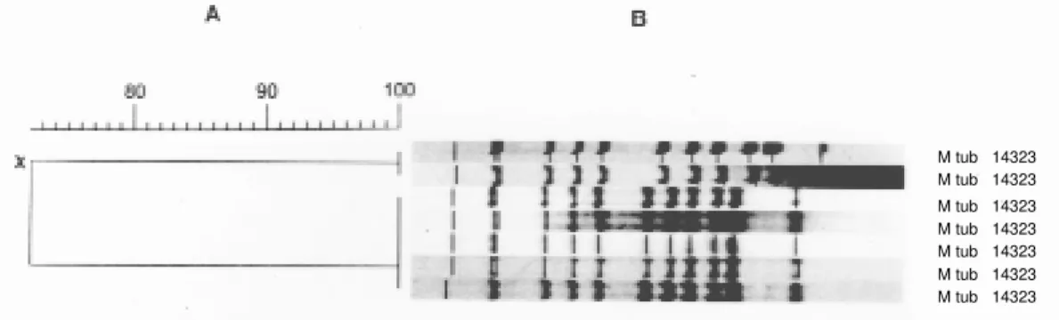
is shown in Fig. 3B, confirming the reduced dis-criminative power of this system. As a quality control for reproducibility of the RFLP method, a reference strain called Mt14323 is included in each gel. Upon normalization of the banding patterns by computer using the internal markers (Fig. 3C), the reproducibility of the fingerprinting can be determined through comparison of the fingerprints of the reference strains from different gels (Fig. 4). As an illustration, Mt14323 patterns from dif-ferent gels with or without normalization were compared, demonstrating both intra-laboratory
311 311 311 311 311 Mem Inst Oswaldo Cruz, Rio de Janeiro, Vol. 92(3), May/Jun. 1997
producibility and necessity for normalization and use of internal markers. This made it possible to compare fingerprints generated in our laboratory with fingerprints from strains from Rio Grande do Sul that were analyzed in The Netherlands (Suffys et al. 1996) and with a fingerprint database gener-ated under the EC Concerted Action on the Epi-demiology of Tuberculosis (unpublished). Further-more, our preliminary results indicate a higher rate of clustering in strains from AIDS patients from a hospital at the Oswaldo Cruz Institute (Rio de Janeiro), as compared with strains from TB patients without AIDS from Rio de Janeiro (Ivens de Araujo et al. 1995, Ivens de Araujo et al. manuscript in preparation). Although highly reproducible and of great use in specific epidemiologic studies, the standardized protocol is labor-intensive and the time lag between isolation of the strain and gen-eration of its fingerprint is long. We therefore started using the PCR-based technique called spoligotyping and are currently comparing finger-prints generated with both techniques.
CONCLUSIONS
The WHO estimates that, if the effectiveness of TB control does not improve substantially, the number of TB cases will reach 10 million in the year 2000, due to a combination of demographic factors, population movements, the expanding HIV epidemic and increasing drug resistance (Kochi 1994). Some drastic control measures should be taken both to protect individuals from TB and to interrupt the cycle of transmission. Improvement of socio-economic conditions in developing coun-tries should have a drastic effect in reducing the disease while a better case finding and treatment will reduce considerably the risk of transmission (Rodrigues & Smith 1990). Research priorities should be focused on improved epidemiological studies, on better prevention and diagnosis, on de-velopment of more efficient chemotherapeutic
regi-mens, on better program design and on understand-ing of the interaction of HIV and TB (Murray et al. 1990).
Molecular strain typing can be used as an addi-tional tool in epidemiological investigations in or-der to gain a better unor-derstanding of factors that influence TB transmission, for identification of risk factors of TB transmission in a community and for evaluation of regional control programs, permit-ting as such a rational design of more adequate control measures. Wenger et al. (1995) showed that transmission of MDR-TB can be stopped when CDC guidelines are fully implemented, and that molecular fingerprinting has been shown to be use-ful for decision making within control programs. The methodology used for molecular typing will strongly depend on the questions one needs to an-swer; a standardized technique should be used for comparison of strains between laboratories, re-gions, countries and continents and for the under-standing of global migration of M. tuberculosis
(Small 1995). Creation of national and interna-tional networks for fingerprinting of TB could stimulate the use and spreading of the “know how” of the technique. The creation of a European Com-munity-supported network of laboratories from Europe is an example of such an effort and more than 6 000 fingerprints are now available for com-parison (Herre Heersma, personal communication). An effort for the construction of a network of Latin-American and European laboratories is done un-der supervision of the Latin-American Network for TB (RELACTB), supported by the United Nations University. In New York, a proper network has been created (TBNetwork) with the construction of its own TB fingerprint database. If one needs more rapid identification of strain spreading, an amplification-based fingerprinting method can be used. Although several of these methods have been developed, their applicability still has to be proven by comparison with the standardized method on a
Fig. 4: reproducibility of the standardized RFLP-based strain typing and importance of pattern normalization. Hybridization patterns of the reference strain Mycobacterium tuberculosis 14323 generated from five different gels were compared through construction of a dendrogram after normalization with GelCompar. Two additional reference patterns from another gel were included before normalization (X).
312 312 312 312
312 Molecular Epidemiology of TB • PN Suffys et al.
large scale; preliminary results with some of these methods seem promising and, hopefully, it will be possible in the near future to routineously type M. tuberculosis directly in clinical samples. Finally, fingerprinting could also help resolve questions on pathogenesis of M. tuberculosis, on the relation between drug- and MDR-resistance and virulence, on influence of BCG on TB, on interactions be-tween M. tuberculosis and its host under specific circumstances and on the relation between popu-lation genetics and susceptibility to TB.
REFERENCES
Abed Y, Bolted C, Mica P 1995a. Identification and strain differentiation of Mycobacterium species on the basis of DNA 16S-23S spacer region polymor-phism. Res Microbiol 146: 405-413.
Abed Y, Davin-Reglia A, Bollet C, De Micco P 1995b. Efficient discrimination of M. tuberculosis strains by 16S-23S spacer region-based random amplified polymorphic DNA analysis. J Clin Microbiol 33:
1418-1420.
Alland D, Kalkut GE, Moss AR, McAsdam RA, Hahn JA, Bosworth W, Drucker E, Bloom BR 1994. Transmission of tuberculosis in New York City. N Engl J Med 330: 1710-1716.
Bass PF, Farer LS, Hopewell PC, Jacobs RF, Snider DE 1990. Diagnostic standards and classification of tuberculosis. Am Rev Resp Dis 142: 725-735. Baess I 1979. Deoxyribonucleic acid relatedness among
species of slowly-growing mycobacteria. Acta Pathol Microbiol Scand 87: 221-226.
Bates JH, Fitzhugh JK 1967. Subdivision of the species
M. tuberculosis by mycobacteriophage typing. Am Rev Resp Dis 96: 7-10.
Beck-Sagué C, Dooley SW, Hutton MD, Otten J, Breeden A, Crawford JT, Pitchenik AE, Woodley C, Cauthen G, Jarvis WR 1992. Hospital outbreak of multidrug-resistant M. tuberculosis infection. Factors in transmission to staff and HIV-infected patients. JAMA 268: 1280-1286.
Bercovier H, Kafri O, Sela S 1986. Mycobacteria pos-sess a surprisingly small number of ribosomal RNA genes in relation to the size of their genome. Biochem Biophys Res Commun 136: 1136-1141.
Bloom BR 1992. Back to a frightening future. Nature 358: 538-539.
Boletim de Pneumologia Sanitária, Número Especial 1993. ISSN 0103460X. Pneumologia sanitária -Periódicos. I. Brasil. Ministério da Saúde/FNS/ Centro de Referência Prof. Hélio Fraga.
Cave MD, Eisenach KD, Templeton G, Salfinger M, Mazurek G, Bates J, Crawford JT 1994. Stability of DNA fingerprint pattern produced with IS6110 in strains of M. tuberculosis. J Clin Microbiol 32: 262-266.
Chevrel-Dellagi D, Abderrahman A, Haltiti R, Koubaji H, Gicquel B, Dellagi K 1993. Large-scale DNA fingerprinting of M. tuberculosis strains as a tool for epidemiological studies of tuberculosis. J Clin Microbiol 31: 2446-2450.
Collins DM, Lisle GW 1984. DNA restriction endonu-clease analysis of M. tuberculosis and Mycobacte-rium bovis BCG. J Gen Microbiol 130: 1019-1021. Collins DM, Stephens DM 1991. Identification of an insertion sequence, IS1081, in Mycobacterium bovis. FEMS Microbiol Lett 83: 11-16.
Collins CH, Yates MD, Grange JM 1982. Subdivision of M. tuberculosis in five variants for epidemio-logical purposes: methods and nomenclature. J Hyg Camb 89: 235-242.
Coronado VG, Beck-Sague CM, Hutton MD, Davis BJ, Nicholas P, Villareal C, Woodley CL, Kilburn JO, Crawford JT, Frieden TR, Sinkowitz RL, Jarvis WR 1993. Transmission of multidrug-resistant M. tu-berculosis among persons with Human Immunode-ficiency Virus infection in an urban hospital: epide-miologic and restriction fragment length polymor-phism analysis. J Infect Dis 168: 1052-1055. Cousins DV, Williams SN, Ross BC, Ellis TM 1993.
Use of a repetitive element isolated from M. tuber-culosis in hybridization studies with Mycobacterium bovis: a new tool for epidemiological studies of bo-vine tuberculosis. Vet Microbiol 37: 1-17. Das S, Chan SL, Allen BW, Mitchison DA, Lowrie DB
1993. Application of DNA fingerprinting with IS986 to sequential mycobacterial isolates obtained from pulmonary tuberculosis in Hong Kong before, dur-ing and after short-course chemotherapy. Tubercle Lung Dis 74: 47-51.
Das S, Paramasivan CN, Lowrie DB, Prabhakar R, Narayanan PR 1995. IS6110 RFLP typing of clini-cal isolates of M. tuberculosis from patients with pulmonary tuberculosis in Madras, South India.
Tubercle Lung Dis 76: 550-554.
Dale JW 1995. Mobile genetic elements in mycobacte-ria. Eur Respir J 8 S20: 633s-648s.
Daley CL, Small PM, Schecter GF, Schoolnik GK, McAdam RA, Jacobs Jr WR, Hopewell PC 1992. An outbreak of tuberculosis with accelerated pro-gression among persons infected with the human immunodeficiency virus. N Engl J Med 326: 231-235.
De Wit D, Steyn S, Shoemaker S, Sogin M 1990. Di-rect detection of M. tuberculosis in clinical speci-mens by DNA amplification. J Clin Microbiol 28: 2437-2441.
Dolin, PJ, Raviglione MC, Kochi A 1994. Global tu-berculosis incidence and mortality during 1990-2000. Bull WHO 72: 213-220.
Doran TJ, Hodson AL, Davies JK, Radford AJ 1993. Characterization of a highly repeated DNA sequence from Mycobacterium bovis. FEMS Microbiol Lett 111: 147-152.
Dutt AK, Meht B, Whitaker BJ, Westmoreland H 1994. Outbreak of tuberculosis in a church. Chest 107: 447-452.
Dwyer B, Jackson K, Raios K, Sievers A, Wilshire E, Ross B 1993. DNA restriction fragment analysis to define an extended cluster of tuberculosis in home-less men and their associates. J Infect Dis 167: 490-494.
Will-313 313 313 313 313 Mem Inst Oswaldo Cruz, Rio de Janeiro, Vol. 92(3), May/Jun. 1997
iams J, Sordillo EM, Ong KR, Kilburn JO, Dooley SW, Castro KG, Jarvis WR, Holmberg SD 1992. An outbreak of multidrug-resistant tuberculosis among hospitalized patients with the acquired im-munodeficiency syndrome. N Engl J Med 326:
1514-1521.
Eisenach KD, Cave MD, Bates JH, Crawford JT 1990. Polymerase chain reaction amplification of a repeti-tive DNA sequence specific for M. tuberculosis. J Infect Dis 161: 977-981.
Eisenach KD, Crawford JT, Bates JH 1986. Genetic relatedness among strains of the M. tuberculosis
complex. Am Rev Respir Dis 133: 1065-1068. Eisenach KD, Crawford JT, Bates JH 1988. Repetitive
DNA sequences as probes for M. tuberculosis. J Clin Microbiol26: 2240-2245.
Fadinho FC, Kritski AL, Conde H, Fonseca LS 1995. Drug susceptibility of Mycobacterium tuberculosis iso-lated from HIV infected and non infected in Rio de Janeiro (Brazil). Tubercle Lung Dis 76: S2, 94. Fine PE 1989. The BCG story: lessons from the past
and implications for the future. Rev Infect Dis 11: S353-S359.
Fomukong NG, Dale JW 1993. Transpositional activ-ity of IS986 in Mycobacterium smegmatis. Gene 130: 99-105.
Fomukong NG, Tang TH, AL-Maamary S, Ibrahim WA, Ramayah S, Yates M, Zainuddin ZF, Dale JW 1994. Insertion sequence typing of M. tuberculosis: char-acterization of a widespread subtype with a single copy of IS6110. Tubercle Lung Dis 75: 435-448. Friedman CR, Stoeckle MY, Johnson JR. WD, Riley
LW 1995b. Double-repetitive-element PCR method for subtyping M. tuberculosis clinical isolates. J Clin Microbiol 33: 1383-1384.
Friedman CR, Stoeckle MY, Kreiswirth BN, Johnson Jr WD, Manoach SM, Berger J, Sathianathan K, Hafner A, Riley LW 1995a. Transmission of multidrug-resistant tuberculosis in a large urban setting. Am J Respir Crit Care Med 152: 355-359.
Frothingham R 1995. Discrimination of M. tuberculo-sis strains by PCR. J Clin Microbiol 33: 2801. Frothingham R, Hills HG, Wilson KH 1994. Extensive
DNA sequence conservation throughout the M. tu-berculosis complex. J Clin Microbiol 32: 1639-1643.
Genewein A, Telenti A, Bernasconi C, Mordasini C, Weiss S, Maurer A-M, Rieder HL, Schopfer K, Bodmer T 1993. Molecular approach to identifying route of transmission of tuberculosis in the commu-nity. The Lancet 342: 841-844.
Gillespie SH, Kennedy N, Ngowi FI, Fomukong NG, Al-Maamar YS, Dale JW 1995. Restriction frag-ment length polymorphism analysis of M. tubercu-losis isolated from patients with pulmonary tuber-culosis in Northern Tanzania. Trans R Soc Trop Med Hyg 89: 335-338.
Glennon M, Smith T 1995. Can random amplified poly-morphic DNA analysis of the 16S-23S spacer re-gion of M. tuberculosis differentiate between iso-lates? J Clin Microbiol 33: 3359.
Godfrey-Faussett P, Stoker NG 1992b. Aspects of
tu-berculosis in Africa. 3. Genetic ‘fingerprinting’ for clues to the pathogenesis of tuberculosis. Trans R Soc Trop Med Hyg 86: 472-475.
Godfrey-Faussett P, Mortimer PR, Jenkins PA, Stoker NG 1992a. Evidence of transmission of tuberculo-sis by DNA fingerprinting. Brit Med J 305: 221-223.
Godfrey-Faussett P, Stoker NG, Scott JA, Pasvol G, Kelly P, Clancy L 1993. DNA fingerprints of M. tuberculosis do not change during the development of rifampicin resistance. Tubercle Lung Dis 74: 240-243.
Grange JM, Laszlo A 1990. Serodiagnosis tests for tu-berculosis: a need for assessment of their predictive accuracy and acceptability. Bull WHO 68: 571-576. Grange JM, Stanford JL 1994. Dogma and innovation in the global control of tuberculosis. J R Soc Med 87: 272-275.
Griffith DE, Hardeman JL, Zhang Y, Wallace RJ, Mazurek GH 1995. Tuberculosis outbreak among healthcare workers in a community hospital. Am J Respir Crit Care Med 152: 808-811.
Groenen PM, Van Bunschoten AE, Van Soolingen D, Van Embden JD 1993. Nature of DNA polymor-phism in the direct repeat cluster of M. tuberculosis, application for strain differentiation by a novel method. Mol Microbiol 10: 1057-1065.
Grzybowski S 1991. Tuberculosis in the third world.
Thorax 46: 689-691.
Grzybowski S, Enarson DA 1978. Results in pulmo-nary tuberculosis patents under various treatment program conditions. Bull Int Union Against Tub 53: 70-75.
Haas WH, Butler WR, Woodley CL, Crawford JT 1993. Mixed-linker polymerase chain reaction: a new method for rapid fingerprinting of isolates of the M. tuberculosis complex. J Clin Microbiol 31: 1293-1298.
Hermans PW, Messadi F, Guebrexabher H, Van Soolingen D, De Haas PE, Heersma H, De Neeling H, Ayoub A, Portaels F, Frommel D, Zribi M, Van Embden J 1995. Analysis of the population struc-ture of M. tuberculosis in Ethiopia, Tunesia and the Netherlands: usefulness of DNA typing for global tuberculosis epidemiology. J Infect Dis 171: 1504-1513.
Hermans PW, Schuitema AR, Van Soolingen D, Verstynen CP, Bik EM, Thole JE, Kolk AH, Van Embden JD 1990a. Specific detection of M. tuber-culosis complex strains by polymerase chain reac-tion. J Clin Microbiol 28: 1204-1213.
Hermans PW, Van Soolingen D, Dale JW, Schuitma AR, McAdam R, Catty D, Van Embden JD 1990b. In-sertion element IS986 from M. tuberculosis: a use-ful tool for diagnosis and epidemiology of tubercu-losis. J Clin Microbiol 28: 2051-2058.
Hermans PW, Van Soolingen D, Bik EM, De Haas PE, Dale JW, Van Embden JD 1991. Insertion element IS987 from Mycobacterium bovis BCG isolated in a hot-spot integration region for insertion elements in