○ ○ ○ ○ ○ ○ ○ ○ ○ ○ ○ ○ A B S T R A C T○ ○ ○ ○ ○ ○ ○
C
ase Repor
t
Total occlusion of the common carotid artery is an important cause of brain ischemia, but it is not as frequent as carotid bifurcation stenosis. Moore et al. (1967) found it in 5% of their patients.1 In our own much smaller experience we have found similar rates: out of 65 patients operated on consecutively for ce-rebral ischemia, there were three with com-mon carotid occlusion, representing 4.6% (Figure 1).2
Surgical treatment for occlusive disease of the supra-aortic trunk is not done very often. There is controversy about access: trans-tho-racic or extra-thotrans-tho-racic and, in the last few de-cades, endovascular treatment has been pre-ferred in certain cases.3-5
Our purpose was to report on three cases that demonstrate the feasibility of ring-stripping retrograde common carotid endarterectomy.
○ ○ ○ ○ ○ ○ ○ ○ ○ ○ ○ ○CASE REPORT○ ○ ○ ○ ○ ○ ○ ○
Case 1: G.S.S., an 81-year-old female
patient who was diabetic, hypertensive and a non-smoker, presented critical ischemia of the right lower limb with gangrene of the extrem-ity of the foot. Popliteal and distal pulses were absent from both lower limbs. The right ca-rotid pulse was absent and a bruit was heard at the level of the left carotid bifurcation. Ar-teriography revealed total obstruction of the right common carotid, but the right carotid bifurcation was not adequately seen. There was 60% stenosis at the left internal carotid ori-gin and bilateral femorotibial obstruction with only the peroneal artery as a run-off on the right limb. Exploration of the right carotid bifurcation, common carotid endarterectomy
and femoral-peroneal bypass were scheduled to be done at same time. The indication for exploration of the right carotid artery bifur-cation was based on the premise that, if right internal carotid artery flow could be restored, the risk of brain ischemia after femorotibial reconstruction would be lower: there were no cerebral ischemic symptoms.
On November 19, 1992, she underwent surgery. Surgical exploration of the carotid bi-furcation revealed total occlusion of the in-ternal and common carotid arteries. The en-darterectomy of the external carotid origin was performed via an arteriotomy at the carotid bulb and, using the same cleavage plane, the ring-stripper was introduced back up through the common carotid artery until it reached the brachiocephalic trunk (Figure 2). At this point, resistance to the passage of the ring-stripper decreased abruptly, as the core was spilled out through the arteriotomy by means of the blood flow. The ring was introduced again to deal with any debris, and the arteri-otomy was sutured. The pulse was restored to the external and common carotid arteries. This operation was followed by a femoral-peroneal bypass, but the saphenous vein was not long enough and a superficial femoral endarterec-tomy was done in association with the super-ficial femoral-peroneal bypass.
This restoration occluded on the follow-ing day and thigh amputation was performed eight days later. On the second postoperative day, the patient presented a transient ischemic attack, characterized by left hemiparesis that regressed totally. The patient refused left ca-rotid endarterectomy after recovering, and was discharged from hospital. After a seven-year • Eduardo Toledo de Aguiar
• Alex Lederman
• Patrícia Matsunaga
Ring-stripping retrograde
common carotid
endarterectomy: case report
Hospital Sírio-Libanês and Disciplina de Cirurgia Vascular,
Departamento de Cirurgia, Universidade de São Paulo, São Paulo, Brazil
CONTEXT: Total occlusion of the common carotid is rare and the indications and techniques for surgi-cal treatment are still a matter of controversy.
OBJECTIVE: To demonstrate the feasibility of retrograde common carotid endarterectomy.
DESIGN: Retrospective case report study.
SETTING: Tertiary care private hospital.
PARTICIPANTS: Three patients underwent ring-strip-ping retrograde common carotid endarterectomy. Their ages were 81, 68 and 65 years. All were hypertensive with generalized atherosclerosis, two had diabetes mellitus, and one had undergone coronary artery bypass some years earlier and had non-dialytic chronic renal insufficiency. Symp-toms of brain ischemia were present in two pa-tients. All patients had total occlusion of the com-mon carotid, extending from the origin to the bi-furcation and localized in the right common rotid in two cases. In two cases the internal ca-rotid artery was also occluded.
MAIN MEASUREMENTS: Postoperative early mor-tality and stroke rate, and the medium and long-term endarterectomy patency.
RESULTS: There were no deaths. One patient had a transient ischemic attack. All endarterectomies were patent after eight months, four years and seven years of follow-up.
CONCLUSION: There is low mortality, and the pro-cedure can be done through only one cervical incision. Tandem lesions of the carotid arteries can be treated together. It is suitable for long total occlusions of the common carotid, and long-term patency.
KEY WORDS: Carotid artery. Arteries. Endarte-rectomy. Atherosclerosis. Cerebrovascular disease.
São Paulo Medical Journal - Revista Paulista de Medicina
155
follow-up the patient is doing well and has not had any stroke or transient ischemic at-tack. A duplex-scan has shown some irregu-larities on the right common carotid artery, but without any significant stenosis.
Case 2: A.T.B., a 65-year-old male patient,
was hypertensive, non-diabetic and a smoker. He was a businessman taken off his duties due to memory loss and disorientation. The physi-cal examination revealed absence of the left tem-poral and common carotid pulses. All other pulses were normal. There was bruit at the level of the right carotid bifurcation. Computerized tomography of the brain revealed multiple is-chemic lesions in both hemispheres and arteri-ography showed total occlusion of the left com-mon carotid artery and 80% stenosis of the right internal carotid artery. The left carotid bifurca-tion was not adequately seen.
He was operated on November 17, 1993. Exploration of the left carotid bifurcation re-vealed totally occluded internal and common carotid arteries and severe stenosis of the left external carotid artery origin. As described above, the arteriotomy at the carotid bulb al-lowed endarterectomy of the external carotid artery to be performed, and the introduction of the ring-stripper back up through the com-mon carotid until the resistance decreased and the core spilled out. After suturing the arteri-otomy, pulsatile blood flow was restored in the left common and external carotid arteries.
One week later, right carotid bifurcation endarterectomy was done in the normal man-ner. The patient was discharged from hospital on the 5th postoperative day after this second operation with no complications. He was fol-lowed up for four years, until April 1997, by which time he had partially returned to his pro-fessional activities. The duplex-scan that was performed showed some irregularities on both carotid arteries, but with no significant stenosis.
Case 3: A.J.D. was a 68-year-old diabetic
and hypertensive patient, who eight years ear-lier had undergone a coronary artery bypass. At that time, he quit his smoking habit and had manifestations of leprosy, which were cured. From that time onwards, he suffered from non-dialytic renal insufficiency. The symptoms reported were one episode of left hemiparesis that lasted for 20 minutes and disappeared completely one-and-a-half months before coming to us, and an episode of loss of consciousness that led him to a car crash 15 days before coming to us. He also complained of right-eye vision blurring. The temporal and right carotid pulses were not palpable, nor were the dorsalis pedis and pos-terior tibial on the right lower limb. An
is-chemic lesion in the right brain hemisphere was detected by computerized tomography. A duplex-scan revealed obstruction of the right common carotid, with pervious internal and external carotid arteries. Angiotomography confirmed the duplex-scan findings.
On February 25, 2001, the patient un-derwent retrograde common carotid endart-erectomy using the ring-stripper. After explo-ration of the right carotid bifurcation, the in-ternal and exin-ternal carotid arteries were found to be patent. The common carotid artery was totally occluded and there was an atheroma plaque at the internal carotid artery origin. Endarterectomy of the carotid bifurcation and common carotid artery was performed as de-scribed earlier.
The patient was discharged from hospital after 24 hours with no complications. All symptoms have disappeared except for a sen-sation of dizziness that occurs episodically. Control duplex-scans have revealed full-length dilatation (1.5 cm) of the common carotid artery and 50% to 70% stenosis at its origin. The patient continues under observation.
○ ○ ○ ○ ○ ○ ○ ○ ○ ○ ○ ○ DISCUSSION○ ○ ○ ○ ○ ○ ○ ○
Obstructive disease of the common ca-rotid artery is not often seen. Its incidence
varies from 1% to 5%.6 Controversy still re-mains with regard to the indications and tech-niques for surgical treatment.
The symptoms of brain ischemia may be caused by two factors: hemodynamic changes and embolism. Hemodynamic factors may be dominant, with brain and eye hypoperfusion being responsible for transient ischemic attacks or infarcts in both the carotid and vertebro-basilar regions. Embolic events may occur even when there is total common carotid occlusion. Some surgeons believe that occlusions origi-nate at the end of thrombosed segments of com-mon carotid artery at the carotid bifurcation or at the end of the internal carotid artery at the circle of Willis.7 Moore et al. (1967) stated that total occlusion of the common carotid artery originates from atheroma plaque located at one of the following four points: 1. the origin of the common carotid; 2. the innominate artery or aortic arch; 3. the middle segment of the com-mon carotid (between the origin and the bifur-cation); or 4. immediately before the bifurtion and origin of the internal and external ca-rotid arteries. Moore et al. (1967) also found the most frequent lesions provoking total occlu-sion of the common carotid to be those located at the bifurcation, with thrombosis progressing as far as the common carotid origin at the in-nominate artery or aortic arch.
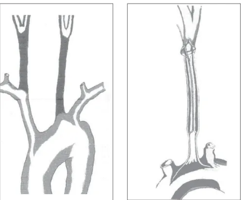
Figure 1. Site of common carotid obstruetions. Right eqmmon carotid - 2 cases. Left common carotid - 1 case.
Figure 2. Ring-stripping retrograde common carotid endarterectomy technique.
São Paulo Medical Journal - Revista Paulista de Medicina
156
Atheroma plaques located at the origin of the common carotid artery rarely progress to total occlusion.1 Frequently, the atherosclerotic lesions on supra-aortic trunks are multiple. Vogt et al. (1982) studied brachiocephalic ar-terial reconstruction in 97 patients and found that 57 of them had multiple stenoses greater than 50%.8 The extent and multiple locations of atheroma plaques and obstructions cause the symptoms to be varied and related both to the carotid and vertebro-basilar regions. In such patients, atherosclerotic disease is very often generalized, with coronary artery dis-ease found in 45% and peripheral arterial ob-structive disease (PAOD) in 27%. Hyperten-sion is also present in 50% of such patients.2,7 All the patients presented here were hy-pertensive and had diffuse atherosclerotic dis-ease compromising the coronary and periph-eral arteries. One patient had diabetes melli-tus. All had atheroma plaques at the carotid bifurcation, in continuity with total occlusion of the common carotid artery. Typical symp-toms of extracranial arterial obstructive dis-ease occurred in case 3, and the ischemic brain lesion uncovered by the CT scan was ipsilat-eral to common carotid obstruction. The pa-tient in case 2 presented symptoms of demen-tia, but the CT scan revealed multiple ischemic lesions in both brain hemispheres, and so it is possible that the symptoms were ischemic in origin, especially because of the patient’s im-provement after surgical therapy. In case 1, the indication was prevention of stroke in a patient undergoing major surgery: this is ab-solutely a doubtful indication. Restorative surgery of supra-aortic trunks has been indi-cated in symptomatic patients.10-12
Vessel patency distal to arterial occlusion (runoff vessels) is important for surgical plan-ning. Arteriography of aortic arch catheteriza-tion sometimes fails to demonstrate runoff. In earlier days, the patency of internal or external carotid arteries or both was only revealed upon surgical exploration, as happened in cases 1 and 2. More recently, colored duplex-scan and angiotomography have enabled surgeons to iden-tify distal runoff, as happened in case 3.8
Total occlusion of the common carotid artery is frequently associated with internal
carotid occlusion, leaving only the external carotid artery patent. The importance of this artery as the origin of collateral circulation to the brain has already been emphasized.13-15 AbuRhama et al. (1998), when studying the natural history of total occlusion of the bilat-eral internal carotid in a group of patients, showed that after surgical brain revascu-larization patients presented long-term mor-tality and stroke rates that were lower than for those treated conservatively. Most of them had their blood flow restored through the ex-ternal carotid artery.16
The three patients presented here illustrate this fact quite well. Two of them only had the external carotid artery as a distal runoff. Lamberth (1983) demonstrated the impor-tance of restoring blood flow to the external carotid artery, especially when there are mul-tiple supra-aortic trunk obstructions.17
Many techniques for surgical treatment of obstructive disease of the common carotid have been proposed. The mortality resulting from aorto-carotid bypasses done through sternotomy or thoracotomy used to be as high as 20%, but has fallen to 6% or less nowa-days.3,18 Thirty-four patients were operated on consecutively for aorto-carotid and/or subcla-vian bypasses with no mortality, between 1985 and 1990, in the Vascular Surgery Service of the University of São Paulo Medical School’s Clinical Hospital.19 Although mortality has fallen remarkably, the transthoracic repair of supra-aortic arteries is followed by high mor-bidity related to thoracic drainage and the multiple cervical incisions needed to complete the procedure, which implies a long postop-erative stay in hospital. Extra-thoracic recon-struction has led to lower mortality and mor-bidity. Its high long-term patency rate and capacity for alleviating symptoms has made subclavian-carotid bypass the treatment of choice for common carotid obstructions.4,20 But even with these good results, a problem still remains: the multiple incisions needed for performing this bypass. Endovascular therapy offers the opportunity to treat such patients without any incision. An early success rate of 92.3% has been reported among these pa-tients, andthere are surgeons who are
com-bining carotid bifurcation endarterectomy with intraoperative dilation of proximal com-mon carotid stenosis to treat these lesions in tandem.9,21 The endovascular technique has a limitation: not infrequently, the guide wire will not cross stenosing or total occlusive lesions, especially when these are long and compro-mise the whole length of the common carotid, as in the cases described here.
Retrograde ring-stripping common ca-rotid endarterectomy, as introduced by Moore, Blaisdell and Hall in 1967 and described in this paper, is an alternative for cases similar to those reported here, i.e. atherosclerotic ob-struction of the full length of the common carotid artery. It is important for surgeons to practice ring endarterectomies in other regions like the abdominal aorta or the femoral or iliac arteries, so that they know exactly what resis-tance is offered by subintimal tissue.22
There are few reports dealing with this technique, and we were only able to find 14 retrograde endarterectomies performed as de-scribed here by different surgeons. There were two deaths: one case in which saline endarter-ectomy was used to treat a long internal ca-rotid artery occlusion in addition to a com-mon carotid retrograde endarterectomy, and another patient who died two weeks after the operation because of congestive heart fail-ure.8,12,18 Some surgeons believe the risk of per-foration or aortic dissection is high and that this technique should be banished. This does not seem to be the case, but stenosing residual plaques in the proximal common carotid have been reported, as happened in case 3.11 Moore et al. (1967) stated that this lesion rarely progresses to total obstruction.1 But it may be a source of emboli, although this has not of-ten been reported in the literature.
In conclusion, with the results presented here and those from other services, it may be taken that the procedure can be done through only one cervical incision, and tandem lesions of the carotid arteries can be treated together. It is suitable for long total occlusions of the common carotid, and long-term patency is maintained. Bearing in mind the limitations imposed by the small sample size, it is thought that mortality is low.
São Paulo Medical Journal - Revista Paulista de Medicina
157
1. Moore WS, Blaisdell W, Hall AD. Retrograde thrombectomy for chronic occlusion of the common carotid artery. Arch Surg 1967;95:664-73.
2. Aguiar ET, Lederman A, Higutchi C, Schreen G. Early and late results of carotid endarterectomy: retrospective study of 70 op-erations. São Paulo Med J Med 2001;119:206-11. 3. Berger R, Morasch MD, Kline RA Transthoracic repair of
in-nominate and common carotid artery disease: immediate and long-term outcome for 100 consecutive surgical reconstructions. J Vasc Surg 1998;27:34-42.
4. Owens LV, Tinsley Jr. EA, Criado E, Burnham SJ, Keagy BA. Extra-thoracic reconstruction of arterial occlusive disease in-volving the supra-aortic trunks. J Vasc Surg 1995;22:217-22. 5. Pernes JM, Marjelle J, Auguste M, et al. Techniques
endoluminales de traitement des lésions de troncs supra-aortiques (a l’exception de la carotide interne). In: Techniques chirurgicales - chirurgie vasculaire. Encycl Méd Chir. Paris: Elsevier 1999;43-128:14p.
6. Hass WK, Fields WS, North RR, Kricheff II, Chase NE, Bauer RB. Joint study of extracranial carotid occlusion. II: Arteriography techniques, sites and complications. JAMA 1968;201:961-8. 7. Belkin M, Mackey WC, Pessin MS, Caplan LR, O’Donnel TF.
Common carotid artery occlusion with patent internal and
ex-○ ○ ○ ○ ○ ○ ○ ○ ○ ○ ○ ○ ○ ○ ○ ○ ○ ○ ○ ○ ○ ○ ○ ○ ○ ○ ○ ○ ○ ○ ○ ○ ○ ○ ○ ○ ○ ○ ○ ○ ○ ○ ○ ○ ○ ○ ○ ○ ○ ○ ○ ○ ○ ○ ○ ○ REFERENCES○ ○ ○ ○ ○ ○ ○ ○
ternal carotid arteries: Diagnosis and surgical management. J Vasc Surg 1993;17:1019-28.
8. Vogt DP, Hertzer NR, O’Hara PJ, Beven EG. Brachiocephalic arterial reconstruction. Ann Surg 1982;196:541-52. 9. Queral LA, Criado FJ. The treatment of focal aortic and branch
lesions with Palmaz stents. J Vasc Surg 1996;23:368-75. 10. Riles TS, Imparato AM, Posner MP, Eikelboom BC. Common
carotid occlusion. Assessment of distal vessels. Ann Surg 1983;199:363-6.
11. Kiefer E. Chirurgie des troncs supra-aortiques. In: Techniques chirurgicales - chirurgie vasculaire. Encycl Méd Chir. Paris: Elsevier 1987;43-120:16p.
12. Moore WS, Malone JM, Goldstone J. Extra-thoracic repair of branch occlusions of the aortic arch. Am J Surg 1976;132:249-57. 13. Longo PH, Zaclis J. Anastomose entre artéria carótida externa e artéria vertebral em dois casos de trombose de artéria carótida interna. Arq Neuropsiq 1958;16:353-60.
14. Almeida GM, Longo PH. Anastomose carotidobasilar: persistência da artéria trigeminal primitiva (6 casos) e da primeira artéria segmentar cervical (1 caso). Rev Paul Med 1965;67:195-202. 15. Zaclis J, Longo PH, Matosinho-França LC. Distribuição do
contraste em angiografias cerebrais: I - Angiografias carotídeas com enchimento do tronco basilar. Arq Neuropsiq 1957;15:97-124.
16. AbuRhama AF, Copeland SE. Bilateral internal carotid artery occlusion: natural history and surgical alternatives. Cardiovasc Surg 1998,6:579-83.
17. Lamberth WC. External carotid endarterectomy: indications, operative technique and results. Surgery 1983;93:57-63. 18. Crawford ES, DeBackey ME, Morris Jr. GC, Howell JF.
Surgi-cal treatment of occlusion of innominate, common carotid and subclavian arteries: a 10-year experience. Surgery 1969;65:17. 19. Aguiar ET, Mariño JCS. Surgery of the brachiocephalic arter-ies. In: Anais IMAGO 94 - Third International Congress of Angioradiology and Vascular Intervention, Miami, USA, Sep-tember 11-15, 1994.
20. Fry WR, Martin JD, Clagett GP, Fry WJ. Extra-thoracic ca-rotid reconstruction: the subclavian-caca-rotid artery bypass. J Vasc Surg 1992;15:83-9.
21. Sidhu PS, Morgan MBF, Walters HL, Baskerville PA, Fraser SCA. Combined carotid bifurcation endarterectomy and intra-operative transluminal angioplasty of proximal common carotid artery stenosis: an alternative to extra-thoracic bypass. Clin Radiol 1998;53:444-7.
22. Aguiar ET. Trombendarterectomia aortilíaco-femoral com anéis. Professorial thesis. Faculdade de Medicina da Universidade de São Paulo. 1999. 93p.
CONTEXTO: Obstruções da carótida comum
são pouco freqüentes, e as indicações e técni-cas de tratamento cirúrgico são motivo de controvérsia.
OBJETIVO: Apresentar a aplicabilidade da
endarterectomia retrógrada da carótida co-mum com anel.
TIPO DE ESTUDO: Estudo retrospectivo de
relato de casos.
LOCAL: Hospital de cuidados terciários privado
da cidade de São Paulo.
PARTICIPANTES: Três doentes operados para
endarterectomia retrógrada da carótida co-mum com anel. As idades foram 81, 68 e 65 anos, todos hipertensos com aterosclerose generalizada, dois diabéticos, um com ante-cedentes de revascularização do miocárdio e insuficiência renal não-dialítica. Dois apre-sentavam sintomas compatíveis com isquemia cerebral. As obstruções arteriais se estendiam da origem à bifurcação da carótida comum
○ ○ ○ ○ ○ ○ ○ ○ ○ ○ ○ ○ ○ ○ ○ ○ ○ ○ ○ ○ ○ ○ ○ ○ ○ ○ ○ ○ ○ ○ ○ ○ ○ ○ ○ ○R E S U M O○ ○ ○ ○ ○ ○
Eduardo Toledo de Aguiar, MD, PhD. Associate Profes-sor of Vascular Surgery, Departamento de Cirurgia, Faculdade de Medicina, Universidade de São Paulo; Vascular Surgeon at the Hospital Sírio-Libanês, São Paulo, Brazil.
Alex Lederman, MD. Resident in Vascular Surgery, De-partamento de Cirurgia, Faculdade de Medicina, Universidade de São Paulo, São Paulo, Brazil.
Patricia Matsunaga, MD. Resident in the Departamento de Cirurgia, Faculdade de Medicina, Universidade de São Paulo, São Paulo, Brazil.
Sources of funding: Not declared
Conflict of interest: Not declared
Date of first submission: 5 December 2001
Last received: 7 March 2002
Accepted: 11 March 2002
Address for correspondence
Eduardo Toledo de Aguiar
Rua Padre João Manuel, 222 - conjunto 40 São Paulo/SP - Brasil – CEP 01411-000 Tel./Fax (+55-11) 853-3894 E-mail: [email protected]
COPYRIGHT©2002, Associação Paulista de Medicina ○ ○ Publishing information○ ○ ○ ○ ○ ○ ○ ○ ○ ○ ○ ○ ○ ○ ○ ○ ○ ○
nos três casos, localizadas na carótida direita em dois casos e acompanhadas de obstrução total da carótida interna em dois casos.
VARIÁVEIS ESTUDADAS: Mortalidade
imedi-ata, número de acidentes vasculares cerebrais isquêmicos imediatos e função da restaura-ção arterial a médio e longo prazos.
RESULTADOS: Não houve óbitos, um doente
apresentou ataque isquêmico transitório no período pós-operatório imediato e todas as restaurações arteriais estão pérvias após seg-mentos de oito meses, quatro anos e sete anos.
CONCLUSÃO: Mortalidade ébaixa, pode ser feito
por meio de apenas uma incisão cervical, obstru-ções concomitantes da bifurcação e da carótida comum podem ser tratadas simultaneamente, pode ser empregada em oclusões totais longas da carótida comum e a durabilidade é longa.
PALAVRAS-CHAVE: Artéria carótida. Artérias.
Aterosclerose. Endarterectomia. Doença cé-rebro-vascular.