Impulsivity and Concussion in Juvenile Rats:
Examining Molecular and Structural Aspects
of the Frontostriatal Pathway
Harleen Hehar1, Keith Yeates2, Bryan Kolb3, Michael J. Esser1, Richelle Mychasiuk1* 1Alberta Children’s Hospital Research Institute, University of Calgary, Faculty of Medicine, Calgary, Canada,2Alberta Children’s Hospital Research Institute, University of Calgary, Department of Psychology, Calgary, Canada,3Canadian Centre for Behavioural Neuroscience, University of Lethbridge, Lethbridge, Canada
Abstract
Impulsivity and poor executive control have been implicated in the pathogenesis of many developmental and neuropsychiatric disorders. Similarly, concussions/mild traumatic brain injuries (mTBI) have been associated with increased risk for neuropsychiatric disorders and the development of impulsivity and inattention. Researchers and epidemiologists have therefore considered whether or not concussions induce symptoms of attention-deficit/ hyperactivity disorder (ADHD), or merely unmask impulsive tendencies that were already present. The purpose of this study was to determine if a single concussion in adolescence could induce ADHD-like impulsivity and impaired response inhibition, and subsequently determine if inherent impulsivity prior to a pediatric mTBI would exacerbate post-concussion symptomology with a specific emphasis on impulsive and inattentive behaviours. As these behaviours are believed to be associated with the frontostriatal circuit involving the nucleus accumbens (NAc) and the prefrontal cortex (PFC), the expression patterns of 8 genes (Comt,Drd2,Drd3,Drd4,Maoa,Sert,Tph1,and Tph2) from these two regions were exam-ined. In addition, Golgi-Cox staining of medium spiny neurons in the NAc provided a neuro-anatomical examination of mTBI-induced structural changes. The study found that a single early brain injury could induce impulsivity and impairments in response inhibition that were more pronounced in males. Interestingly, when animals with inherent impulsivity experi-enced mTBI, injury-related deficits were exacerbated in female animals. The single concus-sion increased dendritic branching, but reduced synaptic density in the NAc, and these changes were likely associated with the increase in impulsivity. Finally, mTBI-induced impulsivity was associated with modifications to gene expression that differed dramatically from the gene expression pattern associated with inherent impulsivity, despite very similar behavioural phenotypes. Our findings suggest the need to tailor treatment strategies for mTBI in light of an individual’s premorbid characteristics, given significant differences in molecular profiles of the frontostriatal circuits that depend upon sex and the etiology of the behavioural phenotype.
OPEN ACCESS
Citation:Hehar H, Yeates K, Kolb B, Esser MJ,
Mychasiuk R (2015) Impulsivity and Concussion in Juvenile Rats: Examining Molecular and Structural Aspects of the Frontostriatal Pathway. PLoS ONE 10 (10): e0139842. doi:10.1371/journal.pone.0139842
Editor:Ryan K Bachtell, University of Colorado,
UNITED STATES
Received:July 15, 2015
Accepted:September 17, 2015
Published:October 8, 2015
Copyright:© 2015 Hehar et al. This is an open
access article distributed under the terms of the Creative Commons Attribution License, which permits unrestricted use, distribution, and reproduction in any medium, provided the original author and source are credited.
Data Availability Statement:All relevant data are
within the paper and its Supporting Information files.
Funding:The authors would like to thank the Alberta
Children’s Hospital Research Institute (ACHRI), the Alberta Children’s Hospital Foundation, the Canadian Institute for Health Research (CIHR) and the Markin USRP for Health and Wellness for their financial support.
Competing Interests:The authors have declared
Introduction
Many developmental disorders, such as attention-deficit/hyperactivity disorder (ADHD) are believed to have multifactorial etiologies involving alterations to several neural pathways [1]. These variations in pathogenesis contribute to substantial heterogeneity in symptom presenta-tion and diagnosis [2,3]. In essence however, ADHD is a disorder of executive function that is characterized by a pervasive pattern of inattention, impulsivity, and hyperactivity that are developmentally inappropriate [4]. Although all symptoms are potentially debilitating, impul-sivity is now recognized as having a central role in the pathogenesis of many neuropsychiatric disorders [5]. In the broadest of terms, impulsivity is characterized by poor self-control and reflected in rapid decision-making that lacks foresight and anticipation of future consequences. Two main pathways of executive function, response inhibition and reward signaling, seem to be critical for impulse control and the ability to delay gratification. Alterations to these path-ways, which involve the prefrontal cortex and dorsal striatum, are hypothesized to result in dysfunctional processing of anticipated rewards and a reduction in cognitive flexibility [6]. Research has articulated complex and interdependent roles for dopamine and serotonin sys-tems in the etiology of impulsivity. These roles have been corroborated by efficacious drugs that act upon dopamine receptors in the nucleus accumbens (NAc) (for review see [5]). In addition, extensive connections exist between serotonergic neurons and many of the brain regions involved in the regulation of impulse control, such as the NAc, prefrontal cortex (PFC), ventral tegmental area (VTA), and hippocampus [7,8].
Owing to the strong hereditary influence on ADHD prevalence (71% of variance accounted for by genetic differences), many researchers have attempted whole genome linkage studies and genome wide association studies (GWAS) (for review see [1]). Unsurprisingly, strong cor-relations were consistently found for many dopaminergic and serotonergic genes, but a single common genetic variant is unlikely to be responsible for individual presentations of impulsiv-ity. Researchers have therefore begun to postulate that cell architecture and function may play a more significant role than originally believed [1]. In support of this, resting-state functional connectivity MRI (rs-fcMRI) studies have found positive linear relationships between NAc— PFC connectivity and impulsivity [6]. Moreover, a meta-analysis of 55 different studies demon-strated that the executive dysfunction associated with impulsivity and inattention resulted fromhypoactivationof the frontoparietal network, whereas distractibility was linked to hyper-activationof the ventral attention network [9]. To our knowledge no studies have looked at dendritic morphology with respect to impulsivity, but neurotransmitter modulation has been investigated extensively (for review see [5]). For example, mice lacking the dopamine trans-porter 1 (Dat1) exhibit hyperactivity and risk taking behaviours that reflect impulsivity [10], while specific humanDat1genotypes have been linked to striatal activation patterns in ADHD individuals [11]. A novel study by Dalley and colleagues [12] showed that impulsivity in rats was associated with a significant reduction in dopamine 2 and dopamine 3 receptors in the ventral striatum.
reductions in tract integrity associated with tearing and shearing of white matter [22–25], it stands to reason that the complex circuits involved in reward processing and response inhibi-tion are also likely to be at risk. Circumstantial differences in direcinhibi-tionality and severity of the acceleration forces involved in the brain injury, in conjunction with pre-morbid characteristics, could therefore contribute to the heterogeneity of symptom presentation [7,25–27].
With this in mind, the purpose of the current study was two fold. First, the study sought to examine the effects of an adolescent mTBI on aspects of the reward pathway, with specific emphasis on the frontostriatal circuit. Investigation into this pathway involved implementation of multiple techniques, including behavioural analysis of impulsivity and response inhibition, Golgi-Cox staining of medium spiny neurons in the NAc, and determination of gene expres-sion changes in both the PFC and NAc. The second objective of the study was to determine how pre-existing impulsivity moderated outcomes from an early mTBI, with an emphasis on the frontostriatal reward circuit. Two behavioural paradigms capitalized on Go/No-Go training to examine impulsivity, response inhibition, and strategy perseveration, while gene expression changes in dopamine receptors, catecholamine transporters, and neurotransmitter signaling were used to study aspects of the frontostriatal circuit in impulsive and standard rats after a concussion. Based upon epidemiological data [28], we hypothesized that the adolescent brain injury would disrupt normal patterning of the frontostriatal pathway, and that premorbid impulsive behaviours would exacerbate mTBI-induced deficits of attention and inhibition
Materials and Methods
Study Design
The study was divided into two distinct but related experiments. As both experiments used the same protocols and procedures, they will only be described once. The first experiment was designed to examine the effects of a concussion on the frontostriatal pathway in standard juve-nile rats. Male and female pups were trained on the paradigms below, received an early brain injury, underwent testing, and were sacrificed for molecular and neuroanatomical analysis. The second experiment was designed to investigate the effects of pre-morbid impulsivity on mTBI-related modification to the frontostriatal pathway. Male and female pups from the stan-dard and impulsive cohorts were trained on the paradigms below, received an early brain injury, underwent testing, and were sacrificed for molecular analysis.
Subjects and Breeding Procedures
sex-matched cage with 3 other animals from the same dam type (STD vs. IMP). A total of 48 rat pups (12 male STD, 12 male IMP: 12 female STD, 12 female IMP) were used for this study.
Hyperactivity
Animals were tested in the Open Field paradigm on P26, P33, P40, P47, and P55 to measure general locomotor activity. On each of these days, rats were placed in the center of a circular arena (diameter 135 cm) and permitted to explore the environment for 10 minutes. An over-head camera running Noldus Ethovision XT 10.0 software was used to track and analyze the rat’s overall movement and distance travelled. The arena was cleaned with Virkon1
between each testing session.
5-Choice Serial Reaction Test (Go/No-Go Task & Extinction Paradigm)
All animals began training in the 5-Choice Serial Reaction test (Go/No-Go task) on P27. For the duration of Go/No-Go training and testing, rats were food restricted in an effort to increase their motivation to obtain the food rewards associated with the task (weight loss did not exceed 20% of normal body weight). The training protocol used was similar to that of Bari et al [30], but modified by this laboratory [31] for younger rats. The protocol ran on two identical Habit-est Modular 5-Hole Operant Conditioning Chambers (28 cm x 29 cm x 24 cm–W x H x D) (Harvard Apparatus, QC Canada). The training was divided into two stages. The first step was designed to teach the rats that a reward (a banana flavored 45g precision-weight food tablet (BioServ, Product #F0059)) would be provided when they correctly nose-poked an illuminated hole (Go stimuli) and that 3 reward pellets would be provided if they resisted nose-poking an illuminated hole when all of the holes were illuminated (No-Go stimuli). In summary, Stage 1 training sessions were 20 minutes long. They began with an illumination of the house light and reward magazine where a reward pellet had been left. Once the rat retrieved the reward pellet, the session commenced with the house light and reward magazine light being extinguished and the Go stimulus initiated. The Go stimulus in this stage consisted of illumination of 1of the 5 holes; this hole remained illuminated until the rat poked the specific hole. Upon nose-poking the correct hole, the hole light extinguished and the reward magazine/house lights illu-minated at the same time that a banana pellet was dispensed into the reward magazine. During Stage 1 of the training, the rat was not penalized for nose-poking the wrong hole; it was simply just not rewarded, i.e. nose poking of the other 4 dark holes would not dispense a banana pellet nor would it extinguish the Go stimulus. Once the rat correctly nose-poked and retrieved its banana pellet, a second hole was illuminated (the order of the Go stimuli illumination was ran-domly designed) and the process continued. No-Go stimuli were ranran-domly presented (~ every 5–7 stimuli), interspersed among the Go stimuli. A No-Go stimulus was characterized by simultaneous illumination of all 5 holes. The rat had to learn to abstain from nose poking under these conditions. If the rat was able to abstain from responding in the No-Go stimulus, the lights in the 5 holes were extinguished, the reward magazine and house lights were illumi-nated, and 3 banana pellets were dispensed. An incorrect response on the No-Go stimuli (nose-poking any of the illuminated holes) resulted in a 20 second time-out (a period of dark-ness where the rats were unable to poke for rewards). Stage 1 training sessions occurred once/ day for 10–14 consecutive days. When rats were proficient at Stage 1, Go stimuli, animals were switched to Stage 2 training.
now required to nose poke within the 5s illumination period to receive a reward. If the animal nose-poked within the limited period, the illumination of the hole was extinguished, the house light and reward magazine were illuminated, and the banana pellet dispensed. Conversely, if the rat failed to nose poke the correct hole in the 5 s illumination period, it received a 5 s time-out. Once the rat retrieved the reward or the time-out ended, a second Go stimulus was illumi-nated (again the Go stimuli were delivered in a random order that differed from Stage 1). No-Go stimuli were also randomly injected into the Stage 2 training session, approximately every 5–7 stimuli. The No-Go stimuli procedure did not differ from that described in Stage 1. Stage 2 sessions were also 20 minutes long and occurred over 21 consecutive days.
Stage 3 was an Extinction protocol that was delivered over 3 consecutive days. This proce-dure was designed to determine if the animals would continue to nose poke in the absence of a reward. The rats were again placed in the Habitest Modular 5-Hole Operant Conditioning chambers for 20-minute sessions. The Extinction procedure consisted of only Go stimuli (5 s illuminations of a single hole). However in these circumstances when the rat correctly nose-poked, no banana pellet rewards were dispensed, i.e. the illumination would be extinguished, but there would be no subsequent illumination of the reward magazine and house light or deliverance of any pellets. After a 5 s delay, another Go stimuli would be illuminated. If cor-rectly nose-poked in the 5 s period, the light would again extinguish but not be followed by a reward. If the rat did not correctly nose poke in the 5 s period, the light would extinguish, there would be a 5 s delay and another light would illuminate. There were no No-Go stimuli in the Extinction stage.
All data for the Go/No-Go test and Extinction paradigm was collected and analyzed with Graphic State 4 software (Coulburn Instruments, QC Canada). Data was collected for the Go/ No-Go test at two distinct time-points, the day prior to the mTBI (7 days of Stage 2 training had been completed) and on the last day of Stage 2 training. Data for the Extinction paradigm was collected on Day 1 and Day 3 of Extinction training. Rats were scored on 5 measures; A) theiraccuracylevel determined by their ability to detect the illumination and respond correctly by nose-poking the appropriate hole, B) their level ofinaccuracydetermined by nose-poking the wrong hole during the 5 s illumination period; given that illumination remained for 5 s or until the rat correctly nose-poked, rats could incorrectly nose poke multiple wrong holes for any given Go stimulus, C)impulsivity—additional nose-pokes into any of the holes that occurred during a time-out or premature nose-pokes that occurred in the 5 s delay after reward retrieval and before the next light could be illuminated, D)response inhibition—the latency to nose poke on the No-Go stimuli, and E) the number ofmissed lightswhich measured the num-ber of lights that were illuminated for 5 s but not correctly nose-poked.
mTBI Procedure and Validation
Following the injury, all animals were scored for theirtime-to-right. This was measured as the time each rat took to wake and right its self, i.e. flip from the supine position to a prone or standing position. This was used a measure of loss of consciousness that differentiates between average time to wake from just the anesthetic (control animals) and time to wake after the anesthetic plus the injury (mTBI animals). Animals that had experienced a mTBI took signifi-cantly longer to wake and right themselves than animals in the sham group, regardless of sex or cohort,F(1, 41) = 26.19,p<.01. [Male IMP mTBI; 33.31 ±2.6 sec, Male IMP sham; 16.67
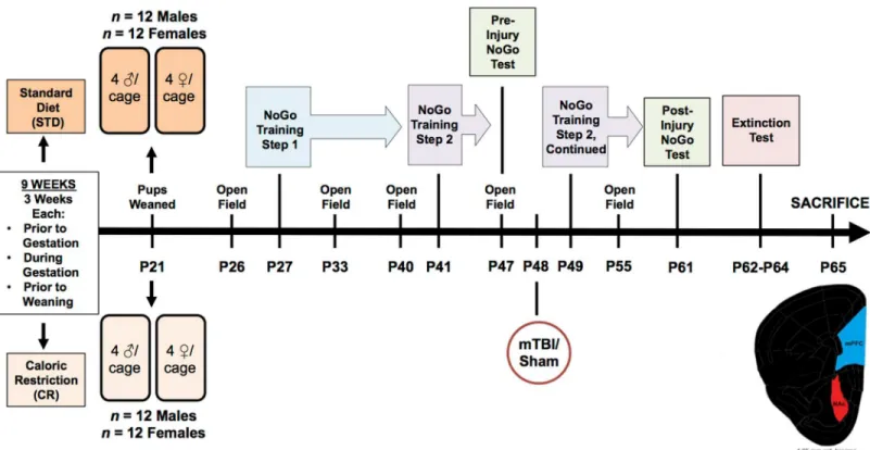
±2.6 sec, Male STD mTBI, 26.82 ± 3.0 sec, Male STD sham; 18.32 ± 3.0 sec, Female IMP mTBI; 31.27 ±2.6 sec, Female IMP sham; 22.9 ±2.6 sec, Female STD mTBI; 34.71 ±3.0 sec, Female STD sham; 19.46 ±3.0 sec]. This finding is consistent with prior studies in our laboratory and is used to validate the presence of a brain injury [32,34]. SeeFig 1for an overview of the exper-imental design along with a timeline of important testing dates.
Neuroanatomical and Molecular Analysis
Following all behavioural testing, rats were sacrificed at P65. A portion of the rats (n =28) were subjected to isoflourane inhalation, weighed, and rapidly decapitated. Using the Zilles atlas [35] tissue from the Cg3 and IL (the PFC) and NAc was removed, flash frozen on dry ice, and stored at -80°C for molecular profiling. The remainder of the rats (n= 20) were adminis-tered an overdose of sodium pentobarbital, weighed, and intracardially perfused with 0.9% saline. The brains were removed and placed in dark bottles containing Golgi-Cox solution where they were stored for 14 days.
For the molecular analysis, total RNA was extracted from the brain tissue with the Allprep RNA/DNA Mini Kit according to manufacturer protocols (Qiagen, Germany). The
Fig 1. Illustrative representation of the experimental paradigm, which includes the age that each rat underwent behavioural testing, mTBI, and sacrifice.
concentration and purity of samples were measured with a NanoDrop 2000 (Thermo Fisher Scientific, USA). Purified RNA (2μg) was reverse transcribed into cDNA using the oligo(dT)20
Superscript III First-Strand Synthesis Supermix Kit (Invitrogen, USA) according to manufac-turer protocols.
Genes were selected based on their prior use in studies of impulsivity, with specific emphasis on the frontostriatal pathways. Eight genes were selected: Catachol-O-methyltransferase (Comt), Dopamine receptors 2, 3, and 4, (Drd2,Drd3,Drd4), Monoamine oxidase A, (Maoa), Serotonin transporter (Sert), and Tryptophan hydroxylase 1, and 2 (Tph1,Tph2). Multiple attempts were made to examine the Dopamine transporter (Dat1), but expression levels were below detectable ranges. In addition,Drd3expression was determined for the NAc, but fell below detection in the PFC and was therefore omitted for that specific brain region.
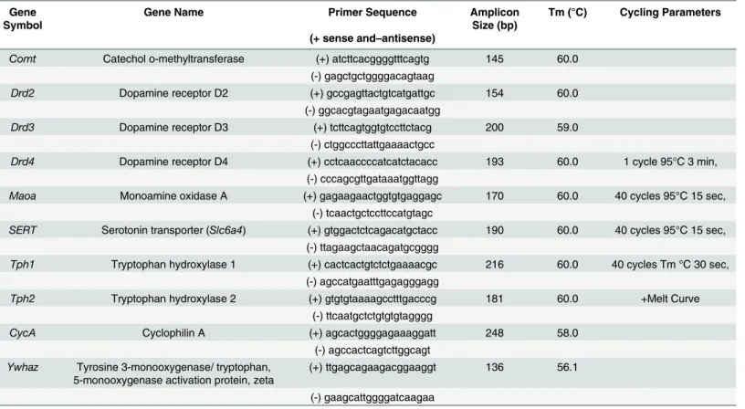
All primers for the qRT-PCR were designed in-house by a research technician using Primer3 (http://bioinfo.ut.ee/primer3) and were then purchased from IDT (Coralville, USA). SeeTable 1for primer sequences and optimal cycling parameters for the 8 genes. Each sample was run in duplicate and two separate research analysts processed all target genes. qRT-PCR was performed and analyzed with the CFX Connect Real-Time PCR detection system (Bio-Rad, Hercules, USA) with 10ng of cDNA, 0.5μM of the forward and reverse primers, and 1X
SYBR Green FastMix with Rox. Relative target gene expression was determined by normaliza-tion to two housekeeping genes, CycA and Ywhaz [36] using the 2-ΔΔCtmethod as previously described by Pfaffl [37].
To complete the histological processing, brains were transferred to 30% sucrose solution, following the 14-day Golgi-Cox impregnation period. Brains remained in sucrose for at least 3 day prior to being cut at 200μm on a Vibratome. Brain slices were mounted on gelatin coated
Table 1. Primer information for the 8 genes examined with relative qPCR.
Gene Symbol
Gene Name Primer Sequence Amplicon
Size (bp)
Tm (°C) Cycling Parameters
(+ sense and–antisense)
Comt Catechol o-methyltransferase (+) atcttcacggggtttcagtg 145 60.0
(-) gagctgctggggacagtaag
Drd2 Dopamine receptor D2 (+) gccgagttactgtcatgattgc 154 60.0
(-) ggcacgtagaatgagacaatgg
Drd3 Dopamine receptor D3 (+) tcttcagtggtgtccttctacg 200 59.0
(-) ctggcccttattgaaaactgcc
Drd4 Dopamine receptor D4 (+) cctcaaccccatcatctacacc 193 60.0 1 cycle 95°C 3 min,
(-) cccagcgttgataaatggttagg
Maoa Monoamine oxidase A (+) gagaagaactggtgtgaggagc 170 60.0 40 cycles 95°C 15 sec,
(-) tcaactgctccttccatgtagc
SERT Serotonin transporter (Slc6a4) (+) gtggactctcagacatgctacc 190 60.0 40 cycles 95°C 15 sec, (-) ttagaagctaacagatgcgggg
Tph1 Tryptophan hydroxylase 1 (+) cactcactgtctctgaaaacgc 216 60.0 40 cycles Tm °C 30 sec, (-) agccatgaatttgagagggagg
Tph2 Tryptophan hydroxylase 2 (+) gtgtgtaaaagcctttgacccg 181 60.0 +Melt Curve
(-) ttcaatgctctgtgtgtagggg
CycA Cyclophilin A (+) agcactggggagaaaggatt 248 58.0
(-) agccactcagtcttggcagt
Ywhaz Tyrosine 3-monooxygenase/ tryptophan, 5-monooxygenase activation protein, zeta
(+) ttgagcagaagacggaaggt 136 56.1
(-) gaagcattggggatcaagaa
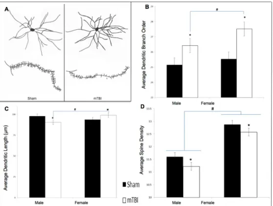
slides and then stained according to the Golgi-Cox procedure described elsewhere [38,39]. Individual neurons were selected from the NAc and traced at 250x using a camera lucida mounted on a microscope. A total of 10 cells (5/hemisphere) were traced from the NAc of each animal. The mean of cells from each hemisphere comprised the data points for the statistical analyses. SeeFig 2Bfor a representative drawing of the cells and spines from the NAc. Neuro-anatomical analyses included: 1) dendritic branch order, an overall estimation of the dendritic complexity based on the number of times the branches bifurcate, 2) Sholl analysis, a measure of dendritic length derived from the number of dendritic branches that intersect concentric cir-cles spaced 20μm from the cell body, and 3) spine density, a measure of synaptic prevalence
that is calculated by increasing the microscope to a 1000x magnification and counting the number of spine protrusions for every 10μm. A research technician blinded to all experimental
conditions drew the neurons under investigation, and another research technician completed the analysis.
Statistical Analysis
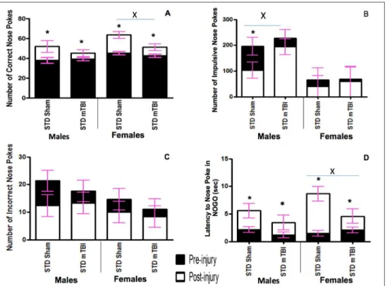
All statistical analyses were carried out with SPSS 22.0 for Mac. Repeated measures ANOVAs were conducted with the pre-injury and post-injury testing points (days) treated as within-sub-jects variables and sex (male/female), injury (mTBI/sham), and cohort (IMP/STD) treated as between-subject factors. Dependent variables were drawn from the Go/No-Go test. Repeated Measures ANOVAs were also conducted with extinction day 1 and extinction day 3 (days) treated as within-subjects variables and sex (male/female), injury (mTBI/sham), and cohort (IMP/STD) treated as between-subject factors with outcomes of the Extinction paradigm Fig 2. Graphical representation of Go/No-Go results.A) Accuracy, B) Impulsivity, C) Inaccuracy, and D) Response Inhibition, for male and female animals from the STD cohorts at the pre-injury (black bars) and post-injury (white bars) test sessions (x significant effect of mTBI,*significant effect of testing period, allp<
.05).
treated as dependent variables. A repeated measures ANOVA for the 5 testing days with sex (male/female), injury (mTBI/sham), and cohort (IMP/STD) as factors was run to analyze hyperactivity in the open field. Three-way ANOVAs with sex (male/female), injury (mTBI/ sham), and cohort (IMP/STD) as factors were run for the measures of injury validation and changes in gene expression. Finally, a two-way ANOVA with sex (male/female) and injury (mTBI/sham) as factors was run for the neuroanatomical variables. In all cases ap<.05 was
considered statistically significant.
Results
Experiment #1. Pediatric mTBI and the Frontostriatal Pathway
Go/No-Go Paradigm. Accuracy: Accuracy was used to measure the animal’s spatial atten-tion and ability to quickly and accurately respond to the illuminaatten-tion of a random hole. Ani-mals were tested in the Go/No-Go paradigm prior to the injury and approximately 2 weeks after the mTBI. All animals improved from the first testing period to the second, but the improvement in accuracy was significantly greater for sham animals than for mTBI animals. The repeated measures ANOVA demonstrated a significant day x injury interaction,F(1,19) = 9.12,p= .01, with a significant between-subjects effect of injury,F(1, 19) = 9.47,p= .01, sex,F
(1, 19) = 7.61,p= .02, but not a significant interaction,p>.05. SeeFig 2A.
Impulsivity: The number of‘impulsive’nose-pokes (nose-poking prior to activation of the Go stimulus, or during a timeout) was measured for both testing sessions. A mTBI induced a significant increase in the number of impulsive nose-pokes committed by male animals at the second testing session. No differences were observed in the number of impulsive nose-pokes for females. The repeated measures ANOVA demonstrated a significant between-subjects effect of sex,F(1, 19) = 45.53,p<.01, but not of injury,F(1, 19) = 2.13,p= .18. SeeFig 2B.
Inaccuracy: Inaccuracy was a measure of the number of holes that were poked incorrectly while the cued light was on. There were no significant differences between sham and mTBI ani-mals at the second testing session. The repeated measures ANOVA demonstrated no signifi-cant effects or interactions,p’s>.05. SeeFig 2C.
Response Inhibition: Animals were scored on their ability to inhibit a response during the No-Go stimuli. Early in the training procedure (test day 1) all animals scored poorly on this task, regardless of sex. Similar to the accuracy testing, sham animals exhibited greater improve-ments from the pre-injury test to the post injury session than mTBI animals. The repeated measures ANOVA demonstrated a significant day effect,F(1, 19) = 26.41,p<.01, and a day x
injury interaction,F(1,19) = 4.94,p= .05. SeeFig 2D.
Neuroanatomical Analysis. Golgi-Cox analysis of neurons in the NAc was only carried out for animals in the standard cohort with a mTBI or sham injury. The analysis did not include IMP animals.
Dendritic Branch Order: Analysis of the medium spiny neurons in the NAc demonstrated that both male and female animals with an early mTBI had significantly more dendritic branches when compared to animals with a sham injury. The two-way ANOVA showed a main effect of injury,F(1, 39) = 12.09,p<.01, but not of sex,F(1, 39) = 2.54,p= .12, nor a
sig-nificant interaction,F(1, 39) = 0.60,p= .45. SeeFig 3B.
Spine Density: Examination of synapse density in the NAc found that males and females dif-fered at baseline, but both experienced a significant reduction following the mTBI. The two-way ANOVA demonstrated a significant main effect of sex,F(1, 39) = 73.27,p<.01, and of
injury,F(1, 39) = 4.97,p =.03, but the interaction was not significant,F(1, 39) = 0.07,p= .80. SeeFig 3D.
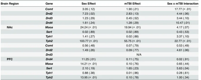
Molecular Analysis. Results from the ANOVAs for the 8 genes examined can be found in Table 2. In summary, a majority of the genes examined in the PFC and NAc exhibited signifi-cant sex-differences or sex x mTBI interactions whereby the mTBI resulted in opposing changes in gene expression for females and males. SeeFig 4.
Experiment #2. Impulsivity, mTBI, and the Frontostriatal Circuit
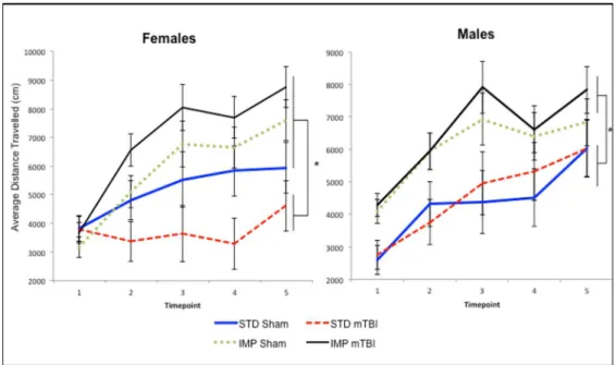
Hyperactivity. Across all time-points measured, IMP animals were significantly more active in the open field than STD animals, both before and after administration of the mTBI. The repeated measures ANOVA, demonstrated a significant test day x sex x cohort effect,
F(4, 36) = 3.46,p= .01. In addition, the test of between subjects effects found a significant main effect of cohort,F(1, 41) = 21.88,p<.01. SeeFig 5.
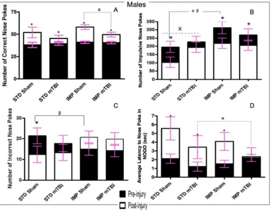
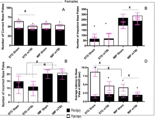
Go/No-Go Paradigm. Results from the Go/No-Go paradigm are split betweenFig 6 (Males) andFig 7(Females).
Accuracy: The repeated measures ANOVA found a significant test day x injury effect,F(1, 36) = 12.21,p<.01; that although all animals improved their accuracy from the original test
day to the 2ndtesting session, the improvement was much greater for sham animals than mTBI
Fig 3. Neuroanatomical findings from STD animals for the NAc medium spiny neurons.A) exhibits a representative tracing of average medium spiny neurons and dendritic spines from the NAc in sham and mTBI animals; B) graphical representation of dendritic branch order from Golgi-Cox stained neurons; C) representation of average dendritic length, and D) is illustrative analysis of dendritic spine density; (*main effect of mTBIp<.05; # main effect of sexp<.05).
Table 2. Summary of results from the two-way ANOVAs for changes in expression of each of the genes examined in the NAc and PFC of animals with a sham or mTBI F(p).
Brain Region Gene Sex Effect mTBI Effect Sex x mTBI Interaction
Comt 3.05 (.12) 1.90 (.21) 17.77 (<.01)
Drd2 7.23 (.02) 2.83 (.13) 4.44 (.06)
Drd3 1.23 (.29) 0.45 (.52) 3.44 (.10)
Drd4 1.61 (.24) 1.28 (.29) 10.47 (.01)
NAc Maoa 24.24 (<.01) 19.94 (<.01) 4.17 (.07)
Sert 0.02 (.89) 0.02 (.89) 0.43 (.53)
Tph1 1.41 (.27) 0.02 (.88) 3.37 (.10)
Tph2 103.77 (<.01) 55.75 (<.01) 22.77 (<.01)
Comt 0.56 (.48) 0.07 (.79) 0.53 (.49)
Drd2 1.49 (.26) 0.09 (.77) 4.61 (.06)
Drd3 N/A
PFC Drd4 11.25 (.01) 0.11 (.75) 0.02 (.91)
Maoa 14.21 (<.01) 0.10 (.76) 0.65 (.44)
Sert 2.10 (.19) 1.65 (.23) 5.63 (.04)
Tph1 0.88 (.38) 0.01 (.96) 0.28 (.61)
Tph2 13.95 (<.01) 0.10 (.78) 1.00 (.34)
doi:10.1371/journal.pone.0139842.t002
Fig 4. Changes in gene expression for STD male and female animals with a mTBI.The centerline represents typical expression (STD-shams) with the bars exhibiting percent changes from normal (*main effect of mTBIp<.05).
animals across both cohorts and sexes. All animals performed similarly during the first test ses-sion, but animals with a sham injury nose-poked significantly more accurately than animals that had experienced a mTBI in the second session. Figs6Aand7A.
Impulsivity: The number of‘impulsive’nose-pokes (nose-poking prior to activation of the Go stimulus, or during a timeout) was measured for both testing sessions. At the first test ses-sion, there were no significant differences in impulsivity were found between sham and mTBI animals. However, significant differences were seen for the IMP and STD cohorts, whereby the IMP animals were significantly more impulsive than the STD animals. In addition, for all groups except IMP males, impulsivity increased in the 2ndsession for animals with a mTBI, but not for those with a sham injury. See Figs6Band7B. The repeated measures ANOVA found a significant test day x sex x cohort effect,F(1,36) = 2.59,p= .02, and a significant test day x injury effect,F(1, 36) = 2.85,p= .02.
Inaccuracy: Inaccuracy was a measure of the number of holes that were poked incorrectly while the cued light was on. While animals in the STD cohort exhibited significant improve-ment on the task from the first test session to the 2nd, male animals in the IMP cohort actually performed worse at the second session, whereas IMP females remained static across time points. There was no effect of injury on inaccuracy scores. The repeated measures ANOVA found a significant test day x cohort effect,F(1, 36) = 8.44,p<.01. In addition, the between
subjects test found a significant main effect of cohort,F(1, 41) = 6.71,p= .01, and a significant sex by cohort interaction,F(1, 41) = 3.88,p= .05. See Figs6Cand7C.
Response Inhibition: Animals were scored on their ability to inhibit a response during the No-Go stimuli. Early in the training procedure (test day 1), all animals scored poorly on this task, regardless of cohort or sex. Similar to the accuracy, sham animals exhibited greater improvements from the pre-injury test to the post injury session than mTBI animals. The time-dependent improvements in response inhibition were also greater for animals in the STD cohort than the IMP cohort. The repeated measures ANOVA (sphericity assumed)
Fig 5. Graphical representation of the average activity level for A) Males, and B) Females,
demonstrating that the IMP cohorts were significantly more active than the STD animals at each of the 5 distinct time-periods with STD-mTBI females also showing significant reductions in activity when compared to STD-shams (*main effect of cohort,p<.05).
demonstrated a significant test day x cohort effect,F(1, 36) = 9.81.p<.01, and a significant
test day x injury effect,F(1, 36) = 11.42,p<.01. See Figs6Dand7D.
Extinction Paradigm
Accuracy and Missed Lights. The accuracy and missed lights measures were used in the extinction paradigm to determine how motivated the rats were to continue the task properly despite the absence of a reward. This would provide a measure of task-switching ability and the degree of habituation that the rats developed to the paradigm. On both accuracy and the num-ber of missed lights, male and female STD shams performed significantly better than all other groups. SeeFig 8A and 8C. When focused on male animals, results demonstrated that STD shams were quicker to learn that their behaviour was no longer being rewarded; as they had less accurate lights and more missed lights than all other male animals at the first time point. Although animals in the other groups (STD-mTBI, IMP-Sham, IMP-mTBI) improved over the testing period, STD-Sham animals still missed significantly more lights on the final day of extinction testing. Examination of the females also showed that STD-Shams missed signifi-cantly more lights and performed better on the accuracy test at both time points when com-pared to all other groups. The only exception to this was accuracy levels for STD-mTBI Fig 6. Graphical representation of Go/No-Go results.A) accuracy, B) Impulsivity, C) Inaccuracy, and D) Response Inhibition, formaleanimals from the STD and IMP cohorts at the pre-injury (black bars) and post-injury (white bars) test sessions. Overall accuracy increased across all cohorts from the pre-post-injury session to the post-injury session, but the magnitude of change was greater for sham animals. Males in the STD cohort exhibited a reduction in incorrect nose pokes with increased training but animals in the IMP cohort actually exhibited an increase in incorrect nose pokes. Cohort and mTBI increased impulsivity when compared to STD-sham animals. Finally, all males except IMP-mTBIs demonstrated improvements in latency to nose poke on the NOGO trials, but again the magnitude was greatest for STD-shams (αsignificant effect of IMP at pre-injury,βsignificant effect of IMP at post-injury,xsignificant effect of mTBI,
*significant effect of testing period, allp<.05).
animals at the 2ndtest period. The repeated measures ANOVA for accuracy demonstrated sig-nificant effects for cohort, sex, injury, cohort x sex, and cohort x injury (p’s= .00, .02, .02. .04, and .02, respectively) in the between-subjects analysis. The repeated measures ANOVA for missed lights, demonstrated main effects of cohort (p<.01), injury (p= .02), and a cohort x
injury interaction (p= .02) in the between-subjects analysis.
Impulsivity. Impulsivity was also measured during the extinction trials to determine if rats were more or less impulsive when the reward was removed from the task. Similar to the accuracy and missed lights measure, STD-Sham animals (both male and female) were cantly less impulsive than animals from all other groups on the first day of testing. No signifi-cant differences in impulsivity were seen between male IMP-mTBIs and STD-mTBIs, but differences were significant between IMP-mTBIs and STD-mTBIs females. Females from the IMP cohort were twice as impulsive as the STD cohort on the first extinction trial. On the last day of extinction testing, no differences in impulsivity were found for males; surprisingly, all ani-mals had low levels of impulsivity. In contrast, female aniani-mals exhibited an alternating pattern of impulsivity reduction whereby IMP-Sham>STD-mTBI>IMP-mTBI>STD-Sham on the last
day of extinction testing. SeeFig 8E and 8F. The repeated measures ANOVA found a significant day x cohort interaction,F(1, 36) = 5.68,p= .03, and a significant day x sex x cohort interaction, Fig 7. Graphical representation of Go/No-Go results.A) Accuracy, B) Impulsivity, C) Inaccuracy, and D) Response Inhibition, forfemaleanimals from the STD and IMP cohorts at the pre-injury (black bars) and post-injury (white bars) test sessions. Overall accuracy increased across all cohorts from the pre-injury session to the post-injury session, but the magnitude of change was greater for sham animals. Similar to males, the STD cohort exhibited a reduction in incorrect nose pokes with increased training but animals in the IMP cohort did not exhibit a change in the number of incorrect nose pokes. STD females exhibited
significantly fewer impulsive nose pokes than IMP females at both time points. Finally, also like males, all animals except IMP-mTBIs demonstrated improvements in latency to nose poke on the NOGO trials, but the magnitude was greatest for the STD-shams (αsignificant effect of IMP at pre-injury,βsignificant effect of IMP at post-injury, x significant effect of mTBI,*significant effect of testing period, allp<.05).
F(1, 36) = 3.91,p =.05. The between-subjects test revealed a significant effect of cohort (p= .05), and a cohort x sex interaction (p= .05).
Molecular Analysis
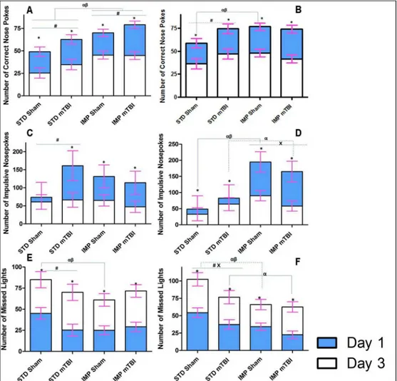
Results from the three-way ANOVAs for changes in gene expression can be found inTable 3. Because 8 distinct genes (Comt,Drd2,Drd3,Drd4,Maoa,Sert,Tph1, andTph2) were analyzed in two brain regions, the data are represented in table format to simplify presentation and com-prehension. Briefly, in the PFC significant main effects of sex were demonstrated in the major-ity of the genes, whereby males and females would display significant but opposing changes in expression when comparing standard shams to impulsive shams and in response to the mTBI. Sex differences were also present in the NAc with males and females displaying opposing Fig 8. Illustrative representation of the results from the extinction portion of the behavioural testing for STD and IMP animals; blue bars represent the initial testing day and white bars represent the scores after the 3rdconsecutive day of no reward in the 5 choice serial reaction task.In this task, rats
are no longer rewarded for completing the task as previously trained; therefore learning would entail switching strategies and reducing the number of correct nose pokes. The upper panels (A & B) demonstrate the number of correct lights with STD-shams learning the task quicker; the middle panels (C & D) show that the average number of impulsive nose-pokes (premature or inappropriate) decreased when the reward was removed; and finally (E & F) represent the number of lights that the rats chose to ignore with STD-shams again outperforming all other groups (αsignificant effect of IMP at Day 1,βsignificant effect of IMP at Day 3, x significant effect of mTBI,*significant effect of testing period, allp<.05).
changes in gene expression. In the NAc changes inDrd4expression were the best predictor of a mTBI for both sexes; but in the PFC of females,Drd4changes were indicative of the IMP. Changes inMaoaandTph1expression in the PFC were the best predictors of impulsivity for males. Illustrative demonstration of the epigenetic modifications can be found inFig 9for the PFC andFig 10for the NAc.
Discussion
As demonstrated in previous study that was conducted our laboratory [31], exposure to a mTBI/concussion during the juvenile period was associated with increased impulsivity, deficits in spatial attention, and reduced response inhibition. Building upon our prior study, the cur-rent experiment demonstrated that the mTBI-induced behavioural changes were linked to alterations in spiny neuron morphology (increased dendritic arbourization and reduced spine density) and gene expression changes in the PFC and NAc. The cohort of animals that were impulsive prior to the injury exhibited a differential pattern of change in the dopamine- and serotonin-related genes, when compared to controls and mTBI animals, in addition to an exacerbation of mTBI-induced deficits in response inhibition and cognitive flexibility.
Pediatric mTBI and the Frontostriatal Pathway
Impairment in executive functioning is often reported in children after an early brain injury, with specific complaints regarding inhibitory control and impulsivity [40,41]. Using the 5-choice serial reaction task and the Go/No-Go paradigm, the current study demonstrated that similar to humans, animals exposed to a single mTBI, exhibited decreased spatial attention, increased impulsivity, and impaired response inhibition. Male mTBI animals were significantly more likely than sham animals to exhibit impulsive behaviours, whereas both male and female animals with an early injury demonstrated deficits in spatial attention and a reduced ability to correctly nose poke the illuminated hole. The brain injury also induced neuroanatomical changes in the medium spiny neurons of the NAc. The NAc is a key structure involved in the
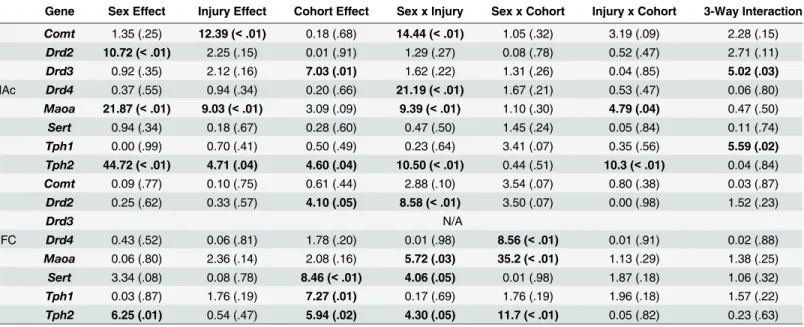
Table 3. Summary of Three-Way ANOVA results for genes examined in the nucleus accumbens and prefrontal cortex of impulsive and standard rats with and without a mTBI (F (p), bold text denotes significant effects).
Gene Sex Effect Injury Effect Cohort Effect Sex x Injury Sex x Cohort Injury x Cohort 3-Way Interaction
Comt 1.35 (.25) 12.39 (<.01) 0.18 (.68) 14.44 (<.01) 1.05 (.32) 3.19 (.09) 2.28 (.15)
Drd2 10.72 (<.01) 2.25 (.15) 0.01 (.91) 1.29 (.27) 0.08 (.78) 0.52 (.47) 2.71 (.11)
Drd3 0.92 (.35) 2.12 (.16) 7.03 (.01) 1.62 (.22) 1.31 (.26) 0.04 (.85) 5.02 (.03)
NAc Drd4 0.37 (.55) 0.94 (.34) 0.20 (.66) 21.19 (<.01) 1.67 (.21) 0.53 (.47) 0.06 (.80)
Maoa 21.87 (<.01) 9.03 (<.01) 3.09 (.09) 9.39 (<.01) 1.10 (.30) 4.79 (.04) 0.47 (.50)
Sert 0.94 (.34) 0.18 (.67) 0.28 (.60) 0.47 (.50) 1.45 (.24) 0.05 (.84) 0.11 (.74)
Tph1 0.00 (.99) 0.70 (.41) 0.50 (.49) 0.23 (.64) 3.41 (.07) 0.35 (.56) 5.59 (.02) Tph2 44.72 (<.01) 4.71 (.04) 4.60 (.04) 10.50 (<.01) 0.44 (.51) 10.3 (<.01) 0.04 (.84)
Comt 0.09 (.77) 0.10 (.75) 0.61 (.44) 2.88 (.10) 3.54 (.07) 0.80 (.38) 0.03 (.87)
Drd2 0.25 (.62) 0.33 (.57) 4.10 (.05) 8.58 (<.01) 3.50 (.07) 0.00 (.98) 1.52 (.23)
Drd3 N/A
PFC Drd4 0.43 (.52) 0.06 (.81) 1.78 (.20) 0.01 (.98) 8.56 (<.01) 0.01 (.91) 0.02 (.88)
Maoa 0.06 (.80) 2.36 (.14) 2.08 (.16) 5.72 (.03) 35.2 (<.01) 1.13 (.29) 1.38 (.25)
Sert 3.34 (.08) 0.08 (.78) 8.46 (<.01) 4.06 (.05) 0.01 (.98) 1.87 (.18) 1.06 (.32)
Tph1 0.03 (.87) 1.76 (.19) 7.27 (.01) 0.17 (.69) 1.76 (.19) 1.96 (.18) 1.57 (.22)
Tph2 6.25 (.01) 0.54 (.47) 5.94 (.02) 4.30 (.05) 11.7 (<.01) 0.05 (.82) 0.23 (.63)
cortical circuits required for top-down frontostriatal mediation of reward and inhibition [7]. A previous study conducted in our laboratory found that the single mTBI also altered dendritic morphology and spine density of pyramidal neurons in the mPFC [42]. Changes to the struc-ture of neurons in these two brain regions would alter the functionality and developmental tra-jectory of the reward and attentional neural circuits. Although a causal relationship was not established, it is possible that the reductions in NAc spine density may have been associated with the mTBI-induced impulsivity. Spontaneously hypertensive rats that are often used to Fig 9. Changes in PFC gene expression for STD animals with a mTBI and IMP animals from both groups.Males are in the upper panel (A) and females in the lower panel (B). The centerline of each graph represents typical expression (STD-shams) with the bars exhibiting percent changes from normal (*effect of mTBI,α
effect of cohortp<.05).
model inattention and impulsivity in the lab also exhibit reduced NAc spine density [43] and rats bred for impulsive traits display reductions in DA receptors localized to NAc neurons [12], also suggesting a reduction in spine density. Interestingly, our study also demonstrated that the NAc neurons exhibited significant increases in dendritic arbourization, which may be a com-pensatory mechanism to accommodate the reduction in spine density. Stimulant exposure increases both dendritic branching and spine density in the NAc [44] suggesting that stimu-lant-induced impulsivity is functionally different from the impulsive behaviours associated with mTBI. In addition,
Fig 10. Changes in NAc gene expression for STD animals with a mTBI and IMP animals from both groups.Males are in the upper panel (A) and females in the lower panel (B). The centerline of each graph represents typical expression (STD-shams) with the bars exhibiting percent changes from normal (*effect of mTBI,α
effect of cohort,p<.05).
The comprehensive examination of the dopaminergic and serotonergic genes of the frontos-triatal pathway predicted to be involved in impulsivity following mTBI clearly illustrate signifi-cant sex and region dependent differences. In both male and female mTBI animals,
significantly more genes were altered in the NAc (5/8 and 6/8 respectively) when compared to those altered in the PFC (2/8 and 3/8). More specifically, expression of the dopamine receptors,
Drd2,Drd3, andDrd4were significantly affected by mTBI. Prior studies in male rats examining distinct forms of impulsivity identified similar reductions in PFCDrd2mRNA [45]. However, we identified mTBI-induced reductions in NAcDrd3, but increases inDrd2, whereas other studies of impulsivity have noted decreases in both for male animals [12,45]. These differences however, may reflect distinctions between inherent impulsivity and induced impulsivity. Con-versely, the female mTBI animals in this study exhibited opposing changes, characterized by increasedDrd2in the PFC, increasedDrd3in the NAc and reducedDrd4in the NAc. To our knowledge no other studies have examined impulsivity-related changes in gene expression for females, making it difficult to draw comparisons, but sex-differences in epigenetic responses are not uncommon [46].
The serotonergic connections between the NAc and PFC appear to play a larger role in the response to mTBI for females than for males. Females exhibited significant enhancement of
Tph1/2(genes involved in the synthesis of 5HT) in the NAc that was not present in males, in addition to modifications in theirSertandTph2expression levels in the PFC. Experimental manipulations in rats that reduce serotonin function have been shown to impair response inhi-bition [5], using the task in this study that exhibited the largest mTBI-effect in females. Studies have indicated that serotonin involvement is likely critical for certain aspects of impulsivity and executive function [5]. Finally, expression of the two genes involved in metabolism of dopamine, serotonin, and norepinephrine (ComtandMaoa) were modified in the NAc but unaltered in the PFC. Similar to the DA receptors, changes in expression ofComtwere opposite for males and females, whereasMaoawas only reduced in males. Given a strong association between catecholamine degradation and the bioavailability of their receptors, the changes in receptor levels and metabolic enzymes reflect systematic dysfunction of critical feedback loops. The sex-dependent changes inComtand the serotonergic pathways may contribute to differ-ences in symptom presentation and treatment response. For example, methylphenidate, which acts uponComtmediated dopamine transmission, is the most commonly prescribed drug for ADHD but is ineffective for a large portion of children, with some studies finding differential efficacy rates based upon the sex of the individual [47,48]. Greater understanding of symptom etiology and sex differences may aid in the development of more efficacious therapeutic strategies.
Impulsivity, mTBI, and the Frontostriatal Circuit
inhibition deficits. While impulsive male animals also exhibited worsening of response-inhibi-tion deficits, the changes were not as substantial. The differences in baseline abilities for males and females may be responsible for the distinct patterns identified in the IMP group; standard male animals were less proficient on the No-Go stimuli and much more impulsive at baseline (sham and mTBI) than standard females. The latter findings are consistent with human litera-ture exemplifying much higher baseline rates (10:1) of impulse control problems and ADHD in boys as compared to girls [51].
When examining results from the extinction training, both IMP and mTBI animals demon-strated impairments in cognitive flexibility, as they were less able to stop performing the task when no longer being rewarded; they maintained high levels of correct nose pokes. A study by Belin et al, [52] also found that highly impulsive rats were unable to switch strategies even when they were being punished rather than rewarded. This result may reflect alterations to the reward system that are believed to contribute to ADHD phenotypes. Studies demonstrate that ADHD children tend to habituate more quickly and intensely to rewarding stimuli, but exhibit reduced physiological responses to extinction stimuli [53]. This impaired activation to the pre-sentation of extinction stimuli may help explain an interesting finding in our study, namely that improper and premature responding (impulsive nose pokes) decreased in all groups when the reward had been removed for consecutive days. This is counter-intuitive, as one would expect increased impulsivity when the rules of the paradigm switch without warning. Discon-tinuation of reinforcing rewards however generally starts an extinction process that is associ-ated with decreased tonic dopamine activity [54]. As impulsivity is believed to be, at least in part attributable to reductions in basal levels of dopamine, further reductions associated with extinction could cause a‘floor’effect [54]. The additive effects of these two processes may have been beneficial for this aspect of the test as depletion of dopamine from specific neural circuits such as the striatum and PFC has been shown to improve attentional control [55].
The examination of the serotonergic and dopaminergic genes in the IMP cohort demon-strated that even prior to a mTBI, animals in this group displayed significant dysregulation of the frontostriatal pathway. Serotonin appears to play a greater role in this model of inherent impulsivity; both male and female animals in the IMP cohort displayed some alteration to the expression levels ofSert,Tph1, andTph2in both the NAc and PFC. The modifications in expression of these genes suggest reduced 5-HT levels, which has been associated with increased impulsivity and premature responding on the Go/No-Go paradigm [56]. The only DA receptor altered in IMP females was theDrd4in the NAc, with IMP males exhibiting reductions inDrd2(NAc) andDrd3(PFC).Maoawas reduced in the NAc of both males and females, but displayed opposing alterations in the PFC (up in females, down in males). This is interesting, not only becauseMaoainhibitors have sex-dependent rates of efficacy for neuro-psychological impairments, but also because the sameMaoahaplotypes have been demon-strated to confer protection or risk for ADHD depending on the sex of the individual [57].
When combining the mTBI with the IMP phenotype, we found an exacerbation ofComt,
differed with respect to the underlying pathophysiology, both patterns of dysfunction could explain why impulsive/ADHD individuals fare worse after a mTBI [50].
Conclusion
Our results demonstrate that early mTBI/concussions can induce a secondary-ADHD like phe-notype characterized by impulsivity and inattention. This behavioural phephe-notype is associated with modifications to the structure of medium spiny neurons in the NAc, as well as changes in expression of dopaminergic and serotonergic genes critical to the function and communica-tional abilities of the frontostriatal circuit. In addition, we found that inherent impulsivity exac-erbated behavioural and epigenetic alterations associated with early mTBI/concussions, but these outcomes were sex-dependent and related to differential pathogenic processes. Impor-tantly the study has shown that despite similar behavioural phenotypes, the underlying causes of impulsivity can vary significantly. In addition, the sex differences and etiological differences in the serotonergic and dopaminergic modifications exemplify the need to tailor treatment strategies to the specifications of the premorbid characteristics of the individual.
Supporting Information
S1 File. Data for each of the rats with respect to performance on the 5-Choice Serial Reac-tion task for the testing dates and changes in gene expression at the time of sacrifice. (XLS)
S2 File. Data for dendritic branch order, dendritic length and spine density based upon Golgi-Analysis of brain tissue collected at the time of sacrifice.
(XLS)
Acknowledgments
The authors would like to thank Rose Tobias, Irene Ma, and Yilin Li for their help and exper-tise. They would also like to thank the Alberta Children’s Hospital Research Institute (ACHRI), the Alberta Children’s Hospital Foundation, the Canadian Institute for Health Research (CIHR) and the Markin USRP for Health and Wellness for their financial support.
Author Contributions
Conceived and designed the experiments: HH KY BK ME RM. Performed the experiments: HH RM. Analyzed the data: RM HH. Contributed reagents/materials/analysis tools: BK ME. Wrote the paper: HH KY BK RM.
References
1. Purper-Ouakil D, Ramoz N, Lepagnol-Bestel A, Gorwoodand P, Simonneau M. Neurobiology of atten-tion deficit/hyperactivity disorder. Pediatric Research, 2011. 69(5): p. 69R–76R. doi:10.1203/PDR. 0b013e318212b40fPMID:21289544
2. Halperin J, Healey D. The influence of environmental enrichment, cognitive enhancement, and physical exercise on brain development: Can we alter the developmental trajectory of ADHD? Neuroscience and Biobehavioral Reviews, 2011. 35: p. 621–634. doi:10.1016/j.neubiorev.2010.07.006PMID: 20691725
3. Sonuga-Barke E. Causal models of attention-deficit/hyperactivity disorder: From common simple defi-cits to multiple developmental pathways. Biological Psychiatry, 2004. 57: p. 1231–1238.
5. Dalley J, Roiser J. Dopamine, serotonin and impulsivity. Neuroscience, 2012. 215: p. 42–58. doi:10. 1016/j.neuroscience.2012.03.065PMID:22542672
6. Costa Dias T, Wilson V, Bathula D, Iyer S, Mills K, Thurlow B, et al. Reward circuit connectivity relates to delay discounting in children with attention-deficit/hyperactivity disorder. European Neuropsycho-pharmacology, 2013. 23: p. 33–45. doi:10.1016/j.euroneuro.2012.10.015PMID:23206930
7. Dalley J, Everitt BJ, Robbins T. Impulsivity, compulsivity, and top-down cognitive control. Neuron, 2011. 69: p. 680–694. doi:10.1016/j.neuron.2011.01.020PMID:21338879
8. Hayes DJ, Greenshaw AJ. 5-HT receptors and reward-related behaviour: A review. Neuroscience and Biobehavioral Reviews, 2011. 35: p. 1419–1449. doi:10.1016/j.neubiorev.2011.03.005PMID: 21402098
9. Cortese S, Kelly C, Chabernaud C, Proal E, Di Martino A, Milham M, et al. Toward systems neurosci-ence of ADHD: A meta-analysis of 55 fMRI studies. American Journal of Psychiatry, 2012. 169(10): p. 1038–1055. doi:10.1176/appi.ajp.2012.11101521PMID:22983386
10. Itohara S, Kobayashi Y, Nakashiba T. Genetic factors underlying attention and impulsivity: mouse mod-els of attention-deficit/hyperactivity disorder. Current Opinion in Behavioral Sciences, 2015. 2: p. 46– 51.
11. Durston S, Fossella J, Mulder M, Casey B, Ziermans T, Vessaz M, et al. Dopamine transporter geno-type conveys familial risk of attention-deficit/hyperactivity disorder through striatal activation Journal of the American Academy of Child and Adolescent Psychiatry, 20098. 47: p. 61–67. doi:10.1097/chi. 0b013e31815a5f17PMID:18174826
12. Dalley J, Fryer T, Brichard L, Robinson E, Theobald D, Laane K, et al. Nucleus accumbens D2/3 recep-tors predict trait impulsivity and cocaine reinforcement. Science (New York, N.Y.), 2007. 315: p. 1267– 1270.
13. Max J, Lindgren S, Knutson C, Pearson C, Ihrig D, Welborn A. Child and adolescent traumatic brain injury: Correlates of disruptive behavior disorders. Brain Injury, 1998. 12(1): p. 41–52. PMID:9483336
14. Max J, Schachar R, Levin H, Ewing-Cobbs L, Chapman S, Dennis M, et al. Predictors of secondary Attention-Deficit/Hyperactivity disorder in children and adolescents 6 to 24 months after traumatic brain injury. Journal of the American Academy of Child and Adolescent Psychiatry, 2005. 44(10): p. 1041– 1049. PMID:16175109
15. Pinto P, Meoded A, Poretti A, Tekes A, Huisman T. The unique features of traumatic brain injury in chil-dren. Review of the characteristics of the pediatric skull and brain, mechanisms of trauma, patterns of injury, complications and their imaging findings—Part 2. Journal of Neuroimaging, 2012. 22: p. e18– e41. doi:10.1111/j.1552-6569.2011.00690.xPMID:22303964
16. Faul M, Xu L, Wald M, Coronado V. Traumatic brain injury in the United States: Emergency department visits, hospitalizations and deaths 2002–2006. 2010, Centers for Disease Control and Prevention: Atlanta, GA.
17. Barlow K, Crawford S, Stevenson A, Sandhu S, Belanger F, Dewey D. Epidemiology of postconcussion syndrome in pediatric mild traumatic brain injury. Pediatrics, 2010. 126(2): p. e374–381. doi:10.1542/ peds.2009-0925PMID:20660554
18. Howell D, Osternig L, van Donkelaar P, Mayr U, Chou L. Effects of concussion on attention and execu-tive funciton in adolescents. Medicine & Science in Sports and Exercise, 2013. 45(6): p. 1030–1037.
19. Covassin T, Schatz P, Swanik B. Sex differences in neuropsychological function and post-concussion symptoms of concussed collegiate athletes. Neurosurgery, 2007. 61(2): p. 345–351. PMID:17762747
20. Ryan L, Warden D. Post concussion syndrome. International Review of Psychiatry, 2003. 15: p. 310– 316. PMID:15276952
21. Hooper S, Alexander J, Moore D, Sasser H, Laurent S, King J, et al. Caregiver reports of common symptoms in children following a traumatic brain injury. NeuroRehabilitation 2004. 19: p. 175–189. PMID:15502252
22. Palasis S. Traumatic pediatric white matter injury. Journal of Pediatric Neuroradiologys, 2013. 2: p. 87–96.
23. Smits M, Houston G, Dippel D, Wielopolski P, Vernooij M, Koudstaal P, et al. Microstructural brain injury in post-concussion syndrome after minor head injury. Neuroradiology, 2011. 53: p. 553–563. doi:10. 1007/s00234-010-0774-6PMID:20924757
24. Maxwell W, Watt C, Graham D, Gennarelli T. Ultrastructural evidence of axonal shearing as a result of lateral acceleration of the head in non-human primates. Acta Neuropathologica, 1993. 86: p. 136–144. PMID:7692693
26. Martel M, Nikolas M, Jernigan K, Friderici K, Waldman I, Nigg J. The dopamine receptor D4 gene (Drd4) moderates family environmental effects on ADHD. Journal of Abnormal Child Psychology, 2011. 39: p. 1–10. doi:10.1007/s10802-010-9439-5PMID:20644990
27. Sagiv S, Epstein J, Bellinger D, Korrick S. Pre- and postnatal risk factors for ADHD in nonclinical pediat-ric population. Journal of Attention Disorders, 2013. 17(1): p. 47–57. doi:10.1177/1087054711427563 PMID:22298092
28. Yeates K, Armstrong K, Janusz J, Taylor H, Wade S, Stancin T, et al. Long-term attention problems in children with traumatic brain injury. Journal of the American Academy of Child and Adolescent Psychia-try, 2005. 44(6): p. 574–584. PMID:15908840
29. Mychasiuk R, Hehar H, Maand I, Esser MJ. Dietary intake alters behavioural recovery and gene expression profiles in the brain of juvenile rats that have experienced a concussion. Frontiers in Behav-ioral Neuroscience, 2015. 9(Article 17): p. 1–16.
30. Bari A, Dalley J, Robbins T. The application of the 5-choice reaction time task for the assessment of visual attentional processes and impulse control in rats. Nature Protocols, 2008. 3(5): p. 759–767. doi: 10.1038/nprot.2008.41PMID:18451784
31. Mychasiuk R, Hehar H, Esser MJ. A mild traumatic brain injury (mTBI) induces secondary attention-def-icit hyperactivity disorder-like symptomology in young rats. Behavioural Brain Research, 2015. 286: p. 285–292. doi:10.1016/j.bbr.2015.03.010PMID:25771208
32. Mychasiuk R, Farran A, Esser MJ. Assessment of an experimental rodent model of pediatric mild trau-matic brain injury Journal of Neurotrauma, 2014. 31: p. 1–9.
33. Kane M, Angoa-Perez M, Briggs D, Viano D, Kreipke C, Kuhn D. A mouse model of human repetitive mild traumatic brain injury. Journal of Neuroscience Methods, 2012. 203: p. 41–49. doi:10.1016/j. jneumeth.2011.09.003PMID:21930157
34. Mychasiuk R, Farran A, Angoa-Perez M, Briggs D, Kuhn D, Esser MJ. A novel model of mild traumatic brain injury for juvenile rats. JoVE, 2014. 8(94): p. e51820.
35. Zilles K. The cortex of the rat: A stereotaxis atlas. 1985, Berlin: Springer-Verlag.
36. Bonefeld B, Elfving B, Wegener G. Reference genes for normalization: A study of rat brain tissue. Syn-apse, 2008. 62(4): p. 302–309. doi:10.1002/syn.20496PMID:18241047
37. Pfaffl M. A new mathematical model for relative quantification in real-time RT-PCR. Nucleic Acids Research, 2001. 29(9): p. e45. PMID:11328886
38. Gibb R, Kolb B. A method for vibratome sectioning of Golgi-Cox stained whole rat brain. Journal of Neu-roscience Methods, 1998. 79: p. 1–4. PMID:9531453
39. Mychasiuk R, Gibb R, Kolb B. Visualizing the effects of a positive early experience, tactile stimulation, on dendritic morphology and synaptic connectivity with Golgi-Cox staining. Journal of visualized Experi-ments, 2013. 79: p. e50694. doi:10.3791/50694PMID:24121525
40. Sinopoli K, Dennis M. Inhibitory control after traumtic brain injury in children. International Journal of Developmental Neuroscience, 2012. 30: p. 207–215. doi:10.1016/j.ijdevneu.2011.08.006PMID: 22100363
41. Konrad K, Gauggel S, Manz A, Scholl M. Inhibitory control in children with traumatic brain injury (TBI) and children with attention deficit/hyperactivity disorder (ADHD). Brain Injury, 2000. 14(10): p. 859– 875. PMID:11076133
42. Mychasiuk R, Hehar H, Ma I, Kolb B, Esser MJ. The development of lasting impairments: A mild pediat-ric brain injury alters gene expression, dendritic morphology, and synaptic connectivity in the prefrontal cortex of rats. Neuroscience, 2015. 288: p. 145–155. doi:10.1016/j.neuroscience.2014.12.034PMID: 25555930
43. Sanchez F, Gomez-Villalobos M, Juarez I, Quevedo L, Flores G. Dendritic morphology of neurons in medial prefrontal cortex, hippocampus, and nucleus accumbens in adult SH rats. Synapse, 2010. 65 (3): p. 198–206.
44. Robinson T, Kolb B. Alterations in the morphology of dendrites and dendritic spines in the nucleus accumbens and prefrontal cortex following repeated treatment with amphetamine or cocaine. European Journal of Neuroscience, 1999. 11(5): p. 1598–1604. PMID:10215912
45. Simon N, Beas B, Montgomery K, Haberman R, Bizon J, Setlow B. Prefrontal cortical-striatal dopamine receptor mRNA expression predicts distinct forms of impulsivity. European Journal of Neuroscience, 2013. 37: p. 1779–1788. doi:10.1111/ejn.12191PMID:23510331
46. McCarthy M, Auger A, Bale T, De Vries G, Dunn G, Forger N, et al. The epigenetics of sex differences in the brain. The Journal of Neuroscience, 2009. 29(41): p. 12815–12823. doi:10.1523/JNEUROSCI. 3331-09.2009PMID:19828794
48. Sonuga-Barke E, Coghill D, Markowitz J, Swanson J, Vandenerghe M, Hatch S. Sex-differences in the response of childrenn with ADHD to once-daily formulations of methylphenidate. Journal of the Ameri-can Academy of Child and Adolescent Psychiatry, 2007. 46(6): p. 701–710. PMID:17513982
49. Clacy A, Sharman R, Lovell G. Risk factors to sport-related concussion for junior athletes. OA Sports Medicine, 2013. 1(1): p. 4–10.
50. White R, Harris G, Gibson M. Attention deficit hyperactivity disorder and athletes. Sports Health: A Mul-tidisciplinary Approach, 2014. 6(149–156).
51. Biederman J, Mick E, Faraone S, Braaten E, Doyle A, Spencer T, et al. Influence of gender on attention deficit hyperactivity disorder in children referred to a psychiatric clinic. The American Journal of Psychi-atry 2002. 159(1): p. 36–42. PMID:11772687
52. Belin D, Mar A, Dalley J, Robbins T, Everitt BJ. High impulsivity predicts the switch to compulsive cocaine-taking. Science (New York, N.Y.), 2008. 320: p. 1352–1355.
53. Iaboni F, Douglas V, Ditto B. Psychophysiological response of ADHD children to reward and extinction. Psychophysiology, 1997. 34: p. 116–123. PMID:9009815
54. Johansen E, Aase H, Meyer A, Sagvolden T. Attention-deficit/hyperactivity disorder (ADHD) behaviour explained by dysfunctioning reinforcement and extinction processes. Behavioural Brain Research, 2002. 130(1–2): p. 37–45. PMID:11864716
55. Scholes K, Harrison B, O'Neill B, Leung S, Croft R, Pipingas A, et al. Acute serotonin and dopamine depletion improves attentional control: Findings from the Stroop task. Neuropsychopharmacology, 2007. 32: p. 1600–1610. PMID:17151596
56. Eagle D, Bari A, Robbins T. The neuropsychopharmacology of action inhibition: Cross species transla-tion of the stop-signal and go/no-go tasks. Psychopharmacology, 2008. 199: p. 439–456. doi:10.1007/ s00213-008-1127-6PMID:18542931