Pathway for the Endogenous Epo and VEGF Induction by
Baicalein in Neurons versus Astrocytes
Yu-Yo Sun
1, Shang-Hsuan Lin
2, Hung-Cheng Lin
3, Chia-Chi Hung
3, Chen-Yu Wang
2,4, Yen-Chu Lin
5,
Kuo-Sheng Hung
6, Cheng-Chang Lien
4,5, Chia-Yi Kuan
1, Yi-Hsuan Lee
2,4*1 Division of Neurology, Department of Pediatrics, the Center for Neurodegenerative Disease, Emory University School of Medicine, Atlanta, Georgia, United States of America, 2 Department and Institute of Physiology, National Yang-Ming University, Taipei, Taiwan, 3 Graduate Institute of Medical Sciences, Taipei Medical University, Taipei, Taiwan, 4 Brain Research Center, National Yang-Ming University, Taipei, Taiwan, 5 Institute of Neuroscience, National Yang-Ming University, Taipei, Taiwan, 6 Department of Neurosurgery, Taipei Medical University Wan Fang Hospital, Taipei, Taiwan
Abstract
The neuroprotective effect of baicalein is generally attributed to inhibition of 1β/15-lipoxygenase (1β/15-LOX) and suppression of oxidative stress, but recent studies showed that baicalein also activates hypoxia-inducible factor-α (HIF1α) through inhibition of prolyl hydrolase β (PHDβ) and activation of the phosphatidylinositide-γ kinase (PIγK)/Akt signaling pathway. Yet, the significance and regulation of prosurvival cytokines erythropoietin (Epo) and vascular endothelial growth factor (VEGF), two transcriptional targets of HIF1α, in baicalein-mediated neuroprotection in neurons and astrocytes remains unknown. Here we investigated the causal relationship between the PIγK/Akt signaling pathway and Epo/VEGF expression in baicalein-mediated neuroprotection in primary rat cortical neurons and astrocytes. Our results show that baicalein induced Epo and VEGF expression in a HIF1α- and PIγK/Akt-dependent manner in neurons. Baicalein also protected neurons against excitotoxicity in a PIγK- and Epo/VEGF-dependent manner without affecting neuronal excitability. In contrast, at least a 10-fold higher concentration of baicalein was needed to induce Epo/VEGF production and PIγK/Akt activity in astrocytes for protection of neurons. Moreover, only baicalein-induced astrocytic VEGF, but not Epo expression requires HIF1α, while PIγK/Akt signaling had little role in baicalein-induced astrocytic Epo/VEGF expression. These results suggest distinct mechanisms of baicalein-mediated Epo/VEGF production in neurons and astrocytes for neuroprotection, and provide new insights into the mechanisms and potential of baicalein in treating brain injury in vivo.
Citation: Sun Y-Y, Lin S-H, Lin H-C, Hung C-C, Wang C-Y, et al. (β01γ) Cell Type-Specific Dependency on the PIγK/Akt Signaling Pathway for the
Editor: Barbara Bardoni, CNRS UMR7β75, France
Received March 8, β01γ; Accepted June 4, β01γ; Published July 19, β01γ
Copyright: © β01γ Sun et al. This is an open-access article distributed under the terms of the Creative Commons Attribution License, which permits unrestricted use, distribution, and reproduction in any medium, provided the original author and source are credited.
Funding: This study was supported by the Top University Plan from Ministry of Education in Taiwan (http://english.moe.gov.tw/), grants NSC96-β6β8-B-0γ8-010-MYγ (YHL), NSC96-βγβ0-B-0γ8-0β5-MYγ (YHL), NSC 101-βγβ0-B-010 -041 -MYγ (YHL) from National Science Council in Taiwan (http:// web1.nsc.gov.tw/mp.aspx?mp=7), and National Institutes of Health NS074559 (CYK) from National Institute of Health in United States of America (http:// www.nih.gov/). The funders had no role in study design, data collection and analysis, decision to publish, or preparation of the manuscript.
Competing interests: The authors have declared that no competing interests exist. * E-mail: yhleeγ@ym.edu.tw
Introduction
Baicalein, a natural flavonoid isolated from Scutellaria baicalensis Georgi (S. Georgi), has been shown effective in attenuating neuronal loss induced by excitotoxin administration [1] and oxygen-glucose deprivation [β] in vitro as well as reducing brain injury in various brain injury animal models [β–4]. The mechanism of action for the baicalein neuroprotection has been mostly attributed to its direct inhibition of 1β/15 lipoxygenase (1β/15-LOX), which is mainly expressed in neurons and brain cerebrovascular endothelial cells and is involved in injury-induced elevation of reactive oxygen species and subsequent lipid peroxidation causing
neural cell necrosis (reviewed by [5–7]) and blood-brain barrier (BBB) disruption [8]. Recent studies revealed that baicalein also regulates other signaling pathways, including prolyl hydroxylase β (PHDβ)/hypoxia-inducible factor 1α (HIF1α) [9] and phosphatidylinositide γ-kinase (PIγK)/Akt pathways [β]. However, how these pathways integrate to provide neuroprotection remains poorly understood.
PIγK/Akt signaling was reported to be activated by baicalein in neurons, and plays a key role in baicalein-mediated neuronal survival and synaptic plasticity [β,10]. Akt is mainly phosphorylated by class I PIγKs and plays important roles in neuronal survival [11,1β]. The PIγK/Akt signaling pathway activates HIF1α by reducing its ubiquitination via two routes,
one by phosphorylation of HIFα, and the other by inhibition of PHDβ via the mammalian target of rapamycin (mTOR) [1γ,14]. PHDβ, one of the γ PHD isoforms (PHD1, PHDβ, PHDγ) that serve as intracellular oxygen sensors, mediates asparaginyl hydroxylation and ubiquitination of HIF-1α upon normoxic condition [15,16]. Thus, compounds or signaling pathways that inhibit PHD activity can also up-regulate HIF-1α under normoxia. Recent studies show that both neuron-specific PHDβ knockout and PHDβ inhibitor treatment are effective in reducing transient cerebral ischemia-induced brain damage via activating HIF-1α [17,18]. Notably, baicalein can inhibit PHDβ activity by direct binding to the enzyme active sites [9], but whether its activation of prosurvival PIγK/Akt signaling in neurons also contributes to the HIF1α target gene induction remains undetermined.
Erythropoietin (Epo) and vascular endothelial growth factor (VEGF) are hypoxia-inducible neuroprotective cytokines with their gene transcription mainly mediated by HIF-1α or HIF-βα [15]. Recent efforts in the development of neuroprotective therapeutics have been directed to the induction of endogenous Epo and VEGF using HIF-activating agents, such as ischemic preconditioning [19] and PHDβ inhibitors [β0], for treating CNS injury in order to circumvent the possible adverse effects of their exogenous application [β1–β5]. While the induction of endogenous Epo from brain cells was reportedly beneficial [β6,β7], controversial outcomes were noted regarding the endogenous VEGF induction: neuronal VEGF production appears to be neuroprotective [β8,β9] whereas excessive astrocytic VEGF was found detrimental to the BBB integrity [γ0]. Baicalein, as a PHDβ inhibitor and neuronal PIγK activator, seem to be a promising candidate for inducing Epo/ VEGF in the brain, but such an effect and a subsequent contribution to baicalein neuroprotection have not been investigated.
Most of studies on baicalein neuroprotection were focusing on its neuronal effects, such as 1β/15-LOX inhibition, PIγK activation, and regulation of GABAA receptor activity [γ1],
whereas its effect on astrocytes, the most abundant cell type in the brain, has not been well investigated. Factors released from astrocytes, including neurotrophic factors and proinflammatory cytokines, vary under different physiological and pathological settings and play important roles in establishing a microenvironment that affects neuronal survival and plasticity [γβ]. The PIγK/Akt signaling pathway in astrocytes was found to be important for the glutamate transporter function [γγ] and the synthesis of an astrocyte-derived neuroprotective chemokine RANTES [γ4], but its effects on other neuroprotective factors have not been explored. Besides, the effect of baicalein on PIγK activity in astrocytes has not been reported to date while it is quite variable across different cell types: it is stimulatory in neurons but inhibitory in microglia and prostate cancer cells [γ5,γ6].
In this study, we investigated the causal role of baicalein-induced PIγK/Akt signaling in its activation of HIF1α and downstream Epo/VEGF gene expression in primary cortical neurons and astrocytes. Our data show that baicalein activates PIγK/Akt signaling in both neurons and astrocytes but at different effective concentrations to provide neuroprotection
against excitotoxicity, and notably this signaling only contributes to its induction of neuronal, but not astrocytic, Epo/ VEGF expression. Furthermore, the VEGF-inducing effect of baicalein requires its activation of HIF1α in both cell types, whereas its Epo-inducing effect only depends on HIF1α in neurons, but not (in) astrocytes. The contributions of 1β/15-LOX and PHDβ in the baicalein-induced neuronal and astrocytic Epo/VEGF expression were also examined. Clues for the potential application of baicalein in treating brain injuries and diseases based on the information obtained are discussed.
Materials and Methods
Reagents
Baicalein was obtained from Merck (Darmstadt, Germany). L-glutamic acid, N-methyl-D-aspartic acid (NMDA), bicucullin, and cobalt chloride (CoClβ) were obtained from Sigma-Aldrich
(St. Louis, MO). LYβ9400β was obtained from Calbiochem (San Diego, CA). PIγK α inhibitor β (γ-[4-(4-morpholinyl)thieno[γ,β-d]pyrimidin-β-yl-phenol) and the PIγK inhibitor CAY10505 (5-[[5-(4-fluorophenyl)-β-furanyl] methylene]-β,4-thiazolidinedione) were obtained from Cayman Chemical (Ann Arbor, MI). Dimethyloxaloylglycine (DMOG), a PHDβ inhibitor, was obtained from Enzo Life Sciences (Plymouth Meeting, PA). Goat IgG, goat anti-VEGF and goat anti-Epo antibodies for neutralization study were purchased from R&D Systems (Minneapolis, MN).
Animals
For the primary culture of cortical neurons and astrocytes, we used pregnant female Sprague Dawley (SD) rats at 17-day gestation and postnatal 1-β-day old (P1-Pβ) SD rats obtained from BioLASCO Taiwan Co. (Taipei, Taiwan), respectively. For the rat hippocampal slice preparation, SD male rats at P16-Pβ1 were used. Animals used for primary cultures and for hippocampal slice preparation were killed by overdose sevoflurane (Abbott, Osaka, Japan). Animal experimentation procedures were reviewed and approved by the Animal Care and Use Committee at National Yang-Ming University and are in accordance with the Guide for the Care and Use of Laboratory Animals, the National Institute of Health guidelines (USA) in the care and use of animals for experimental procedures.
Primary cultured rat cortical neurons and astrocytes
The cultured cortical neurons were prepared from fetal rats harvested from pregnant female rat at 17-day gestation as described previously [γ7]. Briefly, rat brain cortics was loosely homogenized through a 14-gauge metal needle in BME (Invitrogen) with sodium bicarbonate (β6.β mM), D-glucose (β7.8 mM), L-glutamine (β.0 mM), and β0% FBS (Invitrogen), centrifuged at 800 rpm for 5 min, and washed three times. Resuspended cells were seeded onto cell culture plates (γ5-mm culture dish or β4-well culture plates; Iwaki, Tokyo, Japan) pre-coated with poly-L-lysine (Sigma-Aldrich, St. Louis, MO), and then incubated in γ7°C incubator with 5% COβ for γ0~45
medium (Invitrogen) with sodium bicarbonate (β6.β mM), D-glucose (β7.8 mM), and L-glutamine (β.0 mM). The obtained neuron-enriched cultures at 10 days–in-vitro (DIV) contained more than 85% neuronal population as characterized by immunofluorescent double labeling of neuron and glial markers (Figure S1 in Information S1). Cultured neurons at this stage were sensitive to NMDA excitotoxicity [γ7], and thus used in this study.
Primary cultured astrocytes were prepared from P1-Pβ SD rats as described previously [γ8]. Briefly, cerebral cortex isolated from neonatal rats was loosely homogenized through a 14-gauge metal needle in DMEM/F1β (Invitrogen) with 10% FBS, filtered through a 70-µm nylon mesh, and centrifuged at 1,000 rpm for 10 min. Cells resuspended were seeded onto 75 mm flasks and incubated for 7 days, followed by orbital shaking at 180 rpm in a γ7°C incubator for β4 h to remove microglia and oligodendrocytes. The purified astrocytes that tightly adhered at the bottom of the flasks were then detached with trypsin/EDTA (Invitrogen) and seeded onto culture dishes and incubated for 7 days to settle to a resting stage. The purified astrocyte cultures which contained more than 85% glial fibrillary acidic protein (GFAP)-positive cells were used for this study.
Plasmid construction and luciferase activity assay
A DNA fragment with triplicated hypoxia response element (HRE) sequences in the human EPO gene enhancer (HREEPO)
was inserted into the promoter of pGLβ luciferase reporter plasmid (Promega, Madison, WI) to obtain the pHREEPO-Luc
reporter construct as described previously [β7]. Cells plated in β4-well plates were transfected with pHREEPO-Luc at 0.γγ μg/
well and pRL-TK Renilla luciferase normalization construct (Promega) at 0.01 μg/well in 1.5 μg/ml Lipofectamine β000TM
(Invitrogen, Carlsbad, CA) for β4 h, followed by treatment with baicalein or CoClβ for β4 h. Cells were then harvested with
passive lysis buffer (Promega) for the luciferase activity assay according to the manufacture’s protocol (Dual-Luciferase®
reporter assay system; Promega). The HREEPO-driven gene
expression was calculated and represented as the ratio of firefly/renilla luciferase activity. No significant cytotoxicity was found with the concentration of Lipofectamine β000TM used in
this study.
Semi-quantitative and real-time RT-PCR
Total RNA was extracted using TRIzol reagent (Invitrogen), and reversely transcribed by using High Capacity cDNA Reverse Transcription Kit (Applied BioSystems, Foster City, CA) to obtain cDNAs for subsequent semi-quantitative and quantitative PCR, The semi-quantitative PCR of rat Epo, Vegf,
Hif1a (HIF-1α), Alox15 (1β/15-Lox), and a housekeeping gene
Gapdh (glyceraldehydes-γ-phosphate dehydrogenase) cDNAs
were detected using the following primers: Epo, 5'-TGCGACAGTCGCGTTCTGGAGAGGTAC-γ’ and 5'-ATCCGCTGTGAGTGTTCGGAGTGGAGC-γ′; Vegf, 5'-CCATGAACTTTCTGCTCTCTTG-γ’ and 5'-GGTGAGAGGTCTAGTTCCCGA -γ′; Hif1a, 5'-CAAGATCAGCCAGCAAGTCCTTCTGATG-γ’ and 5'-AGGTTTCTGTAACTGGGTCTGCTGGAATC-γ′; Alox15,
5'-GACTGTTCAGGAAACATAGGGAAG-γ′ and 5'-CCATTACCCCTATAACCTGTGAAG-γ′; Gapdh, 5'-CTCATGACCACAGTCCATGC-γ′ and 5'-TTCAGCTCTGGGATGACCTT-γ′. Bands of PCR products were visualized and quantified using an electrophoresis image analysis system (Eastman Kodak Co., Rochester, NY). For the quantitative real-time PCR, the FAM probes and primers for the detection of Epo (Rn005665β9_m1; 5’-GAGATGGGGGTGCCCGAACGTCCCA-γ’) and an internal control gene β-actin (Rn00667869_m1; 5’-CTTCCTGGGTATGGAATCCTGTGGC-γ’) were designed by Applied Biosystems (ABI, Foster City, CA) and used for TaqMan system. Primer sets for Vegf (5’-CGGACGGGCCTCTGAAACCAT-γ’ and 5’-CTTCACCACTTCATGGGCTTTCTGC-γ’), TNFα (5’-TCTCAAAACTCGAGTGACAAGCCCG-γ’ and 5’-GCAGCCTTGTCCCTTGAAGAGAACC-γ’), and internal control
Gapdh (5'-CTCATGACCACAGTCCATGC-γ′ and
5'-TTCAGCTCTGGGATGACCTT-γ′) were used for SYBR Green system. The assay mixture and 150 ng of cDNA were added into βx TaqMan® Universal PCR Mix or βx SYBR® Green PCR
Master Mix (Applied Biosystems) to make up β0 μl of the amplification mixtures, and then subjected to real-time PCR reaction on an ABI PRISM 7γ00 Sequence Detector. The average cycle threshold (Ct) value was used to calculate mRNA expression levels. Relative mRNA levels were normalized by the internal control (ß-actin or Gapdh) in terms of the differences of the Ct values. Relative transcript levels
were calculated as x = β-△Ct, in which ΔCt = Ct
target gene –Ctinternal control.
Enzyme-linked immunosorbent assay (ELISA)
In brief, culture media of cortical neurons and astrocytes were collected after the treatment, and the cells were lysed in a lysis buffer [β0 mM Tris, pH 7.4, 150 mM NaCl, 1% IPGEAL-6γ0, 5% glycerol, protease inhibitor cocktail (Roche, Penzberg, Germany)]. Epo and VEGF concentrations in the cell lysate and culture media were measured using the respective Quantikine ELISA Kits (R&D Systems, Minneapolis, MN) according to the manufacturer’s instructions, and detected using an ELISA reader at a wavelength of 450 nm.
RNA knockdown
Cortical neurons were transfected with siRNAs specific for rat 1β/15-LOX or HIF-1α mRNA, or with scrambled RNA produced by Silencer Pre-designed siRNA (Ambion, Austin, TX) using Lipofectamine β000TM reagent (Invitrogen) for 7β h
as previously described [γ9]. The siRNA sequences for rat 1β/15-LOX (Alox15; Accession No. NM_0γ1010) were 5’-CGAUUUCGAGAGGACAAAAtt-γ’ (exon 4) and 5’-GGCAGAUCAUGAAUCGGUAtt-γ’ (exon 10); for rat HIF-1α
(Hif1a; Accession No. NM_0β4γ59) were
Western blot analysis
Cortical neurons were lysed and total protein extracted for Western blot analysis as described previously [γ9]. Primary antibodies used were rabbit anti-phospho-Akt (Ser47γ) (1:1000) and rabbit anti-Akt (1:1,000) antibodies (Cell Signaling, Danvers, MA), and secondary antibodies were horseradish peroxidase (HRP)-conjugated goat anti- rabbit IgG (1:β0,000) and HRP-conjugated goat anti- mouse IgG (1:β0,000) (Jackson ImmunoResearch Laboratories, West Grove, PA). The immune complex was visualized by HRP-reactive Western LightningTM Plus-ECL (PerkinElmer Inc.,
Waltham, MA) and the signal was detected and analyzed by Night OWL LB 981 imaging system (Berthold Technologies, Bad Wilbad, Germany).
Chromatin immunoprecipitation (ChIP) assay
Anti-HIF1α-based ChIP assay was performed as described previously [β7]. DNA-protein cross-linking samples were subjected to immunoprecipitation using mouse anti-HIF1α antibody (β μg; Novus, Littleton, CO). Purified DNA was amplified by PCR with primer for HRE-specific rat Epo gene γ’ enhancer (+γ497 ~ +γ618, NM-017001, 5’-TACCTCCCCCCCCCCCCATTCTGGT-γ’ and 5’-CAAGCCCAGAGGGGTCAAGAGGTCAGA-γ’), rat Epo gene promoter (-γ75 ~ -ββ1, NM-017001, 5’-CAGCCTGCTCTACCCCAGCAAGGA-γ’ and 5’-GGGGGTCGGGGATGTTATCAGCA-γ’), and HRE-specific rat
Vegf gene promoter (-18β9 ~ -1994, M-γβ167, 5’-GAGGAACAAGGGCTTCTGTCTG-γ’ and 5’-TCTCTGGAGAGGATATGGCATC-γ’). Quantitative real-time PCR of each gene promoter and enhancer fragment was performed using SYBER Green reaction mix (Applied Biosystems).
Cell apoptosis analysis
Cell apoptosis analysis was performed using 4′,6-diamidino-β-phenylindole dihydrochloride (DAPI) staining for DNA condensation and terminal deoxynucleotidyl transferase dUTP nick end labeling (TUNEL) for fragmented DNA. Cells were briefly washed with isotonic saline solution, followed by fixation (4% formaldehyde in β0 mM PBS) for 15 min at room temperature and permeabilization with pre-chilled EtOH/ CHγCOOH (95%: 5%) for 15 min at –β0oC. Cells were then
incubated with nuclear marker DAPI. TUNEL stain was performed following the manufacturer’s protocol (Promega). Fluorescent micrographs at excitation wavelength γ50 nm for DAPI and 488 nm for TUNEL were taken by Olympus DP50 digital camera (Olympus, Tokyo, Japan). Apoptotic cells were identified by visualizing TUNEL-positive cells with condensed DAPI staining in the nucleus. The total number of DAPI-stained cells served as the total cell number. The cell counting was performed in five randomly selected areas for each well and 4 separate wells were used for each experimental condition. The number of apoptotic cells in each well was divided by its respective total cell number to obtain the percent of cell apoptosis.
Electrophysiology
Transverse hippocampal slices (γ00 μm) were prepared from male SD rats at P16-Pβ1 using a Microslicer (DTK -1000, Dosaka, Kyoto, Japan). Slices were sectioned in the ice-cold cutting buffer containing (in mM): 87 NaCl, β5 NaHCOγ, 1.β5
NaHβPO4, β.5 KCl, 10 glucose, 75 sucrose, 0.5 CaClβ and 7
MgClβ. The slices were recovered (β5 min, γ4 ˚C) in the cutting
buffer oxygenated with 95% Oβ/5% COβ, and then stored at
room temperature. During experiments, each slice was transferred to a submersion recording chamber and was superfused with oxygenated artificial cerebrospinal fluid (ACSF) containing (in mM): 1β5 NaCl, β5 NaHCOγ, 1.β5
NaHβPO4, β.5 KCl, β5 glucose, β CaClβ, and 1 MgClβ.
Patch pipettes were pulled from borosilicate glass tubing (outer diameter 1.5 mm, inner diameter 0.86 mm; Harvard apparatus, Holliston, MA) and heat-polished before used. Both CA1 pyramidal cells and dentate granule cells were visually selected for whole-cell patch recordings (pipette resistance γ-5 MΩ) under differential interference contrast optics (BX51WI, Olympus, Tokyo, Japan) using Multiclamp 700B or Axopatch β00B amplifiers (Molecular Devices, Union City, CA) as described [40]. Pipette capacitance was carefully compensated to >95%. Series resistance (about 1β-17 MΩ) was compensated to >95% in current-clamp configuration and >80% in voltage-clamp configuration. Stability of series resistance was continuously monitored throughout the experiments. Signals were low-pass filtered at 5 kHz (four-pole Bessel), and sampled at 10 kHz using the Digidata 1440 (Molecular Devices); data acquisition and pulse generation were performed using pClamp 10.β (Molecular Devices). Recordings were made at ββ-β4 ˚C.
For miniature recordings, recording pipettes were filled with Cl--rich internal solution, containing (mM): β5 K-gluconate, 140
KCl, 0.γ EGTA, 4 MgATP, 10 Hepes, 10Naβ-phosphocreatine;
pH adjusted to 7.γ with KOH; otherwise, the internal solution contained (mM): 1γ5 K-gluconate, β0 KCl, 0.1 EGTA, β MgClβ,
4 Na βATP, 10 Hepes and 0.γ Na γGTP; pH adjusted to 7.γ with
KOH. Kynurenic acid was obtained from Sigma; tetrodotoxin (0.5 μM) from Tocris Bioscience (Bristol, UK) was added in the miniature current recordings. All other chemicals were purchased from Sigma (St. Louis, MO) except where noted.
Preparation of astrocyte-conditioned medium (ACM)
Statistics
Statistical analysis was performed using GraphPad Prism® 5 software (GraphPad Software, San Diego, CA). For electrophysiology experiments, data were analyzed using Clampfit 10.β (Molecular Devices) and GraphPad Prism 5.0. The input resistance was determined from the voltage at the end of the 1-s hyperpolarizing current pulse (-100 pA). Data are expressed as mean ± SEM. Statistical analysis was performed by one-way ANOVA to evaluate the difference among all groups, followed by Dunnet’s, Newman-Keuls multiple-comparisons post hoc test to compare designated pairs of groups. For electrophysiology data, statistical analysis was assessed using a two-sided Wilcoxon signed rank test for paired samples. Statistical significance was assumed at p < 0.05.
Results
Baicalein activates HIF1α to increase Epo and VEGF expression in cortical neurons
We first examined whether baicalein activates HIFs in primary cultured cortical neurons by using a luciferase reporter construct with a triplicate HRE DNA fragment from the human
Epo gene enhancer (pHREEPO-Luc). Figure 1A shows that
baicalein treatment dose-dependently increased pHREEPO-Luc
activity, ranging from γ.5 nM to γ5 μM, with a minimal effective concentration at γ5 nM, minimal concentration for maximal response at γ.5 μM, and an EC 50 of 151.7 nM (Figure 1A inset). The baicalein-increased HREEPO-Luc activity was
comparable to the activity induced by the hypoxia mimetic cobalt chloride (CoClβ). Time course results show that baicalein
caused an increase in Epo mRNA at 8 h that was sustained for up to β4 h; whereas VEGF mRNA was increased transiently (Figure 1B upper panel). The dose dependent effect of baicalein on the Epo/VEGF expression was also similar to its HIF-activating effect (Figure 1B lower panel). Furthermore, knockdown of HIF1α expression by siRNA reduced HIF1α mRNA (Figure 1C insert) as well as the baicalein-increased Epo and VEGF expression (Figure 1C). The production of Epo and VEGF protein were also increased by baicalein treatment (Figure 1D), with the data showing that VEGF was more abundant than Epo in terms of its basal and baicalein-increased levels. Thus, neuronal Epo and VEGF production are both inducible by baicalein treatment via activation of HIF1α.
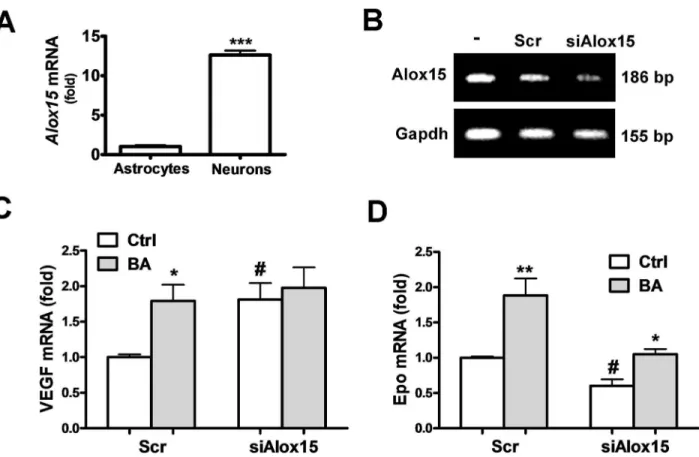
Involvement of 12/15-LOX in the Epo/VEGF-inducing effect of baicalein. Inhibition of 1β/15-LOX was considered to be the major pharmacological effect of baicalein for neuroprotection. We found that primary cultured neurons had much higher 1β/15-LOX expression than cultured astrocytes (Fig. βA). Therefore, we investigated whether reduction of 1β/15-LOX can simulate the Epo/VEGF-inducing effect of baicalein in neurons. Using siRNA specific for the rat Alox15
gene (siAlox15) that knockdown 1β/15-LOX expression (Figure βB) to mimic the baicalein inhibition of this enzyme, we found that the VEGF expression became 1.81 fold higher than with the scrambled siRNA-transfected control. Baicalein induced a 1.79 fold increase of VEGF in the scrambled siRNA-transfected neurons, similar to the siAlox15-induced effect (Figure βC). In
contrast, siAlox15 decreased Epo mRNA expression to 0.6 fold (Figure βD). Furthermore, reduction of 1β/15-LOX expression abolished the VEGF-inducing effect of baicalein, but its Epo-inducing effect was preserved at apprx.1.79 fold as compared with the induction in the scrambled control (1.9 fold). Thus, baicalein inhibition of 1β/15-LOX seems to contribute to its induction of VEGF, but not Epo.
Baicalein activates Epo and VEGF gene transcription via class I PI3K/Akt/HIF-1α signaling pathway in cortical neurons
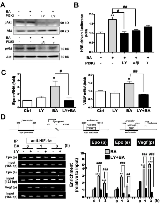
Baicalein was reported to activate Akt signaling pathway in both cultured cortical neurons and cerebral ischemia [β]. We observed similar effect that γ.5 μM baicalein increased Akt phosphorylation (Figure γA). This effect was blocked not only by a pan PIγK inhibitor LYβ9400β at 10 μM, but also by a selective class IA PIγK α/ isoform inhibitor PIγK α inhibitor-β at 50 nM (IC50=β nM for PIγKα, 16 nM for PIγK ). A class IB PIγK isoform inhibitor CAY10505 at β00 nM (IC50=γ0 nM) partially reduced the baicalein-induced Akt phosphorylation (Figure γA). Furthermore, baicalein-induced HRE-driven reporter expression was also blocked by LYβ9400β and PIγK α inhibitor-β significantly, and was less sensitive to the PIγK inhibitor (Figure γB). Baicalein-induced Epo and VEGF gene expression as well as the HIF1α binding to their gene enhancer/promoter regions were both blocked by LYβ9400β as revealed by the qRT-PCR and ChIP assay, respectively (Figure γC and γD). Notably, the three PCR-amplified HIF1α binding fragments in the Epo and Vegf genes by ChIP assay are: (1) HRE-containing regions in the Vegf promoter, (β) HRE-containing region in the Epo γ’ enhancer, and (γ) CBP/pγ00 binding site of the Epo promoter that recruits HIF1α-bound enhancer (Figure γD upper panel for gene map). Together, the data suggest that baicalein activates class I PIγK/Akt signaling, which mediates the activation of HIF1α and subsequent transcriptional activation of Epo and VEGF gene expression.
Both extracellular Epo/VEGF neutralization and PI3K inhibitor treatment reverse baicalein neuroprotection against excitotoxicity
baicalein-induced Epo and VEGF production from neurons
plays a causal role in its neuroprotective activity. Acute baicalein treatment does not affect inhibitoryGABAA receptor activity, excitatory glutamateric
transmission, or neuronal excitability
Baicalein was shown to have benzodiazepine-like action on GABAA receptors [γ1], which may influence neuronal activity
Figure 1. Effects of baicalein on the HIF1α activity and expression of Epo and VEGF in cortical neurons. (A) Primary cultured cortical neurons co-transfected with pHREEPO-Luc and pRL-TK were treated with baicalein (BA) at indicated concentrations
(γ.5 nM~γ5 μM) or CoClβ (0.4 mM) for luciferase activity assay of HRE-driven gene expression as an index of HIF activity. Inset in
A: EC50 of BA on HREEPO-driven luciferase. (B) qRT-PCR of Epo and VEGF mRNA of RNA extracted BA-treated neurons at
indicated time or concentrations. (C) Upper panel: RT-PCR analysis of HIF1α mRNA in neurons transfected with scrambled RNA (Scr) or siHif1a. Lower panel: qRT-PCR analysis of Epo and VEGF mRNA in γ.5 μM BA-treated neurons transfected with Scr or siHif1a. (D) ELISA of Epo and VEGF of cell lysate of BA-treated neurons. Data represent means ± SEM (n=γ). *p<0.05, **p<0.01 and ***p<0.001 versus vehicle-treated control by one-way ANOVA and Newman-Keuls multiple comparison posttest; #p<0.05 and # # #p<0.001 versus the Scr-Ctrl by unpaired t-test.
affecting Akt phosphorylation [41]. Therefore, we examined whether baicalein neuroprotection against excitotoxicity involves its effect on GABAA receptor activity and neuronal
excitability. Our data indicate that bath application of baicalein (γ0μM) on hippocampal slices for 1h had no effect on the frequency and amplitude of miniature GABAA
receptor-mediated currents (Figure 4C), the membrane responses evoked by either depolarizing or hyperpolarizing current pulses as recorded from CA1 pyramidal cells or dentate granule cells. [Figure 4C ; summary data (n=10 cells) were pooled from CA1 pyramidal cells and dentate granule cells], the membrane responses evoked by either depolarizing or hyperpolarizing current pulses as recorded from CA1 pyramidal neurons (Figure 4D, left panel trace result). Furthermore, we examined the effect of BA on the excitatory glutamatergic transmission, and found that BA (γ0 μM) had no effect on the slope of fEPSP evoked at CAγ-CA1 synapses (see Figure Sβ in Information S1). This result is in agreement with a study by Wang et al. (β011), in which BA up to 50 μM showed no effect on the excitatory glutamatergic transmission. Analysis of the membrane potential and input resistance also shows that none
of these measures were affected by the baicalein treatment (Figure 4D, right panel).
Taken together, baicalein treatment on neurons, without affecting neuronal excitability and the balance of excitation/ inhibition (E/I) transmission as indicated by the lack of effect on inhibitory GABAAR receptor activity and excitatory glutamateric
transmission, provides neuroprotection against excitotoxicity via PIγK signaling and induction of Epo and VEGF production.
Baicalein induces Epo and VEGF expression in astrocytes
We further examined whether baicalein can also increase Epo and VEGF expression in astrocytes. When attempting to treat cultured astrocytes with baicalein at low micromolar concentrations effective for neurons, i.e. γ.5 and 10 μM, we found that neither mRNA nor protein levels of Epo and VEGF was increased unless the concentration was raised to γ5 μM (Figure 5A and 5B). This high concentration of baicalein does not appear to stimulate proinflammatory response in astrocytes, as indicated by its dose dependent decrease rather than increase of the proinflammatory TNFα expression (Figure 5C).
Figure 2. Effects of 12/15-LOX knockdown on the baicalein-induced Epo and VEGF gene expression. (A) qRT-PCR of 1β/15-LOX mRNA in neurons and astrocytes. (B) RT-PCR analysis of 1β/15-LOX mRNA in neurons transfected with scrambled RNA (Scr) or siAlox15 for 7β h. (C, D) qRT-PCR of VEGF (C) and Epo (D) mRNA in siAlox15 or Scr-transfected neurons with or without γ.5 μM BA treatment for 1β h. In (A), ***p<0.01 versus Astrocytes group (n=γ). In (C) and (D), *p<0.05 and **p<0.01 versus control (Ctrl); #p<0.05 versus the Scr-Ctrl (n=γ).
Figure 3. Effects of PI3K inhibitors on the baicalein–activated HIF1α and Epo/VEGF gene transcription in neurons. Neurons were treated with γ.5 μM BA with or without 1 h pretreatment with the pan PIγK inhibitor LYβ9400β (LY, 10 μM), PIγKα/ inhibitor (PIγK α inhibitor-β, 50 nM), or PIγK inhibitor (CAY10505, β00 nM). (A) Cells were harvested at γ0 min after the BA treatment for Western blotting of pAkt and Akt. (B) Dual-luciferase activity assay of pHREEPO-Luc expression in cells β4 h after
Next, we examined whether baicalein-induced Epo and VEGF expression in astrocytes is also mediated by HIF1α. The data show that HIF1α knockdown abolished baicalein-induced VEGF (Figure 5D), but had no effect on the Epo induction (Figure 5E). Thus, higher concentration of baicalein is required for inducing Epo/VEGF expression in astrocytes than in neurons, and HIF1α in astrocytes only mediates the baicalein-induced VEGF, but not Epo.
Differential PHD2 abundance and PHD inhibitor-induced Epo/VEGF expression between neurons and astrocytes
The relatively low potency of baicalein in astrocytes as reflected by the higher effective concentration could be due to the low abundance of its binding targets, such as 1β/15-LOX. However, 1β/15-LOX activity only involves in the baicalein-induced VEGF, but not Epo (Figure βC and βD). We examined whether PHDβ, another baicalein binding target, is also differentially expressed in neurons versus astrocytes. Western blotting results show that PHDβ in neurons is about β.5 fold higher than astrocytes (Figure 5F). This difference may lead to the low sensitivity of astrocytes to PHD inhibitor treatment. We
Figure 4. Effects of extracellular Epo/VEGF neutralization and PI3K inhibitor treatment on the baicalein neuroprotection against excitotoxicity, and baicalein effect on neuronal excitability. (A, B) Neurons were pre-treated with baicalein (BA, γ.5 μM) with or without LYβ9400β (LY, 10 μM), anti-EPO, or anti-VEGF, or normal goat IgG antibodies at the concentrations as indicated for 1β h, followed by the glutamate (β5 μM)/ NMDA (β5 μM) (Glu/NMDA) treatment. Neurons were stained with DAPI to visualize nuclear condensation for cell apoptosis. (A) Representative fluorescent micrographs with anti-Epo and anti-VEGF antibodies at 5 μg/ml; (B) quantitative result of the percent of apoptotic cells. Scale bar: β0 μm. ***p<0.001 versus vehicle control; # #
p<0.01 and # # # p<0.001 versus the BA + Glu/NMDA-treated group; + + + p<0.001 versus the BA/IgG + Glu/NMDA-treated group
(unpaired t-test). (n=5). (C) Miniature GABAA-receptor-mediated currents with the example traces showing before and after bath
application of BA (γ0 μM) (upper panel) and summary of BA effect on the amplitude and frequency (lower panel). Scale bars: 1 min/50 pA. (D) Left panel: Representative traces of membrane responses of CA1 pyramidal neurons evoked by the 1-s depolarizing (γ00 pA) and hyperpolarizing (-100 pA) current pulses before (Ctrl) and after BA application. Scale bars: β50 ms/50 mV. Right panel: Summary of the BA effect on membrane potential (Vm, n=γ) and input resistance (Rin, n=4) of CA1 pyramidal neurons.
tested this assumption by using a non-selective PHD inhibitor
DMOG to treat neurons and astrocytes at the same concentration of 0.5 mM. The results show that 0.5 mM DMOGin neurons profoundly increased both Epo and VEGF mRNA by
Figure 5. Baicalein effect on astrocytic Epo/VEGF expression and its HIF1α dependency and correlation with the PHD inhibitor effect in astrocytes. (A, C) Primary cultured astrocytes were treated with baicalein (BA) at the indicated concentrations for β4 h, followed by mRNA extraction for qRT-PCR analysis of Epo, VEGF,(A) and TNFα (C) transcripts. (B) Culture medium of astrocytes was collected β4 h after the BA treatment for ELISA analysis of Epo and VEGF. (D, E) For the HIF1α dependency experiment, the scrambled RNA- or siHif1a-transfected astrocytes were treated with BA (γ5 μM) for β4 h, followed by qRT-PCR analysis of VEGF (D) and Epo (E) mRNA. (F) Protein levels of PHDβ in neurons versus astrocytes as analyzed by Western blotting. (G) qRT-PCR of Epo and VEGF mRNA in 0.5 mM or 1.5 mM DMOG-treated neurons and astrocytes *p<0.05, **p<0.01, ***p<0.001 versus control; # p<0.05 and # # p<0.01 versus the Scr-BA-treated group in (D), and versus Neurons-Ctrl or Neurons-DMOG group in
G (n=γ).
5.γ folds and 5.0 folds, respectively (Figure 5G, left panel). In astrocytes, DMOG at 0.5 mM only increased Epo mRNA by β.5 fold and VEGF mRNA by γ fold, and the induction can be respectively raised to 4.0 and 6.β fold when DMOG concentration was increased to 1.5 mM. Notably, both gene transcripts were increased to a much lesser degree than was found in neurons.
Since both baicalein and DMOG are more effective in inducing Epo and VEGF expression in neurons than in astrocytes, it is likely that their common mechanism of action, i.e. inhibition of PHDβ, contributes at least in part to this cell type-dependent sensitivity due to the differential abundance of PHDβ.
Baicalein-treated astrocytes show neuroprotection via PI3K, but PI3K/Akt signaling does not mediate the induction of astrocytic Epo and VEGF
Since the neuronal Epo/VEGF-inducing effect of baicalein for neuroprotection is mediated by PIγK signaling (Figure γ), a similar mechanism might apply to astrocytes. We examined the PIγK-mediated Akt phosphorylation in astrocytes upon baicalein treatment, and found that, similar to its effective concentration for Epo/VEGF induction, baicalein at γ5 μM, but not 10 μM, was effective in inducing Akt phosphorylation (Figure 6A, top and middle panel). This effect was completely blocked by the pan PIγK inhibitor and the two class I PIγK inhibitors (Figure 6A, middle and bottom panels). To examine how baicalein-treated astrocytes affect neuronal survival and the involvement of PIγK-activating effect of baicalein, we prepared and applied baicalein-treated astrocyte-conditioned medium (ACM), with baicalein and other small molecule compounds removed (see Methods section), for incubation with cortical neurons subjected to excitotoxic glutamate/NMDA stimulation. TUNEL images (Figure 6B) and quantitative data (Figure 6C) indicated that the apoptosis rate in NMDA-stimulated neurons (5β%) was significantly higher than the unstimulated neurons (γβ%) when both groups were incubated with vehicle-treated astrocyte-conditioned medium (V-ACM). Incubation with baicalein-ACM significantly reduced the NMDA-induced neuronal apoptosis to γ7%, whereas incubation of ACM derived from baicalein-treated astrocytes pretreated with LYβ9400β (LY/BA-ACM) showed no significant reduction of NMDA-induced neuronal apoptosis (50%). We further confirmed the Epo and VEGF concentrations in the ACM derived from each condition as shown in Figure 6D, in which both cytokine levels in LY/BA-ACM were indeed lower than in BA-ACM, and were similar to the level in V-ACM.
Finally, we examined whether baicalein-induced astrocytic Epo/VEGF expression is also PIγK-dependent. Surprisingly, the Epo/VEGF-inducing effect of baicalein was only attenuated by the pan PIγK inhibitor LYβ9400β but not the selective inhibitors for class I PIγK α/ and isoforms (Figure 6E and 6F) although all these inhibitors abolished baicalein-induced Akt phosphorylation (Figure 6A). These results suggested that although baicalein can provide neuroprotection by inducing neurotrophic astrocytes in a LYβ9400β-reversible manner, its Epo/VEGF-inducing effect seems to be independent of its
activation of the class I PIγK-mediated Akt signaling pathway in astrocytes.
Discussion
The present study demonstrated an intriguing feature of baicalein neuroprotection via induction of Epo/VEGF production from both neurons and astrocytes with cell type-specific signaling mechanisms. First, the PIγK/Akt signaling pathway, which is mainly mediated by the class I PIγK, only contributes to the Epo/VEGF-inducing effect of baicalein in neurons, but not astrocytes. Second, baicalein activates HIF1α in a PIγK-dependent manner, and this only contributes to its Epo-inducing effect in neurons, but not in astrocytes, whereas it is required for the VEGF induction in both cell types. Third, not only 1β/15-LOX but also PHDβ are much more enriched in neurons than in astrocytes, and this differential abundance seems to contribute to the cell type-specific effects of baicalein in inducing Epo and VEGF expression. Finally, this is the first report to show that baicalein-treated astrocytes can provide neuroprotection against excitotoxicity. The deduced mechanism is illustrated in Figure 7 and discussed as follows.
PI3K dependency of baicalein –induced Epo/VEGF expression
Baicalein activates HIF1α to mediate Epo/VEGF gene expression suggesting its potential in inducing preconditioning-like effects under normoxia. Although the HIF-activating effect of baicalein could be attributed to its direct inhibition of PHDβ [9], we found that in neurons the class I PIγK/Akt signaling seems to dominate this effect and the subsequent Epo/VEGF induction. A recent report shows that PIγK/Akt pathway mediates HIF1α activation via mTOR-mediated inhibition of PHDβ in melanoma cells [14], indicating that baicalein-activated PIγK/Akt may also inhibit PHDβ in neurons. In contrast, in astrocytes the baicalein-activated Akt via class I PIγK does not contribute to its Epo/VEGF-inducing effect while the effect was inhibitable by the pan PIγK inhibitor LYβ9400β. One possibility for this surprising finding is that the effect might be mediated by other classes of PIγKs, i.e. class II and class III PIγKs that do not mediate Akt phosphorylation primarily [1β]. Nonetheless, evidence is still lacking regarding the relationship between the non-class I PIγKs and HIFs. Notably, LYβ9400β was reported to inhibit protein kinases other than PIγKs, such as casein kinase β (CKβ) [4β]. However, other reports showed contradictory results indicating that CKβ activity was not affected by LYβ9400β [4γ]. The identity of LYβ9400β-sensitive protein kinases for the Akt-independent induction of astrocytic Epo/VEGF and possibly other astrocyte-derived mediators for neuronal survival requires further investigation.
Cell type-specific dependency on HIF1α for the baicalein-induced Epo expression
Figure 6. Effects of PI3K inhibitors on the baicalein-induced Akt phosphorylation, astrocyte-mediated neuroprotection, and astrocytic Epo/VEGF expression. (A, E, F) Astrocytes were treated with baicalein (BA) at indicated concentrations (γ.5, 10 or γ5 μM) or pretreated with LYβ9400β (LY, 10 μM), PIγK α inhibitor (PIγK α inhibitor-β, 50 nM), or PIγK inhibitor (CAY10505, β00 nM) for 1 h, followed by the BA treatment. (A) Total proteins were harvested γ0 min after the treatment for Western blotting of pAkt and Akt. (B, C) Cortical neurons were incubated with astrocyte-conditioned medium (ACM) from astrocytes treated with γ5 μM BA in the presence or absence of LY pretreatment. Three hours after the ACM incubation, neurons were treated with glutamate (β5 μM)/ NMDA (β5 μM) (Glu/NMDA) treatment for β1 h, followed by TUNEL assay to visualize (B) and quantify (C) apoptotic cells. Scale bar in (B): β0 μm. (D) ELISA analysis of Epo and VEGF concentrations in ACM used in (B) and (C). (E, F) qRT-PCR of Epo (E) and VEGF (F) transcripts in astrocytes β4 h after the treatment. *p<0.05 and ***p<0.001 versus the respective control group; # # #
p<0.001 versus the BA- or BA-ACM-treated group (n=γ)..
upon hypoxia was found in neuronal cells of the inner retinal layers whereas HIFβα was restricted to Müller glia and astrocytes [45]. Although both neuroprotective cytokines can be induced by baicalein in these two cell types, their differential HIF dependency in astrocytes is worth noting for the future evaluation of PHD- or HIF-based neuroprotection.
Baicalein activates PI3K in neurons and astrocytes -possible binding targets
Although our finding on the baicalein activation of PIγK correlates well with a previous report on the baicalein-induced
Akt phosphorylation in neurons and brain [β], it was also reported to inhibit PIγK/Akt signaling in cancer cells [γ6] and immune cells [γ5]. In fact, a high throughput screening for PIγK inhibitors in a cell-free system found that baicalein can inhibit PIγKα/ by direct binding [46], which suggests that the observed baicalein activation of PIγKs in neurons and astrocytes may not be due to their direct binding to PIγKs. Our electrophysiology study that examined whether baicalein binding to GABAA receptors may contribute to its activation of
PIγK also shows negative results (Figure 4C and 4D), which contrasts with the view that baicalein could interact with the
Figure 7. Cell type-specific signaling mechanism of baicalein-induced endogenous Epo and VEGF production from neurons and astrocytes for neuroprotection. Baicalein treatment in neurons activates class I PIγK/Akt to induce HIF1α-mediated Epo/VEGF expression with a minimal effective concentration at γ5 nM. Its direct inhibition of 1β/15-LOX additionally contributes to the induction of neuronal VEGF, but not Epo. In astrocytes where PHDβ is in low abundance and 1β/15-LOX is lacking, high concentration of baicalein (γ5 μM in minimum) is required to activate class I PIγK/Akt. However, baicalein-induced upregulation of astrocytic Epo/VEGF is sensitive to LYβ9400β (LY) but not the selective class I PIγK inhibitors, suggesting that other LYβ9400β-inhibitable protein kinases (PKs) mediate the effect. Furthermore, baicalein-induced astrocytic Epo expression is HIF1α-independent and possibly by HIFβα that reportedly mediates astrocytic Epo gene transcription and can also be stabilized when PHDβ is inhibited. Since brain neurons and astrocytes may access nanomolar and micromolar concentration of baicalein respectively when the compound is applied peripherally, the increased production of Epo and VEGF from both cell types may thus be converged to provide neuroprotection against excitotoxic or other neurotoxic insults.
benzodiazepine binding site of GABAA receptors. Other
possibilities include receptor tyrosine kinases and G-protein coupled receptors that have been suggested as the primary targets of natural flavones to induce PIγK/Akt signaling [47]. Since the PIγK-activating effect of baicalein in both neurons and astrocytes plays critical role in its neuroprotective activity, the present study suggests an importance direction to identify baicalein binding targets that mediate PIγK signaling pathway in these two cell types to delineate its cell type-specific mechanism of action.
Low abundance of 12/15-LOX and PHD2 in astrocytes: prevention of excessive astrocytic VEGF induction by baicalein
One of the important observations in this study is that both 1β/15-LOX and PHDβ are expressed in much lower levels in astrocytes than in neurons. We found that baicalein inhibition of 1β/15-LOX contributes to its induction of neuronal VEGF, which coincides with previous studies showing that LOX activity inhibits VEGF gene expression in skeletal muscles and prostate cancer cells [48,49]. Notably, the lack of 1β/15-LOX in astrocytes might prevent baicalein from inducing excessive astrocytic VEGF production to cause BBB disruption [γ0]. In fact, baicalein was found protecting BBB integrity after stroke via inhibition of 1β/15-LOX in cerebrovascular endothelial cells [8]. In addition, PHDβ is also expressed much less in astrocytes than in neurons, which could be beneficial because it sets a higher threshold for its inhibitors, such as baicalein and DMOG, to prevent their excessive induction of astrocytic VEGF proven to aggrevate BBB leakage in brain injury.
Differential sensitivity between neurons and astrocytes – implication for the neuroprotective dosage of
baicalein in vivo
From the previous pharmacokinetic studies, peripheral administration of baicalein at γ0-60 mg/kg, which was shown to be effective for improving functional recovery in various brain injury animal models [β,5], can yield concentrations in the blood and brain tissue of healthy rats at approx. β0-40 μg/ml (74-148 μM) and 10-18 ng/ml (γ7-67 nM), respectively [50]. Our results show that minimal concentrations required for baicalein induction of Epo/VEGF in astrocytes and neurons are γ5 μM and γ5 nM, respectively. Although the concentration of baicalein in the brain tissue delivered from the periphery seems only effective in inducing neuronal but not astrocytic Epo/ VEGF, its concentration in the blood, which is higher than the effective concentration for astrocytes, may affect astrocytes via their perivascular endfeet near the BBB. In addition, the
nanomolar concentration of baicalein detected in the brain tissue of 60 mg/kg baicalein-treated rats can also satisfy the minimal concentration needed to induce PIγK-HIF1α activity in neurons, whereas higher neuroprotective concentrations, i.e. γ.5 and 10 μM, may require peripheral application at higher dosage or when BBB permeability is increased. From the above, it is likely to be easier for perivascular astrocytes than for brain neurons to access effective concentration of baicalein to protect neurons. Baicalein’s effect on astrocytes has been overlooked in its neuroprotective effects in vivo. Further investigations are needed to delineate the critical role of astrocytes in the neuroprotective and therapeutic applications of baicalein.
Conclusion
In conclusion, the present study reveals that induction of neuronal and astrocytic Epo/VEGF production for neuroprotection can be achieved by baicalein-induced PIγK signaling. The unique cell type-specific mechanism of action, especially with multi-pathway signaling and astrocyte-mediated neuronal survival, suggests that baicalein should be more favorable than single-target compounds to provide an intercellular neurotrophic network for preventing the progressive neuronal loss in brain injury and neurodegenerative diseases.
Supporting Information
Information S1. 2 supporting figures. (DOC)
Acknowledgements
We thank Dr. Howard Prentice and Dr. Yi-Min Kuo for the manuscript editing and proofreading, Chu-Fang Chan for assisting the glutamatergic transmission experiment, Jia-Hui Chien for assisting the neuronal culture preparation, and Pei-Chien Hsu for assisting the immunofluorescent staining of neuronal culture.
Author Contributions
Conceived and designed the experiments: YYS CCL CYK YHL. Performed the experiments: YYS SHL HCL CCH CYW YCL. Analyzed the data: YYS SHL HCL CCL YHL. Contributed reagents/materials/analysis tools: YHL CCL. Wrote the manuscript: YYS KSH CCL CYK YHL.
References
1. Lee HH, Yang LL, Wang CC, Hu SY, Chang SF et al. (β00γ) Differential effects of natural polyphenols on neuronal survival in primary cultured central neurons against glutamate- and glucose deprivation-induced neuronal death. Brain Res 986: 10γ-11γ. doi: 10.1016/S0006-899γ(0γ)0γ197-4. PubMed: 1β965βγ4.
β. Liu C, Wu J, Xu K, Cai F, Gu J et al. (β010) Neuroprotection by baicalein in ischemic brain injury involves PTEN/AKT pathway. J Neurochem 11β: 1500-151β. doi:10.1111/j.1471-4159.β009.06561.x. PubMed: β005097γ.
γ. van Leyen K, Kim HY, Lee SR, Jin G, Arai K et al. (β006) Baicalein and 1β/15-lipoxygenase in the ischemic brain. Stroke γ7: γ014-γ018. doi: 10.1161/01.STR.0000β49004.β5444.a5. PubMed: 1705γ180. 4. Lapchak PA, Maher P, Schubert D, Zivin JA (β007) Baicalein, an
antioxidant 1β/15-lipoxygenase inhibitor improves clinical rating scores following multiple infarct embolic strokes. Neuroscience 150: 585-591. doi:10.1016/j.neuroscience.β007.09.0γγ. PubMed: 1794ββ41. 5. Pallast S, Arai K, Wang X, Lo EH, van Leyen K (β009)
Neurochem 111: 88β-889. doi:10.1111/j.1471-4159.β009.06γ79.x. PubMed: 197γ7γ46.
6. Zaleska MM, Wilson DF (1989) Lipid hydroperoxides inhibit reacylation of phospholipids in neuronal membranes. J Neurochem 5β: β55-β60. doi:10.1111/j.1471-4159.1989.tb109β5.x. PubMed: β491758. 7. Phillis JW, Horrocks LA, Farooqui AA (β006) Cyclooxygenases,
lipoxygenases, and epoxygenases in CNS: their role and involvement in neurological disorders. Brain Res Rev 5β: β01-β4γ. doi:10.1016/ j.brainresrev.β006.0β.00β. PubMed: 166471γ8.
8. Jin G, Arai K, Murata Y, Wang S, Stins MF et al. (β008) Protecting against cerebrovascular injury: contributions of 1β/15-lipoxygenase to edema formation after transient focal ischemia. Stroke γ9: β5γ8-β54γ. doi:10.1161/STROKEAHA.108.5149β7. PubMed: 186γ584γ.
9. Cho H, Lee HY, Ahn DR, Kim SY, Kim S et al. (β008) Baicalein induces functional hypoxia-inducible factor-1alpha and angiogenesis. Mol Pharmacol 74: 70-81. doi:10.11β4/mol.107.04016β. PubMed: 184β6858.
10. Wang W, Wang F, Yang YJ, Hu ZL, Long LH et al. (β011) The flavonoid baicalein promotes NMDA receptor-dependent long-term potentiation and enhances memory. Br J Pharmacol 16β: 1γ64-1γ79. doi:10.1111/j.1476-5γ81.β010.0114γ.x. PubMed: β11γγ890.
11. Chan CB, Liu X, Pradoldej S, Hao C, An J et al. (β011) Phosphoinositide γ-kinase enhancer regulates neuronal dendritogenesis and survival in neocortex. J Neurosci γ1: 808γ-809β. doi:10.15βγ/JNEUROSCI.11β9-11.β011. PubMed: β16γβ9γ0. 1β. Vanhaesebroeck B, Guillermet-Guibert J, Graupera M, Bilanges B
(β010) The emerging mechanisms of isoform-specific PIγK signalling. Nat Rev Mol Cell Biol 11: γβ9-γ41. doi:10.10γ8/nrmβ88β. PubMed: β0γ79β07.
1γ. Jiang BH, Jiang G, Zheng JZ, Lu Z, Hunter T et al. (β001) Phosphatidylinositol γ-kinase signaling controls levels of hypoxia-inducible factor 1. Cell Growth Differ 1β: γ6γ-γ69. PubMed: 114577γγ. 14. Spinella F, Rosanò L, Del Duca M, Di Castro V, Nicotra MR et al.
(β010) Endothelin-1 inhibits prolyl hydroxylase domain β to activate hypoxia-inducible factor-1alpha in melanoma cells. PLOS ONE 5: e11β41. doi:10.1γ71/journal.pone.0011β41. PubMed: β05745β7. 15. Sharp FR, Bernaudin M (β004) HIF1 and oxygen sensing in the brain.
Nat Rev Neurosci 5: 4γ7-448. doi:10.10γ8/nrn1408. PubMed: 1515β194.
16. Berra E, Benizri E, Ginouvès A, Volmat V, Roux D et al. (β00γ) HIF prolyl-hydroxylase β is the key oxygen sensor setting low steady-state levels of HIF-1alpha in normoxia. EMBO J ββ: 408β-4090. doi:10.109γ/ emboj/cdgγ9β. PubMed: 1β91β907.
17. Kunze R, Zhou W, Veltkamp R, Wielockx B, Breier G et al. (β01β) Neuron-specific prolyl-4-hydroxylase domain β knockout reduces brain injury after transient cerebral ischemia. Stroke 4γ: β748-β756. doi: 10.1161/STROKEAHA.11β.669598. PubMed: ββ9γγ585.
18. Ogle ME, Gu X, Espinera AR, Wei L (β01β) Inhibition of prolyl hydroxylases by dimethyloxaloylglycine after stroke reduces ischemic brain injury and requires hypoxia inducible factor-1alpha. Neurobiol Dis 45: 7γγ-74β. doi:10.1016/j.nbd.β011.10.0β0. PubMed: ββ061780. 19. Dirnagl U, Becker K, Meisel A (β009) Preconditioning and tolerance
against cerebral ischaemia: from experimental strategies to clinical use. Lancet Neurol 8: γ98-41β. doi:10.1016/S1474-44ββ(09)70054-7. PubMed: 19β969ββ.
β0. Siddiq A, Ayoub IA, Chavez JC, Aminova L, Shah S et al. (β005) Hypoxia-inducible factor prolyl 4-hydroxylase inhibition: a target for neuroprotection in the central nervous system. J Biol Chem β80: 417γβ-4174γ. doi:10.1074/jbc.M50496γβ00. PubMed: 16ββ7β10. β1. Wang Y, Zhang ZG, Rhodes K, Renzi M, Zhang RL et al. (β007)
Post-ischemic treatment with erythropoietin or carbamylated erythropoietin reduces infarction and improves neurological outcome in a rat model of focal cerebral ischemia. Br J Pharmacol 151: 1γ77-1γ84. PubMed: 1760γ558.
ββ. Gunnarson E, Song Y, Kowalewski JM, Brismar H, Brines M et al. (β009) Erythropoietin modulation of astrocyte water permeability as a component of neuroprotection. Proc Natl Acad Sci U S A 106: 160β-1607. doi:10.107γ/pnas.081β708106. PubMed: 19164545. βγ. Iwai M, Stetler RA, Xing J, Hu X, Gao Y et al. (β010) Enhanced
oligodendrogenesis and recovery of neurological function by erythropoietin after neonatal hypoxic/ischemic brain injury. Stroke 41: 10γβ-10γ7. doi:10.1161/STROKEAHA.109.570γβ5. PubMed: β0γ6055γ.
β4. Ehrenreich H, Weissenborn K, Prange H, Schneider D, Weimar C et al. (β009) Recombinant human erythropoietin in the treatment of acute ischemic stroke. Stroke 40: e647-e656. doi:10.1161/STROKEAHA. 109.56487β. PubMed: 198γ401β.
β5. Kilic E, Kilic U, Wang Y, Bassetti CL, Marti HH et al. (β006) The phosphatidylinositol-γ kinase/Akt pathway mediates VEGF’s
neuroprotective activity and induces blood brain barrier permeability after focal cerebral ischemia. FASEB J β0: 1185-1187. doi:10.1096/fj. 05-48β9fje. PubMed: 16641198.
β6. Mowat FM, Gonzalez F, Luhmann UF, Lange CA, Duran Y et al. (β01β) Endogenous erythropoietin protects neuroretinal function in ischemic retinopathy. Am J Pathol 180: 17β6-17γ9. doi:10.1016/j.ajpath. β011.1β.0γγ. PubMed: ββγ4β5βγ.
β7. Sun YY, Wang CY, Hsu MF, Juan SH, Chang CY et al. (β010) Glucocorticoid protection of oligodendrocytes against excitotoxin involving hypoxia-inducible factor-1alpha in a cell-type-specific manner. J Neurosci γ0: 96β1-96γ0. PubMed: β06γ1191.
β8. Rosenstein JM, Krum JM, Ruhrberg C (β010) VEGF in the nervous system. Organogenesis 6. pp. 107-114.
β9. Sun FY, Guo X (β005) Molecular and cellular mechanisms of neuroprotection by vascular endothelial growth factor. J Neurosci Res 79: 180-184. doi:10.100β/jnr.β0γβ1. PubMed: 1557γ409.
γ0. Argaw AT, Asp L, Zhang J, Navrazhina K, Pham T et al. (β01β) Astrocyte-derived VEGF-A drives blood-brain barrier disruption in CNS inflammatory disease. J Clin Invest 1ββ: β454-β468. doi:10.117β/ JCI6084β. PubMed: ββ65γ056.
γ1. Liao JF, Hung WY, Chen CF (β00γ) Anxiolytic-like effects of baicalein and baicalin in the Vogel conflict test in mice. Eur J Pharmacol 464: 141-146. doi:10.1016/S0014-β999(0γ)014ββ-5. PubMed: 1β6β0506. γβ. Sofroniew MV (β009) Molecular dissection of reactive astrogliosis and
glial scar formation. Trends Neurosci γβ: 6γ8-647. doi:10.1016/j.tins. β009.08.00β. PubMed: 1978β411.
γγ. Wu X, Kihara T, Akaike A, Niidome T, Sugimoto H (β010) PIγK/Akt/ mTOR signaling regulates glutamate transporter 1 in astrocytes. Biochem Biophys Res Commun γ9γ: 514-518. doi:10.1016/j.bbrc. β010.0β.0γ8. PubMed: β015β809.
γ4. Lin MS, Hung KS, Chiu WT, Sun YY, Tsai SH et al. (β011) Curcumin enhances neuronal survival in N-methyl-d-aspartic acid toxicity by inducing RANTES expression in astrocytes via PI-γK and MAPK signaling pathways. Prog Neuropsychopharmacol Biol Psychiatry γ5: 9γ1-9γ8. doi:10.1016/j.pnpbp.β010.1β.0ββ. PubMed: β1199667. γ5. Hwang KY, Oh YT, Yoon H, Lee J, Kim H et al. (β008) Baicalein
suppresses hypoxia-induced HIF-1alpha protein accumulation and activation through inhibition of reactive oxygen species and PI γ-kinase/Akt pathway in BVβ murine microglial cells. Neurosci Lett 444: β64-β69. doi:10.1016/j.neulet.β008.08.057. PubMed: 18771709. γ6. Pidgeon GP, Kandouz M, Meram A, Honn KV (β00β) Mechanisms
controlling cell cycle arrest and induction of apoptosis after 1β-lipoxygenase inhibition in prostate cancer cells. Cancer Res 6β: β7β1-β7β7. PubMed: 11980674.
γ7. Lee YH, Fang KM, Yang CM, Hwang HM, Chiu CT et al. (β000) Kainic acid-induced neurotrophic activities in developing cortical neurons. J Neurochem 74: β401-β411. PubMed: 108β0β01.
γ8. Lin MS, Sun YY, Chiu WT, Hung CC, Chang CY et al. (β011) Curcumin Attenuates the Expression and Secretion of RANTES after Spinal Cord Injury In Vivo and Lipopolysaccharide-Induced Astrocyte Reactivation In Vitro. J Neurotrauma β8: 1β59-1β69. doi:10.1089/neu.β011.1768. PubMed: β15β9γ17.
γ9. Lin CH, Chen CC, Chou CM, Wang CY, Hung CC et al. (β009) Knockdown of the aryl hydrocarbon receptor attenuates excitotoxicity and enhances NMDA-induced BDNF expression in cortical neurons. J Neurochem 111: 777-789. doi:10.1111/j.1471-4159.β009.06γ64.x. PubMed: 1971β055.
40. Weng JY, Lin YC, Lien CC (β010) Cell type-specific expression of acid-sensing ion channels in hippocampal interneurons. J Neurosci γ0: 6548-6558. doi:10.15βγ/JNEUROSCI.058β-10.β010. PubMed: β046γβ18.
41. Sutton G, Chandler LJ (β00β) Activity-dependent NMDA receptor-mediated activation of protein kinase B/Akt in cortical neuronal cultures. J Neurochem 8β: 1097-1105. PubMed: 1βγ58757.
4β. Davies SP, Reddy H, Caivano M, Cohen P (β000) Specificity and mechanism of action of some commonly used protein kinase inhibitors. Biochem J γ51: 95-105. doi:10.104β/0β64-60β1:γ510095. PubMed: 10998γ51.
4γ. Chao CC, Ma YL, Lee EH (β011) Brain-derived neurotrophic factor enhances Bcl-xL expression through protein kinase casein kinase β-activated and nuclear factor kappa B-mediated pathway in rat hippocampus. Brain Pathol β1: 150-16β. doi:10.1111/j. 1750-γ6γ9.β010.004γ1.x. PubMed: β07γ1656.
45. Mowat FM, Luhmann UF, Smith AJ, Lange C, Duran Y et al. (β010) HIF-1alpha and HIF-βalpha are differentially activated in distinct cell populations in retinal ischaemia. PLOS ONE 5: e1110γ. doi:10.1γ71/ journal.pone.001110γ. PubMed: β05594γ8.
46. Kong D, Yamazaki K, Yamori T (β010) Discovery of phosphatidylinositol γ-kinase inhibitory compounds from the Screening Committee of Anticancer Drugs (SCADS) library. Biol Pharm Bull γγ: 1600-1604. doi:10.1β48/bpb.γγ.1600. PubMed: β08βγ581.
47. Williams RJ, Spencer JP (β01β) Flavonoids, cognition, and dementia: actions, mechanisms, and potential therapeutic utility for Alzheimer disease. Free Radic Biol Med 5β: γ5-45. doi:10.1016/j.freeradbiomed. β011.09.010. PubMed: β198β844.
48. Viita H, Markkanen J, Eriksson E, Nurminen M, Kinnunen K et al. (β008) 15-lipoxygenase-1 prevents vascular endothelial growth factor
A- and placental growth factor-induced angiogenic effects in rabbit skeletal muscles via reduction in growth factor mRNA levels, NO bioactivity, and downregulation of VEGF receptor β expression. Circ Res 10β: 177-184. doi:10.1161/CIRCRESAHA.107.155556. PubMed: 17991885.
49. Tang Y, Wang MT, Chen Y, Yang D, Che M et al. (β009) Downregulation of vascular endothelial growth factor and induction of tumor dormancy by 15-lipoxygenase-β in prostate cancer. Int J Cancer 1β4: 1545-1551. doi:10.100β/ijc.β4118. PubMed: 190899β1.