Involvement of glutamate and reactive
oxygen species in methylmercury
neurotoxicity
1Departments of Pediatrics and Pharmacology, and the Kennedy Center,
Vanderbilt University Medical Center, Nashville, TN, USA
2Department of Clinical Neuroscience, Norwegian University of Science and
Technology, Trondheim, Norway
3Departamento de Análises Clínicas e Toxicológicas, Centro de Ciências da Saúde,
Universidade Federal de Santa Maria, Santa Maria, RS, Brasil
4Departamento de Bioquímica, Instituto de Ciências Básicas da Saúde,
Universidade Federal do Rio Grande do Sul, Porto Alegre, RS, Brasil
5Departamento de Bioquímica, Centro de Ciências Biológicas,
Universidade Federal de Santa Catarina, Florianópolis, SC, Brasil M. Aschner1,
T. Syversen2,
D.O. Souza3,
J.B.T. Rocha4
and M. Farina5
Abstract
This review addresses the mechanisms of methylmercury (MeHg)-induced neurotoxicity, specifically examining the role of oxidative stress in mediating neuronal damage. A number of critical findings point to a central role for astrocytes in mediating MeHg-induced neurotoxicity as evidenced by the following observations: a) MeHg preferentially accumulates in astrocytes; b) MeHg specifically inhib-its glutamate uptake in astrocytes; c) neuronal dysfunction is second-ary to disturbances in astrocytes. The generation of reactive oxygen species (ROS) by MeHg has been observed in various experimental paradigms. For example, MeHg enhances ROS formation both in vivo (rodent cerebellum) and in vitro (isolated rat brain synaptosomes), as well as in neuronal and mixed reaggregating cell cultures. Antioxi-dants, including selenocompounds, can rescue astrocytes from MeHg-induced cytotoxicity by reducing ROS formation. We emphasize that oxidative stress plays a significant role in mediating MeHg-induced neurotoxic damage with active involvement of the mitochondria in this process. Furthermore, we provide a mechanistic overview on oxidative stress induced by MeHg that is triggered by a series of molecular events such as activation of various kinases, stress proteins and other immediate early genes culminating in cell damage. Correspondence
M. Aschner
Department of Pediatrics B-3307 Medical Center North Vanderbilt University School of Medicine
Nashville, TN 37232-2495 USA
Fax: +1-615-322-6541 E-mail:
Research supported in part by Public Health Service Grant ES07331 to M. Aschner.
Received August 21, 2006 Accepted January 16, 2007
Key words
•Methylmercury
neurotoxicity
•Oxidative stress •Glutamate and
selenocompounds
•Reactive oxygen species •Astrocytes
Introduction
Methylmercury (MeHg) is a major neuro-toxicant that continues to pose appreciable risk to human health as evidenced by the tragic epidemics of MeHg poisoning in Japan and Iraq (1,2). All sources of environmental
aberrant central nervous system (CNS) func-tions (3-5). Recent studies in human popula-tions support the earlier findings that maternal exposure to mercury during pregnancy is as-sociated with neurological as well as neuro-psychological deficits detectable in the child at 6 to 7 years of age (6,7). Another recent study has pointed to the selective detrimental effects of MeHg on neurogenesis. However, despite these observations, the issue remains controversial, as exemplified by other studies (8,9) in which no association was noted be-tween MeHg and neurodevelopmental out-comes in children at 66 months of age. Thus, the ultimate effects of MeHg in the human population remain unknown and clearly there is an urgent need to understand the mechan-isms and consequences of MeHg exposure for CNS function. The current literature suggests that no single mechanism can explain the multitude of effects observed in MeHg-in-duced neurotoxicity.
The role of astrocytes in brain function
Among various cell types, astrocytes rep-resent a major cell type occupying approxi-mately 50% of the CNS volume and their dysfunction following toxicant exposure has been implicated as a main cause of the ob-served neurotoxicity (10). The “foot” pro-cesses of these cells are known to be closely associated with synapses, axonal tracts, nodes of Ranvier, and capillaries. Astrocytes per-form important duties and some of their functions include the maintenance of normal extracellular ion concentrations, the uptake of potassium (K+) and the control of
extra-cellular pH. They express a pantheon of receptors and uptake systems for neurotrans-mitters, properties that were formerly thought to be exclusively neuronal. Thus, astrocytes can respond to and prevent the build-up of potentially dangerous levels of neurotrans-mitters (e.g., glutamate) in the extracellular fluid (11). Important roles of astrocytes
dur-ing early brain development include the syn-thesis and elaboration of cues for neuronal migration and the production of neurotrophic factors important for neuronal division and differentiation. Astrocytes also induce the high electrical resistance (tightness) of the blood-brain barrier that limits the transport of noxious substances entering the brain, and modulates optimal transport of nutrients and metabolites (12).
Mechanisms of methylmercury-induced neurotoxicity and glutamate
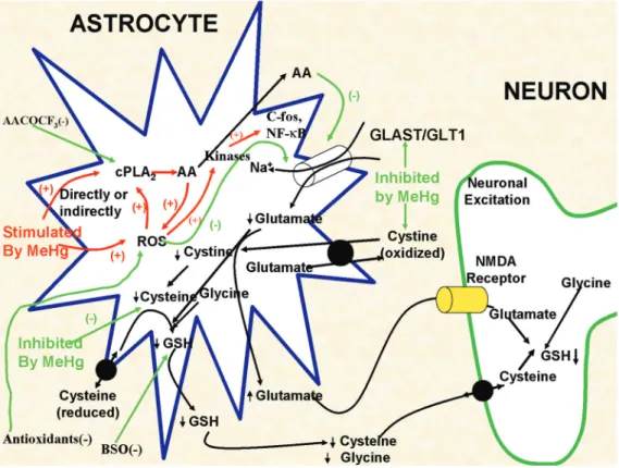
Although not the only cell type to be adversely affected by MeHg, a number of studies have established a key role for astro-cytes in mediating MeHg neurotoxicity: a) astrocytes represent a preferential cellular site for MeHg accumulation (13-16); b) MeHg selectively inhibits astrocytic trans-port of cystine and cysteine (Figure 1), thereby adversely affecting their redox sta-tus and attenuating glutathione (GSH) con-tent (17-21); c) MeHg inhibits astrocytic glutamate (and aspartate) uptake (Figure 1) and stimulates its efflux, thereby increasing glutamate concentrations in the extracellu-lar fluid and sensitizing neurons to excito-toxic injury (17,18,22-24); d) MeHg-induced neuronal dysfunction is secondary to distur-bances in astrocytes (25), and the in vitro co-application of non-toxic concentrations of mercury with glutamate results in the ap-pearance of typical neuronal lesions found with excitotoxic stimulation (26); e) MeHg causes the activation of cytosolic phospholi-pase A2 leading to arachidonic acid release
and further inhibition of the glutamate trans-porter (Figure 1), setting in motion an unim-peded cytotoxic cycle (27,28).
The role of reactive oxygen species in methylmercury-induced
neurotoxicity
physiological role in optimal cell function, excessive oxidative stress has been impli-cated in a variety of neurodegenerative dis-eases, including Alzheimer’s disease, Park-inson’s disease and amyotrophic lateral scle-rosis (29-31). Oxidative stress also plays an important role in other degenerative condi-tions such as autoimmune and inflammatory diseases (i.e., ischemia and rheumatoid ar-thritis), cancer, diabetes mellitus, and ath-erosclerosis (32-34), as well as in metal-induced toxicity (35,36). The balance be-tween oxidative and reductive cellular pro-cesses is known to be adversely affected in these various disorders (37). Oxidative stress is associated with the accumulation of high
levels of toxic reactive species, such as reac-tive oxygen species (ROS), reacreac-tive nitro-gen species, reactive nitronitro-gen oxynitro-gen spe-cies, as well as unbound metal ions (38). Typical ROS include oxygen radicals such as superoxide radical, hydroxyl radical, as well as non-radical derivatives of oxygen including hydrogen peroxide (H2O2).
Reac-tive nitrogen species include nitric oxide radical, and reactive nitrogen oxygen cies include the highly reactive oxidant spe-cies peroxynitrite, which is a product of reaction between nitric oxide and the super-oxide radical. These reactive species are highly oxidizing and potently damaging to cellular redox-sensitive proteins, enzymes
Figure 1. A schematic model of some of the currently proposed as well as some of our previously studied processes resulting in methylmercury (MeHg)-dependent neurotoxicity. Red lines and text indicate processes where MeHg stimulates cellular processes, whereas green lines and text indicate targets for MeHg-induced inhibition. For example, MeHg is known to increase the release of arachidonic acid (AA)from astrocytes and to stimulate glutamate release, increasing its synaptic levels. At the same time MeHg also inhibits glutamate uptake, as well as a number of the amino acids that are associated with the synthesis of astrocytic glutathione (GSH). Combined, these effects lead to a reduction in intracellular GSH levels and increased synaptic glutamate levels, which in turn activate NMDA receptors on adjacent neurons, leading to excitotoxicity. cPLA2 = cytosolic
phospho-lipase A2; ROS = reactive oxygen species; NF-κB = nuclear factor kappa B; BSO = L-buthionine-[S,R]-sulfoximine;
and DNA, and they also cause peroxidation of membranes.
ROS are known to mediate MeHg-induced neurotoxicity in multiple experimental mod-els. For example, MeHg induces ROS forma-tion in vivo (rodent cerebellum), and in vitro
(isolated rat brain synaptosomes) (39), as well as in cerebellar neuronal cultures, a hypotha-lamic neuronal cell line and in mixed reaggre-gating cell cultures (40-42). In addition, an increase in ROS has been observed in a) mito-chondria isolated from MeHg-injected rat brains (36), b) mitochondria isolated in vitro
from rat brain and then exposed to MeHg (43), and c) mitochondria from Hg- and glutamate-exposed astrocytes and neurons (44,45). induced ROS production and MeHg-induced glutamate dyshomeostasis are con-nected phenomena affecting each other. In fact, MeHg-induced inhibition of astrocyte glutamate transporters leads to increased glu-tamate concentrations in the extracellular fluid, causing hyperactivation of N-methyl D-aspar-tate type glutamate receptors and leading to an increase in Na+ and Ca2+ influx (46). Increased
intracellular Ca2+ levels are associated with
the generation of ROS (47). On the other hand, MeHg-induced ROS (mainly H2O2)
produc-tion appears to directly inhibit astrocyte mate transporters, leading to increased gluta-mate concentrations in the extracellular fluid (17,18). This ROS formation resulting from MeHg- and glutamate-induced oxidative stress contributes to mitochondrial dysfunction. Overproduction of ROS is mediated, at least in part, by glutamate, since this toxicity can be attenuated by N-methyl D-aspartate receptor antagonists. The source of glutamate is likely to be astrocytic, given the effect of MeHg on glutamate uptake and release (Figure 1) from astrocytes (17,18,48). Therefore, although the toxic damage caused by MeHg might be most prevalent in neurons, a large body of literature suggests that neuronal damage in response to MeHg most likely represents aberrant control of the extracellular milieu by astrocytes.
Experimental studies have investigated
the potential protective effects of antioxi-dant molecules (i.e., GSH precursors and anti-oxidant selenocompounds) against MeHg-induced neurotoxicity (49,50). Since hydro-gen peroxide has been shown to be an im-portant hazardous molecule involved in MeHg toxicity (17,18,50), such protective effects appear to be related, at least in part, to the ability of these compounds to mitigate the deleterious effects of hydrogen perox-ide. MeHg exhibits a direct inhibitory effect on the activity of glutathione peroxidase in mouse CNS, leading to increased lipid per-oxidation and decreased glutamate uptake into cerebrocortical slices (50,51). Organose-leno compounds with thiol peroxidase activ-ity show protective effects that appear to be related to the maintenance of H2O2 status at
low physiological levels in MeHg-exposed systems (50,52). Although the GSH precur-sor N-acetylcysteine can contribute to the maintenance of GSH intracellular homeo-stasis, which is crucial for the detoxification of hydrogen peroxide by glutathione peroxi-dase, part of the beneficial effects elicited by N-acetylcysteine under in vivo conditions is also related to its ability to accelerate urinary MeHg excretion in poisoned animals (53). However, even though antioxidant molecules have been showing protective effects against MeHg-induced neurotoxicity under experi-mental conditions (21,50), their use as pos-sible therapeutic agents in MeHg poisoning is far from becoming a reality. Currently, the only way to prevent or ameliorate toxicity in MeHg poisoning is to accelerate its elimina-tion from the body. Strategies for removing MeHg include hemodialysis, exchange trans-fusion, and chelation therapy (54,55).
Signaling pathways mediating
methylmercury-induced neurotoxicity
(activator protein-1, etc.)/early response genes (c-fos, c-jun, etc.)). This activation leads to induction of various target genes (i.e., inducible nitric oxide synthase, cyclo-oxygenase II, manganese-superoxide dismu-tase, inducible form (HSP-72), cytokines, etc.), which contribute to cell damage (56-58). Studies have also shown that distinct kinases mediate the metal/toxicant-induced toxicity both downstream and upstream of generated ROS (43). For example, the ROS-generating nicotinamide-adenine dinucle-otide phosphate oxidase enzyme is stimulat-ed by zinc in astrocytes and neurons in a protein kinase C (PKC)-dependent manner. Furthermore, the upstream involvement of tyrosine kinase, PKC, and mitogen-activated protein kinase (MAPK) pathway kinases in MeHg-induced generation of ROS in synap-tosomes has been demonstrated with selec-tive inhibitors (43). The generated ROS re-sulting from oxidative stress (induced by a variety of agents like copper, arsenic, chro-mium, H2O2, angiotensin II, and from
de-generative conditions) causes the down-stream activation of a variety of kinases (p38MAPK, ERK and JNK), as well as trans-cription factors such as NF-κB, leading to a cytotoxic response (59,60).
There are no detailed and systematic stud-ies examining how MeHg-induced ROS for-mation in astrocytes ultimately leads to cy-totoxicity, and what, if any, is the role of various signal transduction pathways in this process. Figure 2 (see legend for a detailed description) depicts various targets that may be affected by MeHg. Future studies with astrocytes should be designed to answer these issues and to determine if the neurotoxic effects of MeHg are, at least in part, due to the generation of ROS and may activate signaling pathways involving distinct kinases (i.e., PKC, tyrosine protein kinase, p38MAPK, and ERK MAPK), phospholipase A2, as well
as immediate early genes (c-fos) and trans-cription factor NF-κB (please refer to Figure 2). Since mitochondria are known mediators
of ROS generation, future studies on the efficacy of mitochondrial permeability tran-sition pore inhibitors and mitochondrial cy-tochrome C release in attenuating MeHg-induced cellular damage should be profit-able. Another consequence of increased oxi-dative stress is the induction of the mito-chondrial permeability transition, a Ca2+
-dependent process characterized by the open-ing of the permeability transition pore in the inner mitochondrial membrane. This causes increased permeability to protons, ions and other solutes ≤1500 Da, in turn, leading to a
Figure 2. A model depicting the role of oxidative stress in methylmercury (MeHg) neurotoxic-ity: 1) by involvement of various enzymes; 2) increased reactive oxygen species (ROS) can potentially be prevented with a) antioxidants, b) nicotinamide-adenine dinucleotide phos-phate (NADPH) oxidase inhibitor (DPI), c) cytosolic phospholipase A2 (cPLA2) inhibitor
(AACOCF3), d) protein kinase C inhibitor (bisindolylmaleimide), e) tyrosine protein kinase
inhibitor (genistein), and e) microsomal triglyceride transfer protein (MTP) inhibitor (CSA); 3,4) release of cytochrome C into cytosol from mitochondria; 5) reactive oxygen species (ROS) formed may lead to the activation of p38 mitogen-activated protein kinase (p38MAPK) and extracellular-signal regulated kinase (ERK) MAPK, which may be inhibited by pretreat-ment with inhibitors SB 202190 and PD 98059; 6) stimulated kinases may mediate the activation of c-fos and nuclear factor kappa B (NF-κB); 7,8) possible induction of target proteins like inducible nitric oxide (iNOS) may be blocked with nitro-L-arginine (L-NNA), an iNOS inhibitor); 9) the diffusion of ROS from astrocytes to adjacent neurons can lead to possible mitochondrial damage in neurons; 10,11) there may be involvement of other signaling (i.e., JNK, c-jun, and AP-1) pathways in modulating MeHg cytotoxicity. SN-50 = inhibitory peptide derived from the p50 protein.
Activation of p38MAPK, ERK MAPK Antioxidants
DPI, AACOCF3, bisindolylmaleimide, genistein, CSA NADPH oxidase, cPLA2,
Protein kinase C, Tyrosine protein kinase, MTP
Activation of c-jun N-terminal kinase (JNK)
c-jun, AP-1 activation
Diffusion to juxtaposed neurons Neurotoxicity
c-fos,
NF-κB activation
iNOS, Cytokines, etc. Cytochrome
C release
Activation of caspases
Cell damage
Target genes induction
(4) (3)
(2) (1)
(9)
(10)
(5) ROS
Cellular and (1)
(11)
(6)
SN-50
L-NNA (8) MeHg
SB 202190 PD 98059
collapse of the mitochondrial inner mem-brane potential (∆Ψm). Loss of the ∆Ψm
results in colloid osmotic swelling of the mitochondrial matrix, movement of metabo-lites across the inner membrane, defective oxidative phosphorylation, cessation of ATP synthesis, and further generation of ROS. The concentration-dependent deleterious ef-fects of MeHg on mitochondrial ∆Ψm in
cultured astrocytes suggest that ∆Ψm is a
very sensitive endpoint for MeHg toxicity, and these effects are consistent with increased (Ca2+)
i triggering ROS formation and
in-creased oxidative stress. What has yet to be established is the temporal sequence of events reported here and whether changes in mem-brane potential precede the changes in oxi-dative stress. This would be consistent with early reports from our laboratory showing that MeHg increases cellular permeability to ions such as Na+ (and K+), and that an
in-crease in Na+ permeability via Na+/H+
ex-change, offsetting K+ loss, is the primary
mechanism in its inhibition of regulatory volume decrease in astrocytes.
References
1. Takeuchi T. Biological reactions and pathological changes in human beings and animals caused by organic mercury contamination. In: Hartung R, Dinman BD (Editors), Environmental mercury contami-nation. Ann Arbor: Ann Arbor Science; 1972. p 247-289.
2. Bakir F, Damluji SF, Amin-Zaki L, Murtadha M, Khalidi A, al-Rawi NY, et al. Methylmercury poisoning in Iraq. Science 1973; 181: 230-241.
3. Kjelstrom T, Kennedy P, Wallis S, Stewart A, Friberg L, Lind B, et al.
Physical and mental development of children with prenatal expo-sure to mercury from fish. Stage II: Interviews and psychological tests at age 6. Solna: National Swedish Environmental Protection Board Report; 1989.
4. Grandjean P, Weihe P, White RF, Debes F, Araki S, Yokoyama K, et al. Cognitive deficit in 7-year-old children with prenatal exposure to methylmercury. Neurotoxicol Teratol 1997; 19: 417-428.
5. Grandjean P, Budtz-Jorgensen E, White RF, Jorgensen PJ, Weihe P, Debes F, et al. Methylmercury exposure biomarkers as indicators of neurotoxicity in children aged 7 years. Am J Epidemiol 1999; 150: 301-305.
6. Steuerwald U, Weihe P, Jorgensen PJ, Bjerve K, Brock J, Heinzow B, et al. Maternal seafood diet, methylmercury exposure, and neo-natal neurologic function. J Pediatr 2000; 136: 599-605.
7. Grandjean P, White RF, Weihe P, Jorgensen PJ. Neurotoxic risk caused by stable and variable exposure to methylmercury from seafood. Ambul Pediatr 2003; 3: 18-23.
8. Myers GJ, Davidson PW, Shamlaye CF, Axtell CD, Cernichiari E, Choisy O, et al. Effects of prenatal methylmercury exposure from a high fish diet on developmental milestones in the Seychelles Child Development Study. Neurotoxicology 1997; 18: 819-829.
9. Davidson PW, Myers GJ, Cox C, Axtell C, Shamlaye C, Sloane-Reeves J, et al. Effects of prenatal and postnatal methylmercury exposure from fish consumption on neurodevelopment: outcomes at 66 months of age in the Seychelles Child Development Study.
JAMA 1998; 280: 701-707.
10. Chen SH, Liu SH, Liang YC, Lin JK, Lin-Shiau SY. Death signaling pathway induced by pyrrolidine dithiocarbamate-Cu(2+) complex in
the cultured rat cortical astrocytes. Glia 2000; 31: 249-261. 11. Aschner M, Kimelberg HK. The role of glia in neurotoxicity. Boca
Raton: CRC Press; 1996.
12. Aschner M, Allen JW, Kimelberg HK, LoPachin RM, Streit WJ. Glial cells in neurotoxicity development. Annu Rev Pharmacol Toxicol
1999; 39: 151-173.
13. Garman RH, Weiss B, Evans HL. Alkylmercurial encephalopathy in the monkey (Saimiri sciureus and Macaca arctoides): a histopatho-logic and autoradiographic study. Acta Neuropathol 1975; 32: 61-74.
14. Charleston JS, Body RL, Bolender RP, Mottet NK, Vahter ME, Burbacher TM. Changes in the number of astrocytes and microglia in the thalamus of the monkey Macaca fascicularis following long-term subclinical methylmercury exposure. Neurotoxicology 1996; 17: 127-138.
15. Aschner M. Methylmercury in astrocytes - what possible signifi-cance? Neurotoxicology 1996; 17: 93-106.
16. Aschner M, Yao CP, Allen JW, Tan KH. Methylmercury alters gluta-mate transport in astrocytes. Neurochem Int 2000; 37: 199-206. 17. Allen JW, Mutkus LA, Aschner M. Methylmercury-mediated
inhibi-tion of 3H-D-aspartate transport in cultured astrocytes is reversed by the antioxidant catalase. Brain Res 2001; 902: 92-100. 18. Allen JW, Shanker G, Aschner M. Methylmercury inhibits the in vitro
uptake of the glutathione precursor, cystine, in astrocytes, but not in neurons. Brain Res 2001; 894: 131-140.
19. Shanker G, Allen JW, Mutkus LA, Aschner M. The uptake of cys-teine in cultured primary astrocytes and neurons. Brain Res 2001; 902: 156-163.
20. Shanker G, Aschner M. Identification and characterization of uptake systems for cystine and cysteine in cultured astrocytes and neu-rons: evidence for methylmercury-targeted disruption of astrocyte transport. J Neurosci Res 2001; 66: 998-1002.
21. Shanker G, Aschner M. Methylmercury-induced reactive oxygen species formation in neonatal cerebral astrocytic cultures is attenu-ated by antioxidants. Brain Res Mol Brain Res 2003; 110: 85-91. 22. Brookes N, Kristt DA. Inhibition of amino acid transport and protein
synthesis by HgCl2 and methylmercury in astrocytes: selectivity and
reversibility. J Neurochem 1989; 53: 1228-1237.
astrocyte cultures. Brain Res 1993; 602: 181-186.
24. Dave V, Mullaney KJ, Goderie S, Kimelberg HK, Aschner M. Astro-cytes as mediators of methylmercury neurotoxicity: effects on D-aspartate and serotonin uptake. Dev Neurosci 1994; 16: 222-231. 25. Brookes N. In vitro evidence for the role of glutamate in the CNS
toxicity of mercury. Toxicology 1992; 76: 245-256.
26. Matyja E, Albrecht J. Ultrastructural evidence that mercuric chloride lowers the threshold for glutamate neurotoxicity in an organotypic culture of rat cerebellum. Neurosci Lett 1993; 158: 155-158. 27. Shanker G, Mutkus LA, Walker SJ, Aschner M. Methylmercury
enhances arachidonic acid release and cytosolic phospholipase A2 expression in primary cultures of neonatal astrocytes. Brain Res Mol Brain Res 2002; 106: 1-11.
28. Shanker G, Syversen T, Aschner M. Astrocyte-mediated methyl-mercury neurotoxicity. Biol Trace Elem Res 2003; 95: 1-10. 29. Kaltschmidt B, Baeuerle PA, Kaltschmidt C. Potential involvement of
the transcription factor NF-kappa B in neurological disorders. Mol Aspects Med 1993; 14: 171-190.
30. Hunot S, Brugg B, Ricard D, Michel PP, Muriel MP, Ruberg M, et al. Nuclear translocation of NF-kappaB is increased in dopaminergic neurons of patients with Parkinson disease. Proc Natl Acad Sci U S A 1997; 94: 7531-7536.
31. Kitamura Y, Shimohama S, Ota T, Matsuoka Y, Nomura Y, Taniguchi T. Alteration of transcription factors NF-kappaB and STAT1 in Alz-heimer’s disease brains. Neurosci Lett 1997; 237: 17-20.
32. Kehrer JP, Smith CV. Free radicals in biology: sources, reactivities, and roles in the etiology of human diseases. In: Frei B (Editor),
Natural antioxidants in human health and disease. San Diego: Aca-demic Press; 1994. p 25-62.
33. Halliwell B. Free radicals and antioxidants: a personal view. Nutr Rev 1994; 52: 253-265.
34. Betteridge DJ. What is oxidative stress? Metabolism 2000; 49: 3-8. 35. LeBel CP, Ischiropoulos H, Bondy SC. Evaluation of the probe 2',7'-dichlorofluorescin as an indicator of reactive oxygen species formation and oxidative stress. Chem Res Toxicol 1992; 5: 227-231. 36. Yee S, Choi BH. Oxidative stress in neurotoxic effects of
methylmer-cury poisoning. Neurotoxicology 1996; 17: 17-26.
37. Sayre LM, Perry G, Smith MA. Redox metals and neurodegenera-tive disease. Curr Opin Chem Biol 1999; 3: 220-225.
38. Davis KL, Martin E, Turko IV, Murad F. Novel effects of nitric oxide.
Annu Rev Pharmacol Toxicol 2001; 41: 203-236.
39. Ali SF, LeBel CP, Bondy SC. Reactive oxygen species formation as a biomarker of methylmercury and trimethyltin neurotoxicity. Neuro-toxicology 1992; 13: 637-648.
40. Sarafian TA, Vartavarian L, Kane DJ, Bredesen DE, Verity MA. bcl-2 expression decreases methyl mercury-induced free-radical gen-eration and cell killing in a neural cell line. Toxicol Lett 1994; 74: 149-155.
41. Park ST, Lim KT, Chung YT, Kim SU. Methylmercury-induced neu-rotoxicity in cerebral neuron culture is blocked by antioxidants and NMDA receptor antagonists. Neurotoxicology 1996; 17: 37-45. 42. Sorg O, Schilter B, Honegger P, Monnet-Tschudi F. Increased
vul-nerability of neurones and glial cells to low concentrations of methyl-mercury in a prooxidant situation. Acta Neuropathol 1998; 96: 621-627.
43. Myhre O, Fonnum F. The effect of aliphatic, naphthenic, and aro-matic hydrocarbons on production of reactive oxygen species and reactive nitrogen species in rat brain synaptosome fraction: the involvement of calcium, nitric oxide synthase, mitochondria, and phospholipase A. Biochem Pharmacol 2001; 62: 119-128. 44. Dugan LL, Sensi SL, Canzoniero LM, Handran SD, Rothman SM,
Lin TS, et al. Mitochondrial production of reactive oxygen species in cortical neurons following exposure to N-methyl-D-aspartate. J Neurosci 1995; 15: 6377-6388.
45. Brawer JR, McCarthy GF, Gornitsky M, Frankel D, Mehindate K, Schipper HM. Mercuric chloride induces a stress response in cul-tured astrocytes characterized by mitochondrial uptake of iron. Neu-rotoxicology 1998; 19: 767-776.
46. Choi DW. Excitotoxic cell death. J Neurobiol 1992; 23: 1261-1276. 47. Lafon-Cazal M, Pietri S, Culcasi M, Bockaert J. NMDA-dependent
superoxide production and neurotoxicity. Nature 1993; 364: 535-537.
48. Aschner M, Mullaney KJ, Wagoner D, Lash LH, Kimelberg HK. Intracellular glutathione (GSH) levels modulate mercuric chloride (MC)- and methylmercuric chloride (MeHgCl)-induced amino acid release from neonatal rat primary astrocytes cultures. Brain Res
1994; 664: 133-140.
49. Ballatori N, Lieberman MW, Wang W. N-acetylcysteine as an anti-dote in methylmercury poisoning. Environ Health Perspect 1998; 106: 267-271.
50. Farina M, Frizzo ME, Soares FA, Schwalm FD, Dietrich MO, Zeni G, et al. Ebselen protects against methylmercury-induced inhibition of glutamate uptake by cortical slices from adult mice. Toxicol Lett
2003; 144: 351-357.
51. Farina M, Franco JL, Ribas CM, Meotti FC, Missau FC, Pizzolatti MG, et al. Protective effects of Polygala paniculata extract against methylmercury-induced neurotoxicity in mice. J Pharm Pharmacol
2005; 57: 1503-1508.
52. Moretto MB, Funchal C, Santos AQ, Gottfried C, Boff B, Zeni G, et al. Ebselen protects glutamate uptake inhibition caused by methyl mer-cury but does not by Hg2+. Toxicology 2005; 214: 57-66.
53. Koh AS, Simmons-Willis TA, Pritchard JB, Grassl SM, Ballatori N. Identification of a mechanism by which the methylmercury antidotes N-acetylcysteine and dimercaptopropanesulfonate enhance urinary metal excretion: transport by the renal organic anion transporter-1.
Mol Pharmacol 2002; 62: 921-926.
54. Clarkson TW, Magos L, Cox C, Greenwood MR, Amin-Zaki L, Majeed MA, et al. Tests of efficacy of antidotes for removal of methylmercury in human poisoning during the Iraq outbreak. J Pharmacol Exp Ther 1981; 218: 74-83.
55. Lund ME, Banner W Jr, Clarkson TW, Berlin M. Treatment of acute methylmercury ingestion by hemodialysis with N-acetylcysteine (Mucomyst) infusion and 2,3-dimercaptopropane sulfonate. J Toxicol Clin Toxicol 1984; 22: 31-49.
56. Kumagai Y, Mizukado S, Nagafune J, Shinyashiki M, Homma-Takeda S, Shimojo N. Post-transcriptional elevation of mouse brain Mn-SOD protein by mercuric chloride. Brain Res 1997; 769: 178-182.
57. Hsieh HJ, Cheng CC, Wu ST, Chiu JJ, Wung BS, Wang DL. In-crease of reactive oxygen species (ROS) in endothelial cells by shear flow and involvement of ROS in shear-induced c-fos expres-sion. J Cell Physiol 1998; 175: 156-162.
58. Goering PL, Fisher BR, Noren BT, Papaconstantinou A, Rojko JL, Marler RJ. Mercury induces regional and cell-specific stress protein expression in rat kidney. Toxicol Sci 2000; 53: 447-457.
59. Rosenberger J, Petrovics G, Buzas B. Oxidative stress induces proorphanin FQ and proenkephalin gene expression in astrocytes through p38- and ERK-MAP kinases and NF-kappaB. J Neurochem
2001; 79: 35-44.