Cristiane Miranda França(a) Fabiana Mesquita Barros(b) Monica Andrade Lotufo(c) Kristianne Porta Santos Fernandes(a)
Ricardo Carneiro Borra(b)
(a) Rehabilitation Science, School of Dentistry, Universidade Nove de Julho, São Paulo, SP, Brazil.
(b) Stomatology, Department of Pathology, School of Dentistry, São Leopoldo Mandic, Campinas, SP, Brazil.
(c) Department of Stomatology, School of Dentistry, Universidade de Guarulhos, São Paulo, SP, Brazil.
Corresponding Author:
Cristiane Miranda França E-mail: [email protected]
Received for publication on Jun 07, 2011 Accepted for publication on Aug 26, 2011
Response of peripheral blood
mononuclear cells to conditioned
medium from cultured oral squamous
cell carcinomas
Abstract: The current study investigated the capacity for tumor factors secreted by head and neck squamous cell carcinoma (HNSCC) cell lines, KB, KB16, and HEP, to induce the secretion of various cytokines from peripheral blood mononuclear cells (PBMCs). PBMCs were isolated from blood samples collected from six healthy volunteers and these cells were incubated for 6, 24, 48, or 72 hours in the presence of 50% conditioned medium collected from cultured cell lines pretreated with, or without, stimulants such as phytohemagglutinin (PHA) or lipopolysaccharides (LPS). Aliquots of each supernatant were then assayed for levels of IFN-γ, vascular endothelial growth factor (VEGF), TNF-α, and IL-4 using en-zyme linked immunosorbent assays (ELISAs). Data collected were ana-lyzed using Student’s t-test, an ANOVA test followed by Tukey’s test, and tests of Pearson’s Correlation. PBMCs cultured with KB16-conditioned medium produced the highest levels of IFN-γ. VEGF was also detected in conditioned media collected from all of the squamous cell carcinoma (SCC) cell lines used, and a signiicant difference in VEGF levels between control and KB- or KB16-conditioned media was observed. TNF-α was secreted by all PBMC groups within 6 hours of receiving conditioned me-dia, and these levels increased up to the 24 hour timepoint, after which levels of TNF-α stabilized. In contrast, none of the supernatant samples contained detectable levels of IL-4. In combination, these data suggest that direct contact between fresh human PBMCs and conditioned media from tumor cells induces the secretion of TNF-α and VEGF by PBMCs, and this represents an initial angiogenic response.
Descriptors: Mouth Neoplasms; Cytokines; In vitro.
Introduction
Patients with head and neck squamous cell carcinoma (HNSCC) manifest signiicant immunosuppression. The extent of this immunosup-pression is often more profound than that observed in patients with other malignancies,1 and has been postulated to occur in a hierarchical
man-ner. For example, in the latter case, genetic alterations2 have been found
to affect the primary tumor region more than draining lymph nodes.1
Moreover, immune effector cells obtained from the peripheral blood of patients with cancer, including HNSCC, have been reported to exhibit Declaration of Interests: The authors
a variety of functional abnormalities that vary in magnitude from patient to patient, as well as in rela-tion to the extent of the disease present.3,4
The phenotype and functional characteristics of tumor-iniltrated lymphocytes isolated from hu-man tumors include expression of activation mark-ers and tumor-associated antigens, impaired defense function,3 decreased cell proliferation in response to
mitogens, and an altered cytokine proile for inter-leukin 2 (IL-2) and interferon γ (IFN-γ). In addition, fresh tumor-iniltrating lymphocytes have been ob-served to be poorly cytotoxic against tumor targets and do not undergo normal signaling in response to T-cell receptor engagement.3
In general, both innate and adaptive immune re-sponses have been found to exert opposing effects on the growth of various experimental and human tu-mors.4 However, the conditions that distinguish
tu-mor promotion from tutu-mor suppression are poorly understood. In an analysis of immune responses to tumor development, the type of tumor analyzed and the cytokine conditions involved have been found to be key factors. For example, transplanted tumors, tumors induced by carcinogens, or tumors that de-velop as a consequence of deregulated apoptosis and clearance mechanisms, can induce different immune responses. More speciically, an ineficient Th2 re-sponse (i.e., a humoral rere-sponse with production of IL-4, IL-5, and IL-10) can be observed instead of a competent Th1 reaction against tumors, with the latter accompanied by a cytotoxic response and production of IFN-γ, IL-1, and IL-6.4,5 Moreover,
the mechanism(s) that drive a Th2-mediated im-mune response are not clear, especially in cases of HNSCC. Therefore, it has been hypothesized that peripheral blood lymphocytes and macrophages that interact with a tumor microenvironment will produce a Th1 or Th2 response, depending on the signaling cytokines that have been released by the tumor cells.6,7
Angiogenesis is an essential step in the develop-ment of many neoplasias. In addition, tumor-associ-ated macrophages can participate in the induction of angiogenesis, and for some types of neoplasms, also be of prognostic value. Vascular endothelial growth factor (VEGF) has also been shown to have a role
in the immunosuppression associated with various cancers. For example, neutralizing antibodies to VEGF, or granulocyte-macrophage colony-stimu-lating factor (GM-CSF), have shown the capacity to partially reverse the inhibitory effects of tumor cell supernatants on the differentiation and function of dendritic cells undergoing maturation.7 In the
pres-ent study, early cytokine responses involving IFN-γ, VEGF, TNF-α, and IL-4 were assayed following the incubation of human PBMCs with medium condi-tioned by HNSCC cell lines in order to determine whether tumor-secreted factors can affect PBMCs.
Methodology
This study was approved by the Ethics Commit-tee of the University, and all participants gave their written consent.
Cell lineages
To obtain PBMCs, 30 mL of peripheral blood was collected from each of six healthy volunteers into EDTA-containing vacutainer tubes (BD, Frank-lin Lakes, USA). PBMCs were then separated using Ficoll-Paque Plus (GE Healthcare Bio-Science AB, Pittsburg, USA) and density gradient centrifugation, according to the manufacturer’s instructions. Cells were cultured in RPMI 1640 (Biogen, Cambridge, USA) supplemented with 10% fetal bovine serum (FBS; Cultilab, Campinas, Brazil), 1% streptomycin (Sigma-Aldrich, St. Louis, USA), and 1% L-gluta-mine (Biogen, São Paulo, Brazil) at 37 °C and 5% CO2. Commercially available tumor cell lines were also obtained which included:
• human tongue carcinoma (KB) cells (Adolfo Lutz Institute, São Paulo, Brazil),
• KB cells infected by adenovirus-2 containing E1A and E1B regions (KB16 cells, Adolfo Lutz Institute), and
• human oropharingeal carcinoma (HEP) cells (Rio de Janeiro Cell Bank, Fundação Oswaldo Cruz, Rio de Janeiro, Brazil).
assays were used to evaluate cell viability. Lipopoly-saccharide (LPS) and phytohemagglutinin (PHA) were purchased from Sigma-Aldrich.
Conditioned medium (CM) from tumoral cells
Subconluent tumor cells were trypsinized, counted, and 1 × 10⁶ cells were transferred into
25 cm² lasks with 6 mL MEM without serum. Af-ter 24 h, the conditioned media was transferred to 1.5 mL tubes, centrifuged (300 × g) to remove cel-lular debris, and the resulting supernatants were il-tered using 0.22 µm disposable TPP Syringe ilters (Techno Plastic Products, Trasadingen, Switzerland).
Experimental groups and cytokine production assays
Fresh PBMCs (2 × 10⁵) were added to 24-well
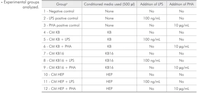
polycarbonate plates and incubated for 6, 24, 48, or 72 h with 50% v/v conditioned media, with or with-out the presence of stimulants. At the timepoints in-dicated, 600 µl of each supernatant was collected, centrifuged, aliquoted, and frozen at −80 °C. The experimental groups generated are listed in Table 1. All experiments were performed in triplicate, with cell viability checked immediately following collec-tion of each supernatant (Trypan Blue exclusion).
Enzyme linked immunosorbent assays (ELISAs)
Levels of IFN-γ, VEGF, TNF-α, and IL-4 were assayed in supernatant samples collected from each experimental group, as well as in each conditioned medium prior to incubation with PBMCs, using ELISAs, according to the manufacturer’s instruc-tions (Invitrogen, Carlsbad, USA). Optical density at 450 nm was measured using a UVM340 spec-trophotometer (Oasys, Asys Hitech, Austria). Blood collection and ELISAs were repeated on three differ-ent occasions.
Statistical analysis
After conirming the normality of data and ho-mogeneity of the variance (SPSS Statistics v. 16.0, SPSS Inc., USA), statistical analyses were performed that included Student’s t-test, ANOVA followed by Tukey’s test, and tests of Pearson’s Correlation. A p-value less than 0.05 was considered statistically signiicant.
Results
Initially, levels of cytokines secreted by the SCC lines used (i.e., KB, KB16, and HEP cells) were de-termined from 1:2 dilutions of culture media col-lected at the 0 h timepoint using ELISAs speciic for detection of IFN-γ, VEGF, TNF-α, and IL-4. Cell
Groupa Conditioned media used (500 µl) Addition of LPS Addition of PHA
1 - Negative control None No No
2 - LPS positive control None 100 ng/mL No
3 - PHA positive control None No 10 µg/mL
4 - CM KB KB No No
5 - CM KB + LPS KB 100 ng/mL No
6 - CM KB + PHA KB No 10 µg/mL
7 - CM KB16 KB16 No No
8 - CM KB16 + LPS KB16 100 ng/mL No
9 - CM KB16 + PHA KB16 No 10 µg/mL
10 - CM HEP HEP No No
11 - CM HEP + LPS HEP 100 ng/mL No
12 - CM HEP + PHA HEP No 10 µg/mL
a: All groups were cultured in 500 µL RPMI 1640 + L-glutamine (2 mM) + 10% FBS.
LPS: lipopolysaccharides; PHA: phytohemagglutinin.
viability for all cell groups was also determined to be 90% at each of the timepoints assayed.
When PBMCs were cultured in the presence of HEP- or KB-conditioned medium, low levels of IFN-γ were detected between 6 and 72 h after the addition of conditioned medium (Figures 1a and 1b). Moreover, these levels were similar to those of the control group in the absence of conditioned me-dium (Figure 1). In contrast, PBMCs cultured with KB16-conditioned medium produced a higher con-centration of IFN-γ than PBMCs cultured in the ab-sence of conditioned medium, or PBMCs cultured in the presence of KB or HEP cells (ANOVA: p < 0.01;
Tukey’s test: KB16 > KB = Hep = control). As a posi-tive control, a parallel culture of each experimental group was stimulated with 10 µg/mL PHA at the 48 and 72 h timepoints. As a result, an increase in IFNγ secretion was detected in all treated cells, and there was no statistically signiicant difference among the groups (ANOVA: p = 0.293).
When VEGF was detected in the conditioned media of all SCC cell lineages (Figures 2a and 2b), a signiicant difference in the levels of VEGF de-tected for control cells versus KB/KB16 conditioned cells was observed (ANOVA, p < 0.01; Tukey’s test, KB > KB16 > Hep = control). Moreover, tests of Pear-Figures 1a and 1b - Production of IFN-γ by PBMCs (n = 6) at various timepoints after PBMCs were cultured in the absence (a) or presence (b) of 10 µg/mL PHA, then incubated with or without (control) conditioned media obtained from KB, KB16, and HEP cell lines. Bars represent mean IFN-γ levels ± standard error of the mean (SEM).
Figures 2a and 2b - Concentrations of VEGF detected at various timepoints in the supernatant of PBMC cultures (n = 6) in the absence (a) or presence (b) of 10 µg/mL PHA, as well as following an incubation with or without (control) conditioned media collected 24 h after culturing KB, KB16, and HEP cells. Bars represent mean VEGF levels ± SEM.
a
a
b
son’s correlation coeficient identiied a positive cor-relation between the levels of VEGF and the time of culture for the KB16 experimental group (r = 0.427, p < 0.05). In contrast, stimulation of PBMCs with PHA decreased the levels of VEGF by the 72 h time-point (Figure 2), particularly in the KB16 experi-mental group (Student’s t-test; p < 0.05).
Secretion of TNF-α was detected in all PBMC experimental groups within 6 h, with levels increas-ing up to 24 h and then stabilizincreas-ing. Furthermore, PBMCs that were cultivated with conditioned me-dia from KB16 and HEP cells produced signiicantly more TNF-α than KB-conditioned PBMCs and con-trol groups (ANOVA: p < 0.01; Tukey’s test: KB16 = Hep > KB = control). Stimulation of PBMCs with PHA and LPS also increased the production of TNF-α similarly in all groups, with no statistically signiicant differences observed between the experi-mental groups (Figures 3a through 3c).
None of the experimental groups produced de-tectable levels of IL-4.
Discussion
In this study, direct contact between fresh hu-man PBMCs and conditioned media collected from tumor cells induced the secretion of TNF-α and VEGF, and this did not contribute to a Th1 or Th2 response.
Previously, most studies of HNSCC immunol-ogy have analyzed patients in the advanced stages of disease in comparison with healthy controls.6,7,8
For example, Lathers et al. reported cytokine levels in HNSCC patients associated with a shift towards
a Th2 bias, with increased levels of Th2 cytokines (i.e., IL-4, IL-6, and IL-10), and decreased levels of the Th1 cytokine, IFN-γ, detected.8 In addition,
Bose et al. compared the immunological status of stage III and IV individuals with HNSCC with those of age-matched controls and found that, in affected patients, the proile of immunocompetent cells and cytokine secretion was dysregulated towards a Th2 response. Moreover, the total leukocyte count was reduced, and the proliferative ability of PBMCs in response to PHA stimulation was signiicantly downregulated.3
The role of Th2 cells in the immune response is gradual, and is related to the tumor stage involved. However, neither the nature of the regulatory fac-tors present, nor the steps involved in the resis-tance of transformed oral keratinocytes, has been fully elucidated.9,10 Correspondingly, the present
study was designed to analyze the early response of healthy donor PBMCs versus PBMCs directly exposed to tumor-derived products. Moreover, re-garding the KB16 cell line that was used and derived from KB cells, it contains the E1A and E1B regions of the Ad2 virus and was chosen for this study as an alternate to analyzing PBMC cytokine secretion in the presence of viral particles.9,10
The results of this study indicate that PBMCs obtained from healthy donors that were incubated with conditioned medium from SCC cell lines did not secrete IFN-γ.In this experimental model, the PBMCs were not activated by dendritic cells, which may decline their function; however the secretion of IFN-γ was expected to some degree since the cells Figures 3a, 3b and 3c - Concentrations of TNF-α detected at various timepoints in the supernatant of PBMC cultures (n = 6) in the absence (a) or presence of 10 µg/mL PHA (b), or 100 ng/mL LPS (c), as well as following an incubation with or without (control) conditioned media collected 24 h after culturing KB, KB16, and HEP cells. Bars represent mean TNF-α levels ± SEM.
from control groups were able to produce this cy-tokine when stimulated by PHA. Thus, it seems that additional factors are needed to induce a Th1 response, with INF-γ production in PBMCs. More-over, conditioned medium did not suppress the se-cretion of IFN-γ by cells stimulated with PHA, in-dicating that this aspect of immunosupression is not deinitive during the initial interactions between PBMCs and tumor cells.
The observation that none of the PBMC experi-mental groups secreted detectable levels of IL-4 may indicate that the contact of PBMCs with SCC prod-ucts does not directly lead to a Th2 response, or that other Th2 cytokine as IL-10 is produced.
It has previously been shown that tumor cells se-crete VEGF,11,12 and after 72 h, PBMCs exposed to
conditioned media from two of the SCC lines (i.e., HEP and KB16) began to secrete VEGF. This ind-ing is of interest, since VEGF production is directly related to metastasis and pro-tumor activities of the immune system.
For all PBMC groups, secretion of TNF-α was detected. However, cells exposed to conditioned me-dia from KB16 and HEP cells produced signiicantly more TNF-α than the other groups. Currently, the role of TNF-α in oral cancers, particularly in spe-ciic immune subsets at particular stages of matu-ration, has not been fully explored.13,14,15 Moreover,
TNF-α is a pleiotropic cytokine that is potently in-duced during the interaction of immune effectors with transformed oral keratinocytes. An interesting characteristic of TNF-α has been its paradoxical role in cancer, where high doses of TNF-α that were locally administered led to the selective destruction of tumor blood vessels and powerful anti-cancer effects.1 However, TNF-α has also been shown to
promote tumor growth by inducing angiogenic tors, including cytokines and ibroblast growth fac-tor. In addition, TNF-α has been shown to be in-volved in stromal development, tissue remodeling, and increased survival, proliferation, and differen-tiation of tumor cells. TNF-α can also induce DNA damage, select resistant tumor clones, and promote metastasis through the conversion and maintenance of the M2 phenotype of monocytes/macrophages, which is favorable to angiogenesis. Furthermore, a
poor prognosis, as well as resistance to therapy, are characteristic of tumors that have been found to re-lease signiicant amounts of TNF-α into the tumor microenvironment.15,16,17,18
An important ethical limitation of the present study was the amount of blood that each donor could donate. Initially, the focus of the study was on macrophage-derived cytokine production, whereby monocytes were separated from whole blood, cul-tured, differentiated into macrophages, and superna-tants were assayed by ELISAs. However, to obtain an adequate number of macrophages suficient to be distributed to all experimental groups, twice the vol-ume of blood collected was needed. Thus, in order to diminish the blood uptake from donors, and not to exclude the possibility of lymphocyte interactions, it was decided that all PBMCs would be analyzed.
The main results of this study indicate that tu-mor cells have the capacity to “crosstalk” with PBMCs and to induce angiogenesis via the secre-tion of VEGF and TNF-α. However, a Th2 pro-ile was not observed, suggesting that development of a Th2 response occurs during a more advanced stage of disease. Alternatively, a patient’s immune system perse may respond to tumor products with a Th2 response, thereby favoring tumoral develop-ment. Overall, the stimulation of PBMCs represents a complex issue, and additional research is needed to fully understand the mechanisms involved, espe-cially regarding the role of TNF-α and VEGF.
Conclusions
The results of this study suggest that direct con-tact of fresh human PBMCs with conditioned media obtained from tumor cells can induce PBMCs to se-crete TNF-α and VEGF. Furthermore, this response represents an angiogenic response, rather than a Th1 or Th2 response.
Acknowledgements
References
1. Balkwill F. Tumour necrosis factor and cancer. Nat Rev Can-cer. 2009 May;9(5):361-71.
2. Abrahao AC, Bonelli BV, Nunes FD, Dias EP, Cabral MG. Immunohistochemical expression of p53, p16 and hTERT in oral squamous cell carcinoma and potentially malignant disorders. Braz Oral Res. 2011 Jan-Feb;25(1):34-41. 3. Bose A, Chakraborty T, Chakraborty K, Pal S, Baral R.
Dys-regulation in immune functions is reflected in tumor cell cy-totoxicity by peripheral blood mononuclear cells from head and neck squamous cell carcinoma patients. Cancer Immun. 2008 Jun;8:10.
4. Chade DC, Borra RC, Nascimento IP, Villanova FE, Leite LC, Andrade E, et al. Immunomodulatory effects of recom-binant BCG expressing pertussis toxin on TNF-alpha and IL-10 in a bladder cancer model. J Exp Clin Cancer Res. 2008 Nov;27:78.
5. Chen Z, Malhotra PS, Thomas GR, Ondrey FG, Duffey DC, Smith CW, et al. Expression of proinflammatory and proan-giogenic cytokines in patients with head and neck cancer. Clin Cancer Res. 1999 Jun;5(6):1369-79.
6. DeNardo DG, Johansson M, Coussens LM. Immune cells as mediators of solid tumor metastasis. Cancer Metastasis Rev. 2008 Mar;27(1):11-8.
7. Jewett A, Head C, Cacalano NA. Emerging mechanisms of immunosuppression in oral cancers. J Dent Res. 2006 Dec;85(12):1061-73.
8. Lathers DM, Achille NJ, Young MR. Incomplete Th2 skew-ing of cytokines in plasma of patients with squamous cell carcinoma of the head and neck. Hum Immunol. 2003 Dec;64(12):1160-6.
9. Kämmerer PW, Toyoshima T, Schöder F, Kämmerer P, Kuhr K, Brieger J, Al-Nawas B. Association of T-cell regulatory gene polymorphisms with oral squamous cell carcinoma. Oral Oncol. 2010 Jul;46(7):543-8.
10. Lee JJ, Lin CL, Chen TH, Kok SH, Chang MC, Jeng JH. Changes in peripheral blood lymphocyte phenotypes distribu-tion in patients with oral cancer/oral leukoplakia in Taiwan. Int J Oral Maxillofac Surg. 2010 Aug;39(8):806-14.
11. Noonan DM, De Lerma Barbaro A, Vannini N, Mortara L, Albini A. Inflammation, inflammatory cells and angiogen-esis: decisions and indecisions. Cancer Metastasis Rev. 2008 Mar;27(1):31-40.
12. Aonuma M, Saeki Y, Akimoto T, Nakayama Y, Hattori C, Yoshitake Y, et al. Vascular endothelial growth factor over-produced by tumour cells acts predominantly as a potent an-giogenic factor contributing to malignant progression. Int J Exp Pathol. 1999 Oct;80(5):271-81.
13. Müller-Hermelink N, Braumüller H, Pichler B, Wieder T, Mailhammer R, Schaak K, et al. TNFR1 signaling and IFN-gamma signaling determine whether T cells induce tumor dormancy or promote multistage carcinogenesis. Cancer Cell. 2008 Jun;13(6):507-18.
14. Romero-Reyes M, Head C, Cacalano NA, Jewett A. Potent in-duction of TNF-alpha during interaction of immune effectors with oral tumors as a potential mechanism for the loss of NK cell viability and function. Apoptosis. 2007 Nov;12(11):2063-75.
15. Saito T, Kuss I, Dworacki G, Gooding W, Johnson JT, White-side TL. Spontaneous ex vivo apoptosis of peripheral blood mononuclear cells in patients with head and neck cancer. Clin Cancer Res. 1999 Jun;5(6):1263-73.
16. Sparano A, Lathers DM, Achille N, Petruzzelli GJ, Young MR. Modulation of Th1 and Th2 cytokine profiles and their association with advanced head and neck squamous cell carci-noma. Otolaryngol Head Neck Surg. 2004 Nov;131(5):573-6. 17. Teruel A, Romero M, Cacalano NA, Head C, Jewett A.
Potential contribution of naïve immune effectors to oral tu-mor resistance: role in synergistic induction of VEGF, IL-6, and IL-8 secretion. Cancer Immunol Immunother. 2008 Mar;57(3):359-66.