Caroline Morini Calil(a) Gisele Mattos Oliveira(b) Karina COGO(c)
antonio Carlos Pereira(d) Fernanda Klein MarCOnDes(b) Francisco Carlos GrOPPO(b)
(a)Halitosis Diagnostic Center - Halicenter, São Paulo, SP, Brazil.
(b)Department of Physiological Sciences, Piracicaba Dental School, University of Campinas - UNICAMP, Piracicaba, SP, Brazil.
(c)Department of Dentistry, Implantology Area, Universidade de Santo Amaro – Unisa, São Paulo, SP, Brazil.
(d)Department of Social and Preventive Dentistry, Piracicaba Dental School, University of Campinas – UNICAMP, Piracicaba, SP, Brazil.
effects of stress hormones on the
production of volatile sulfur compounds
by periodontopathogenic bacteria
abstract: Little is known about the effects of stress hormones on the etiologic agents of halitosis. Thus, the aim of this study was to evaluate
in vitro the effects of adrenaline (ADR), noradrenaline (NA) and corti-sol (CORT) on bacteria that produce volatile sulfur compounds (VSC), the major gases responsible for bad breath. Cultures of Fusobacterium nucleatum (Fn), Porphyromonas endodontalis (Pe), Prevotella intermedia (Pi)
and Porphyromonas gingivalis (Pg) were exposed to 50 µM ADR, NA and CORT or equivalent volumes of sterile water as controls for 12 and 24 h. Growth was evaluated based on absorbance at 660 nm. Portable gas chromatography was used to measure VSC concentrations. Kruskal-Wallis and the Dunn post-hoc test were used to compare the groups. For Fn, ADR, NA and CORT signiicantly reduced bacterial growth af -ter 12 h and 24 h (p < 0.05). All the substances tested increased hydro-gen sulide (H2S) production (p < 0.05). For Pe, all the substances tested
reduced bacterial development after 24 h (p < 0.05), and NA signiicant -ly increased the H2S concentration after 12 h (p < 0.05). In the Pg and
Pi cultures, no effects on bacterial growth were observed (p > 0.05). In the Pi cultures, ADR, NA and CORT increased H2S (p < 0.05).
Catechol-amines and cortisol can interfere with growth and H2S production of
sub-gingival species in vitro. This process appears to be complex and supports the association between stress and the production of VSC.
Keywords: Halitosis; Catecholamines; Hydrocortisone; Gram-Negative
Anaerobic Bacteria.
introduction
Halitosis, also known as bad breath or malodor, is a term used to describe unpleasant, fetid odors present in air exhaled from the mouth.1
Being unpleasant, bad breath is a problem that affects both the patient and the family.1
Studies have indicated that in the majority of cases, malodor is associ-ated with the degradation of sulfur-containing amino acids (methionine, cysteine and cystine) by anaerobic gram-negative bacteria present in the oral cavity, including Porphyromonas gingivalis, Fusobacterium nucleatum,
Prevotella intermedia, Tannerella forsythia and Porphyromonas endodontalis.2,3
Volatile sulfur compounds (VSC), such as hydrogen sulide (H2S), methyl
mercaptan (CH3SH) and dimethyl sulide [(CH3)2S], are generated as a
product of this metabolism.3
Declaration of interests: The authors certify that they have no commercial or associative interest that represents a conflict of interest in connection with the manuscript.
Corresponding author: Karina Cogo
E-mail: [email protected]
DOi: 10.1590/1807-3107BOR-2014.vol28.0008 Epub Jun 02, 2014
Submitted: Aug 20, 2013
Some patients with halitosis do not present clinical evidence of oral pathologies or systemic disorders. Stress, depression and anxiety have been identiied as risk factors for a shift in oral homeostasis.4,5 It has been proposed that emotional
alterations may be a co-factor in halitosis devel-opment. Calil and Marcondes6 reported increased
VSC concentrations in healthy men faced with an anxiety-evoking situation compared with the basal situation, independent of salivary low.
The relationship between stress and the pro-gression of infectious diseases, including periodon-titis, has been the focus of some studies over the last few decades.4,7,8,9,10 However, the mechanisms involved in this process are still not clear. Poten-tially pathogenic microorganisms can recognize stress hormones, a fact that has led to the devel-opment of the concept of “microbial endocrinol-ogy.”11 These microorganisms could use the
hor-mones produced by the host as ‘environmental trails’ to initiate their growth and pathogenic pro-cesses.12,13,14,15,16,17 In one study, the growth rate of
bacteria commonly found in the oral cavity, such as Actinomyces naeslundii, Eikenella corrodens, and
Campylobacter gracilis, was stimulated by exposure to noradrenaline.13 In addition, recent studies show
conlicting results of noradrenaline and adrenaline on the growth of P. gingivalis, with studies showing either no effects of these hormones on growth12,17 or
demonstrating a reduction in viability.13,15 Recently,
it was demonstrated that noradrenaline can stimu-late the production and activity of arg-gingipain B, an important protease of P. gingivalis.15 Thus, the
effects of stress could be mediated by two poten-tially synergistic pathways involving the down-regulation of the host’s defenses and the up-reg-ulation of bacterial pathogenicity.
Because there is clinical evidence that stress is related to halitosis,6 we hypothesized that
periodon-tal bacteria would respond to stress substances by stimulating their growth and their production of VSC. Because there have been no studies on the effects of stress hormones on VSC production, the aim of the present study was to investigate the effects of cat-echolamines and cortisol on the growth and VSC production of periodontopathogens.
Methodology
substances Used
The following substances were tested: adrena -line - ADR (epinephrine bitartrate, Sigma Chemical Co.®, St Louis, USA), noradrenaline – NA (arterenol,
Sigma®) and cortisol - CORT (hydrocortisone, Sigma®).
The 50 µm concentration of ADR, NA13,18 and CORT19 was chosen based on previous studies.The solutions were prepared using distilled water as the solvent and were sterilized using Millex® (Millipore®,
Bill-erica, USA) sterile ilters with a pore size of 0.22 µm.
Bacterial samples and Culture Conditions
The growth patterns and VSC production of four bacterial species (Porphyromonas endodontalis from clinical specimens - Pe, Porphyromonas gingivalis
W83 - Pg, Fusobacterium nucleatum nucleatum ATCC 25586 - Fn and Prevotella intermedia ATCC 25611 -
Pi) were examined. These species were selected based on their ability to produce VSC constitutively. The bacteria were grown statically in brain heart infusion broth (BHI; Difco Co., Detroit, USA). To cultivate Pg and Pe, 5 µg/mL hemin (Sigma®) and
1 µg/mL menadione (Sigma®) were added to the
BHI. Bacterial cultures were grown at 37 °C under anaerobic conditions (10% CO2, 10% H2 and 80% N2;
MiniMacs Anaerobic Workstation, Don Whitley Scientiic®, Shipley, UK).
study design and experimental Groups
Cultures of Pg, Pe, Pi and Fn were exposed to 50 µM ADR, NOR and CORT for 12 and 24 h (early and mid-logarithmic growth phases). Tubes containing bacteria without the tested substances but containing equivalent volumes of distilled water (vehicle) were used as positive control for growth. Negative controls contained only the tested substances and the culture medium. After incubation, bacterial growth and VSC production were assessed as follows. At least two sep-arate experiments were performed for each hormone tested. In each experiment, 8 replicates were tested for each group (ADR, NA, CORT and controls).20
susceptibility Tests (Macrodilution Broth Tests)
5 mL of BHI broth medium (plus 5 µg/mL hemin (Sigma®) and 1 µg/mL menadione (Sigma®) for the
Pg and Pe cultures) were prepared for each experi-mental group. A standardized bacterial suspension was prepared in sterile saline solution (0.9% NaCl) and adjusted with a spectrophotometer (Unico 1100 RS;Unico®, Dayton, USA) to an optical density of 0.5
(8 x 10⁶ CFU/mL). An inoculum of 500 µL was added
to each tube to give a inal concentration of 4 x 10⁶
CFU/mL. The tubes were then incubated at 37°C in anaerobic conditions for 12 or 24 h. After these peri-ods, bacterial growth was assessed spectrophoto-metrically at a wavelength of 660 nm.
Bacterial vsC Production Tests
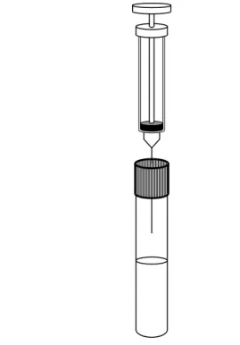
This experiment was conducted as previously described, with some modiications.20 For bacterial
VSC production analyses, the experimental groups and inocula were prepared as described above. After incubation at 37 °C in anaerobic conditions, 0.1 mL of air from the tubes was aspirated using a 1 mL syringe (BD®, Franklin Lakes, USA) coupled
to a needle (BD®) (Figure 1) and injected into Oral
Chroma® (Abimedical, Osaka, Japan). For a period of
8 minutes, the apparatus performed molecule counts in parts per billion (ppb) of hydrogen sulite (H2S)
and methylmercaptan (CH3SH). VSC analyses were
performed at two different time intervals (12 and 24 h). These incubation lengths were chosen because at these periods, the bacteria are growing exponen-tially, and there are more viable cells than dead cells. Gaseous standards of the VSC (H2S and CH3SH)
were prepared as described previously22 by White
Mar-tins (Campinas, Brazil) (RBC-INMETRO n M-32433/10), and the device was calibrated every three months according to Tangerman and Winkel.23
statistical analysis
Levene’s test was used to test the homogeneity of variance, and the normality of distribution was tested by the Shapiro-Wilk’s test. Kruskal-Wallis and the Dunn post-hoc test were used to compare the groups (ADR, NA and CORT compared to control for each bacteria and each time interval separately). Statistical analysis was performed using BioEstat 5.0 (Fundação Mamirauá, Belém, Brazil) at a signiicance level of 5%.
results
effects of adrenaline, noradrenaline and Cortisol on Bacterial Growth
Figure 2 summarizes the effects of these sub-stances on bacterial growth (optical density, 660 nm). For the Fn cultures, ADR, NA and CORT signii -cantly reduced bacterial growth after 12 and 24 h (p < 0.05, Kruskal Wallis test). For Pe, the tested substances reduced bacterial growth, but only after 24 h of exposure (p < 0.05). No statistically signii -cant differences in optical density (p > 0.05) were observed for the Pg and Pi cultures in any of the time periods evaluated.
effects of noradrenaline, adrenaline and Cortisol on Bacterial vsC Production
The median values and interquartile deviations of H2S and CH3SH concentrations (in ppb) for Fn,
Pe, Pg and Pi cultures exposed for 12 and 24 h to ADR, NA and CORT are presented in Figures 3 and 4, respectively.
In Fn, only CORT caused a signiicant increase in
H2S levels at 12 h (Kruskal-Wallis, p < 0.05), while no
changes in H2S concentration were observed at 24 h
(Kruskal-Wallis, p > 0.05). However, ADR and NA did
increase the H2S concentration in Fn cultures after 24
h of exposure (p < 0.05). Only Fn showed a reduction in CH3SH production by CORT after 24 h (p < 0.05).
No other conditions changed the CH3SH levels.
H2S concentrations were signiicantly increased
only in Pe cultures exposed to NA for 12 h (p < 0.05). No difference in H2S production was observed for
the groups exposed to ADR and CORT (p > 0.05). In addition, CH3SH levels were not altered by exposure
of Pe to catecholamines and CORT (p > 0.05). No statistically signiicant differences (p > 0.05) were found in H2S and CH3SH levels among groups and
their respective controls in Pg cultured for 12 or 24 h.
Pi was more sensitive to the effects of catechol-amines and CORT than the other bacterial cultures. In this case, H2S production was up-regulated in the
groups exposed to ADR, CORT (12 and 24 h) and NA
(24 h) (p < 0.05). None of the experimental groups showed differences in CH3SH production compared
to their controls (p > 0.05).
Discussion
Clinical evidence has shown that stress can increase VSC concentrations, thus contributing to halitosis.6
The present study provides additional support for the role of stress substances on halitosis, as it was shown that ADR, NOR and CORT can up-regulate the production of VSC by microorganisms commonly found in sub-gingival bioilms.
This study reveals a variation in the growth rates of four periodontopathogenic microorganisms in response to the addition of stress hormones to their culture media. The growth of Fn and Pe was reduced by stress hormones, primarily after 24 h of expo-sure. In contrast, no signiicant effects on growth Figure 2. Mean and standard deviation of optical density (660 nm) representing the bacterial growth. Statistically significant p
values are shown (*) on the upper side of the columns (p < 0.05, Kruskal Wallis, Dunn).
12 h 24 h
Control
Cortisol 50 µM
Noradrelanine 50 µM
Adrenaline 50 µM
OD 660 nm
1.0
0.5
0.0 1.5 2.0
* *
*
* *
*
* *
*
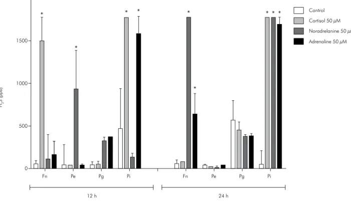
Figure 3. Median and interquartile deviation of H2S concentrations (ppb) representing the VSCs production. Statistically significant
p values are shown (*) on the upper side of the columns (p < 0.05, Kruskal Wallis, Dunn).
Control
Cortisol 50 µM
Noradrelanine 50 µM
Adrenaline 50 µM
Fn Pe Pg Pi Fn Pe Pg Pi
24 h 12 h
2000
1500
1000
500
0
*
H2
S (ppb)
*
*
* *
*
* * *
Figure 4. Median and interquartile deviation of CH3SH concentrations (ppb) representing the VSCs production. Statistically
signi-ficant p values are shown (*) on the upper side of the columns (p < 0.05, Kruskal Wallis, Dunn).
Fn Pe Pg Pi
Fn Pe Pg Pi
24 h 24 h
*
Control
Cortisol 50 µM
Noradrelanine 50 µM
Adrenaline 50 µM
0 200 400 600 800
CH
3
were observed in Pg and Pi for any of the substances tested. Most of these results are in agreement with the literature regarding the effects of catecholamines on bacterial growth. Roberts et al.13 demonstrated an
inhibitory effect of catecholamines on Fn growth and no effect on the growth of Pi cultures, while Belay et al.12 and Jentsch et al.17 showed no change
in Pg growth patterns. However, some of the pres -ent indings are contrary to previous reports. It was reported that the growth rate of Pg was reduced when exposed to catecholamines.13,15 Additionally, cortisol
was demonstrated to stimulate the growth of Pg cul-tures.24 Our results showed no effects of NA, ADR
or CORT on the growth rate of this pathogen. There were several differences in the experimental factors used in these studies, including the period of hor-mone exposure, bacterial strains and culture media used, catecholamine concentration and the method of growth assessment, which may have contributed to the divergent indings.
There are some reports in the literature demon-strating VSC production by periodontopathogenic bacteria. Previous studies revealed that saliva and dental plaque from patients with periodontal disease produces large quantities of VSC, thus contributing to malodor.25,26,27 Our study shows that periodontal
bacteria produce signiicant VSC levels in vitro and that this production is altered by stress hormones. We observed that the major effects of catecholamines and CORT were on H2S levels, mainly in the case of
Fn, Pe and Pi. For these bacteria, exposure to these substances caused an increase in H2S concentration.
Only CORT changed the production of CH3SH, and
this effect was inhibitory in Fn cultures treated with this glucocorticoid. Given these results, it is possi-ble to assume that the main roles of ADR, NA and CORT are in H2S production. To better understand
the effects of stress hormones, it will be necessary to utilize enzymatic, genomic and proteomic approaches.
Interestingly, the growth and VSC production of
Pg was not affected by any of the stress hormones tested. It is possible that Pg is less sensitive to hor-monal influence in the favorable environment of our experiment, which offered a rich-nutrient cul-ture media along with controlled atmosphere and temperature. Previous studies have observed that
adrenaline and noradrenaline have strong effects on bacterial growth mainly when the bacteria are cul-tivated in poor nutrient media.18,28 For this reason, those authors designed a special medium, serum-SAPI minimal medium, to mimic the challenging
in vivo environment. However, this medium was not
used in our study. This could be the reason for our observations of slight effects of hormones on growth and VSC production, especially in the case of Pg. It is possible, for example, that these organisms (and others showing negative growth responses) utilize catecholamines for up-regulation of virulence expres-sion rather than for growth13 and VSC production.
When taking into consideration the times of expo-sure (12 and 24 h) in our study, differing effects were found. In some experiments, signiicant effects of the hormones were noted at both times, while the hor-mone effects were observed at only one time period (12 or 24 h) in other experiments. We expected to observe that a substance that showed an effect at 12 h of exposure would have the same effect at 24 h. As demonstrated, however, this was not always the case. These stress-related substances have short half-lives and could be degraded during the time-period of our experiments. Kennedy et al.29 demonstrated that catecholamines are stable in saliva for up to 2 h at 4 °C. In our study, however, catecholamine con -centrations in the culture medium after 12 and 24 h were not determined, and one can imagine that deg-radation was faster due to the higher temperature. We hypothesized that the bacteria might be able to use this degradation by-product to stimulate VSC production; however, further studies are necessary to elucidate this hypothesis. With regard to the deg-radation of CORT, it is known that this hormone remains stable in saliva at ambient temperature for up to one week or more30 and, therefore, would not
it is possible that these stress-related substances have different cellular mechanisms whereby they affect growth and VSC production. Additionally, consid-ering that fewer bacteria could produce higher lev-els of VSC in the presence of these hormones, it is important to evaluate the clinical importance of this observation. It is possible that these results could be the beginning of an explanation for why patients without advanced periodontal disease or other clini-cal signs of inlammation have increased complaints of halitosis in stressful situations and have higher VSC levels than non-stressed patients.6
Investiga-tions of the exposure of catecholamines and corti-sol in multi-species bioilm models, animal models for halitosis and clinical studies should be carried
out to better understand the relationship of stress and VSC production.
Conclusions
In conclusion, our results indicate that the hormones adrenaline, noradrenaline and cortisol can modulate the growth of periodontopathogenic microorganisms and the production of VSC, thus elucidating another possible mechanism of action for the relationship between stress and the development of halitosis.
acknowledgements
We thank Fundação de Amparo à Pesquisa do Estado de São Paulo (Fapesp) for its support. GMO was the recipient of a Fapesp fellowship (no. 2008/00750-1).
references
1. Aylikci BU, Colak H. Halitosis: From diagnosis to manage
-ment. J Nat Sci Biol Med. 2013 Jan;4(1):14-23.
2. Persson S, Claesson R, Carlsson J. The capacity of subgingival microbiotas to produce volatile sulfur compounds in human
serum. Oral Microbiol Immunol. 1989 Sep;4(3):169-72.
3. Krespi YP, Shrime MG, Kacker A. The relationship between oral malodor and volatile sulfur compound-producing
bac-teria. Otolaryngol Head Neck Surg. 2006 Nov;135(5):671-6.
4. Peruzzo DC, Benatti BB, Ambrosano GM, Nogueira-Filho GR, Sallum EA, Casati MZ, et al. A systematic review of stress and psychological factors as possible risk factors for
periodontal disease. J Periodontol. 2007 Aug;78(8):1491-504.
5. Rosania AE, Low KG, McCormick CM, Rosania DA. Stress, depression, cortisol, and periodontal disease. J Periodontol.
2009 Feb;80(2):260-6.
6. Calil CM, Marcondes FK. Influence of anxiety on the production
of oral volatile sulfur compounds. Life Sci. 2006 Jul 10;79(7):660-4.
7. Ng SK, Keung Leung W. A community study on the relation-ship between stress, coping, affective dispositions and peri-odontal attachment loss. Community Dent Oral Epidemiol.
2006 Aug;34(4):252-66.
8. Ishisaka A, Ansai T, Soh I, Inenaga K, Yoshida A, Shigeyama
C, et al. Association of salivary levels of cortisol and dehydro-epiandrosterone with periodontitis in older Japanese adults.
J Periodontol. 2007 Sep;78(9):1767-73.
9. Huang S, Lu F, Zhang Z, Yang X, Chen Y. The role of psycho -logic stress-induced hypoxia-inducible factor-1alpha in rat
ex-perimental periodontitis. J Periodontol. 2011 Jun;82(6):934-41.
10. Semenoff TA, Rosa Junior A, Borges AH, Porto AN, Caporossi C, Semenoff Segundo A. Effect of chronic stress in newborn rats on the progression of ligature-induced-periodontitis in
adulthood. Acta Cir Bras. 2013 Sep;28(9):652-6.
11. Freestone PP, Sandrini SM, Haigh RD, Lyte M. Microbial
endocrinology: how stress influences susceptibility to infec
-tion. Trends Microbiol. 2008 Feb;16(2):55-64.
12. Belay T, Aviles H, Vance M, Fountain K, Sonnenfeld G. Cat
-echolamines and in vitro growth of pathogenic bacteria:
enhancement of growth varies greatly among bacterial
spe-cies. Life Sci. 2003 Aug 8;73(12):1527-35.
13. Roberts A, Matthews JB, Socransky SS, Freestone PP,
Wil-liams PH, Chapple IL. Stress and the periodontal diseases:
effects of catecholamines on the growth of periodontal
bacte-ria in vitro. Oral Microbiol Immunol. 2002 Oct;17(5):296-303.
14. Roberts A, Matthews JB, Socransky SS, Freestone PP,
Wil-liams PH, Chapple IL. Stress and the periodontal diseases:
growth responses of periodontal bacteria to Escherichia coli stress-associated autoinducer and exogenous Fe. Oral
Micro-biol Immunol. 2005 Jun;20(3):147-53.
15. Saito T, Inagaki S, Sakurai K, Okuda K, Ishihara K. Exposure of P. gingivalis to noradrenaline reduces bacterial growth and elevates
ArgX protease activity. Arch Oral Biol. 2011 Mar;56(3):244-50.
16. Pullinger GD, Carnell SC, Sharaff FF, van Diemen PM, Dziva F, Morgan E, et al. Norepinephrine augments Salmonella enteri-ca-induced enteritis in a manner associated with increased net replication but independent of the putative adrenergic sensor
kinases QseC and QseE. Infect Immun. 2010 Jan;78(1):372-80.
17. Jentsch HF, März D, Krüger M. The effects of stress hormones on growth of selected periodontitis related bacteria.
Anaer-obe. 2013 Dec;24:49-54
18. Lyte M, Ernst S. Catecholamine induced growth of gram
negative bacteria. Life Sci. 1992;50(3):203-12.
19. Plotkin BJ, Roose RJ, Erikson Q, Viselli SM. Effect of
andro-gens and glucocorticoids on microbial growth and
20. Tanabe S, Desjardins J, Bergeron C, Gafner S, Villinski JR, Grenier D. Reduction of bacterial volatile sulfur compound production by licoricidin and licorisoflavan A from licorice.
J Breath Res. 2012 Mar;6(1):016006.
21. Winn Jr WC, Janda WM, Koneman EW, Schreckenberger PC, Procop GW, Woods GL. Koneman’s color atlas and textbook
of diagnostic microbiology. 6th ed. Philadelphia: Lippincott Willians & Wilkins Company; 2006. 1531 p.
22. Winkel EG, Tangerman A. Appropriate sample bags and
sy-ringes for preserving breath samples in breath odor research: a technical note. J Breath Res. 2008 Mar;2(1):017011.
23. Tangerman A, Winkel EG. Extra-oral halitosis: an overview.
J Breath Res. 2010 Mar;4(1):017003.
24. Akcali A, Huck O, Buduneli N, Davideau JL, Kose T, Tenen
-baum H. Exposure of Porphyromonas gingivalis to cortisol increases bacterial growth. Arch Oral Biol. 2014 Jan;59(1):30-4.
25. Coli JM, Tonzetich J. Characterization of volatile sulphur compounds production at individual gingival crevicular
sites in humans. J Clin Dent. 1992;3(4):97-103.
26. Yaegaki K, Sanada K. Biochemical and clinical factors influ-encing oral malodor in periodontal patients. J Periodontol.
1992 Sep;63(9):783-9.
27. Kishi M, Ohara-Nemoto Y, Takahashi M, Kishi K, Kimura S, Aizawa F, et al. Prediction of periodontopathic bacte-ria in dental plaque of periodontal healthy subjects by measurement of volatile sulfur compounds in mouth air.
Arch Oral Biol. 2013 Mar;58(3):324-30.
28. O’Donnell PM, Aviles H, Lyte M, Sonnenfeld G. Enhance -ment of in vitro growth of pathogenic bacteria by
norepi-nephrine: importance of inoculum density and role of trans
-ferrin. Appl Environ Microbiol. 2006 Jul;72(7):5097-9.
29. Kennedy B, Dillon E, Mills PJ, Ziegler MG. Catecholamines
in human saliva. Life Sci. 2001 May 25;69(1):87-99.
30. Liening SH, Stanton SJ, Saini EK, Schultheiss OC. Salivary
testosterone, cortisol, and progesterone: two-week stability,
interhormone correlations, and effects of time of day, men-strual cycle, and oral contraceptive use on steroid hormone