RBCCV 44205-1631 DOI 10.5935/1678-9741.20150006
A propose of pulmonary dysfunction stratiication
after valve surgery by physiotherapeutic
assistance level
Proposta de estratiicação da disfunção pulmonar após cirurgia valvar segundo níveis de assistência
isioterapêutica
Satiko Shimada Franco
1, Ft; Luiz Marcelo Sá Malbouisson
2, PhD; Max Grinberg
1, PhD; Maria
Ignêz Zanetti Feltrim
1, PhD
1Instituto do Coração (InCor) do Hospital das Clínicas da Faculdade de Me-dicina da Universidade de São Paulo (HC-FMUSP), São Paulo, SP, Brazil. 2 Anestesiology Department of Faculdade de Medicina da Universidade de São Paulo (HC-FMUSP), São Paulo, SP, Brazil.
Work carried out at Instituto do Coração (InCor) do Hospital das Clínicas da Faculdade de Medicina da Universidade de São Paulo (HC-FMUSP), São Paulo, SP, Brazil and Anestesiology Department of Faculdade de Medicina da Universidade de São Paulo, São Paulo, SP, Brazil.
No inancial support.
Correspondence address: Satiko Shimada Franco
Instituto do Coração do Hospital das Clínica da Faculdade de Medicina da Universidade de São Paulo InCor-HCFMUSP
Av. Dr. Enéas de Carvalho de Aguiar, 44, 2o andar - Cerqueira Cesar – São Paulo, SP, Brazil
Zip code: 05403-000
E-mail: [email protected]
Article received on September 4th, 2014 Article accepted on January 26th, 2015 Abstract
Objective: a) to propose and implement an evaluation system; b) to classify the pulmonary involvement and determine levels of physical therapy; c) to check the progress postoperatively.
Methods: Patients underwent physiotherapy assessment preoperatively, postoperatively and after 5 days of intervention.
They were classiied into three levels of care: level 1 - low risk of complication; Level 2 - medium risk; Level 3 - high risk. We used analysis of variance and Kruskal-Wallis and analysis of variance for repeated measures or Friedman. Chi-square test or Fisher for proportions. We considered statistical signiicance
level P<0.05.
Results: We studied 199 patients, 156 classiied within level 1, 32 at level 2 and 11 at level 3. Thoracoabdominal motion and auscultation changed signiicantly postoperatively, persisting at levels 2 and 3 (P<0.05). Oxygenation and respiratory rate
changed at levels 2 and 3 postoperatively (P<0.05) with recov
-ery at the end. Signiicant decrease in lung volumes occurred in three levels (P<0.05) with partial recovery at level 1, lung col
-lapse occurred at all levels, with recovery by 56% at level 1, 47% at level 2, 27% at level 3.
Conclusion: The proposed assessment identiied valve sur-gery patients who require differentiated physical therapy. Level 1 patients had rapid recovery, while the level 2 showed
signii-cant changes with functional gains at the end. Level 3 patients,
more committed and prolonged recovery, should receive greater assistance.
Descriptors: Thoracic Surgery. Physical Therapy Modalities. Vital Capacity.
Resumo
Objetivo: a) propor e aplicar um sistema de avaliação; b)
classiicar o comprometimento pulmonar e determinar os níveis de assistência isioterapêutica; c) veriicar a evolução no pós-ope-ratório de cirurgia valvar.
Métodos: Pacientes realizaram avaliação isioterapêutica no pré-operatório, pós-operatório e após 5 dias de intervenção. Foram classiicados em três níveis de atenção: nível 1 - baixo risco de complicação; nível 2 - médio risco; nível 3 - alto ris-co. Utilizou-se Análise de Variância e Kruskal-Wallis e Análise de Variância para medidas repetidas ou Friedmann. Teste
qui-quadradoou Fisher para as proporções. Considerou-se nível de
signiicância estatística P<0,05.
INTRODUCTION
The presence of postoperative respiratory dysfunction in patients after cardiovascular surgery with extracorporeal cir-culation (ECC), under general anesthesia, range from 50% to
100%[1-4]. Alterations in lung mechanics, such as decreased
functional residual capacity, contribute to the occurrence of pulmonary collapse, increased shunt, decreased gas dif-fusion, and consequently, hypoxemia[5,6]. In this context, the
presence of pain and chest tubes are directly implicated in keeping low lung volumes[7,8].
The use of techniques to remove bronchial secretions, as well as respiratory and early mobilization exercises, promote improve-ment of pulmonary function, support the correction of hypox-emia, and stimulate functional independence. However, despite that therapeutic protocols are widely used after cardiac surgery, the beneits of these protocols are not yet well established[9-16].
Evaluation and application of a classiication system based on differentiated levels of physical therapy assistance comprise an alternative strategy for optimizing postoperative patient care[17-19]. The challenge is to differentiate patients
ac-cording to the degree of pulmonary alterations present and to recommend appropriate therapies, with consideration of the available resources and application timing.
The use of physical therapy strategies adjusted to the severity level of respiratory dysfunction in individual patients may be ben-eicial in terms of inhibiting the clinical progression of respiratory dysfunction, and the organization and standardization of physical therapy assistance. Therefore, we designed this study with the fol-lowing objectives: a) to propose and apply a postoperative evalu-ation system for patients undergoing cardiac valve surgery; b) to classify pulmonary impairment and to determine recommendable levels of physical therapy assistance; c) to monitor the postopera-tive clinical progress of patients who have been classiied.
METHODS
Patients and Methods
This study was approved by the Research Ethics
Commit-Abbreviations, acronyms & symbols
ECC Extracorporeal circulation ICU Intensive care unit PA Pulmonary auscultation
PFC Peak low cough
SpO2 Peripheral oxygen saturation TxAbM Thoracoabdominal motion
tee of the Hospital das Clinicas at the Faculty of Medicine, University of São Paulo (approval No. 011/09). The informed
consent was obtained from all of the subjects, who had un-dergone valve surgery.
The inclusion criteria for this study were as follows: pa-tients of both sexes, papa-tients aged 18 to 80 years, and papa-tients hospitalized to undergo elective valve surgery who had no signs or symptoms of respiratory distress. Patients who had dificulty performing the functional tests, who were receiv -ing oxygen therapy, or who required noninvasive ventilation in the preoperative period were excluded, as well as patients whose conditions progressed to cerebrovascular accident, who showed hemodynamic instability and worsening of clinical condition, or who died immediately after operation. Personal, anthropometric, and clinical data were collected from the pa-tients who were hospitalized to undergo elective valve surgery. A physical therapy evaluation that comprised eight pa-rameters was conducted as follows:
1. Thoracoabdominal motion (TxAbM): With the patient placed in the dorsal position, the thoracoabdominal movement was evaluated during 1 minute. The TxbAM was classiied as normal when the abdominal displacement pre -dominated; mixed, when no thoracic or abdominal ment predominated; thoracic, with predominant displace-ment of the rib cage; and paradoxical, when the thoracic or abdominal movements were inverted.
2. Pulmonary auscultation (PA): PA was veriied based on lung sound and presence of adventitious sounds.
3. Mobility: Mobility was classiied according to the degree of independence the patient had while sitting down and moving around.
4. Oxygenation: Peripheral oxygen saturation (SpO2) was measured by using pulse oximetry (Dixtall®), with the
patient breathing environment air, after 5 minutes[15] in the
dorsal position, with the headrest at 45º and the sensor placed in the middle inger of the right hand.
5. Respiratory frequency (f): Respiratory frequency was deined as the number of inspiratory incursions occur -ring in 1 minute, in the dorsal position.
e frequência respiratória se modiicaram nos níveis 2 e 3 no pós-operatório (P<0,05), com recuperação no inal. Diminuição
signiicante dos volumes pulmonares ocorreu nos três níveis
(P<0,05), com recuperação parcial no nível 1. Colapso
pulmo-nar ocorreu em todos os níveis, com recuperação em 56% no nível 1, 47% no nível 2, 27% no nível 3.
Conclusão:A avaliação proposta identiicou pacientes de cirur-gia valvar que necessitam de assistência isioterapêutica diferen-ciada. Pacientes do nível 1 tiveram rápida recuperação, enquanto os do nível 2 mostraram alterações signiicativas, com ganhos fun-cionais no inal. Pacientes do nível 3, mais comprometidos e com
recuperação prolongada, devem receber maior assistência.
Descritores:Cirurgia Torácica. Modalidades de Fisioterapia.
6. Pulmonary function: Pulmonary function was as-sessed by measuring forced vital capacity (FVC) in milli-liters, obtained by using a ventilometer (Wright Mark 8®).
While in the sitting position, the patient was guided to in-hale deeply and, subsequently, to expire as fast as far he/she can through mouth piece, with the nose closed with a clip to prevent air leakage. During the procedure, the patient was encouraged to optimize performance. The procedure was re-peated three times, recording the highest value.
7. Peak low cough (PFC): PFC was measured by using a peak low meter (Assess®), with the patient in the
sitting position and the nose closed with a nose clip. The patient was encouraged to inhale deeply and, subsequently, to cough through the mouth piece. The procedure was re-peated three times, recording the highest value, as long as the difference between the measurements was not greater than 20 L/min.
8. Chest radiography: Chest radiographs were analyzed by a radiologist blinded to the study. Pulmonary collapse was assessed by using the Jenkins scale as follows[11]: 0, without
al-teration; 1, minimum collapse; 2, pronounced collapse or con-solidation at one pulmonary base; and 3, bilateral alteration.
The physical therapy evaluations were performed in the ward unit at the following time points: preoperatively (bas-al), when the patient returned to the unit (postoperatively), and on the ifth day of the study (inal of the protocol).
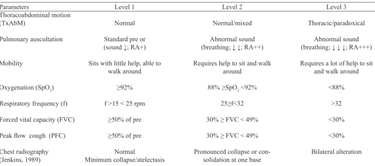
In the postoperative time, the patients who were as-sessed were classiied according to degree of risk of pul -monary impairment by using the criteria shown in Table 1.
Each evaluation parameter corresponded to a point in the column. The preponderance of points in each column deter-mined the type and level of assistance applied to the patient. At level 1, patients with low risk of complications received minimum assistance; at level 2, patients with moderate risk of complications received intermediary assistance; at lev-el 3, patients with high risk of complications received full assistance. In cases when the number of points was equal in 2 columns, the SpO2 criterion was used to differentiate. When paradoxical movement, tachypnea, and hypoxemia were present, level 3 assistance was provided to the patient.
In the postoperative hospitalization period in the inten-sive care unit (ICU), the patient was attended to according to the ICU routine, without inluence of this study. During this period, data on the surgical procedure, times of extracorpo-real circulation (ECC), orotracheal intubation, and length of stay in the ICU were collected.
Level of Physical Therapy Assistance
The patients received differentiated physical therapy as-sistance according to their classiication. Patients with low risk of pulmonary complication (level 1) received physical therapy assistance for 20 minutes, once daily, with direct supervision by the physiotherapist. In this event, 3 series of 10 repetitions of therapeutic breathing exercises were per-formed, followed by coughing. In addition, general mobili-zation and walking exercises were performed. The patient was guided to repeat the breathing exercises every 2 hours, recording the results in a spreadsheet.
Table 1. Parameters for clinical and functional evaluations to deine the degree of pulmonary impairment.
Parameters
Thoracoabdominal motion (TxAbM)
Pulmonary auscultation
Mobility
Oxygenation (SpO2)
Respiratory frequency (f)
Forced vital capacity (FVC)
Peak low cough (PFC)
Chest radiography (Jenkins, 1989)
Level 1
Normal
Standard pre or
(sound ↓; RA+)
Sits with little help, able to walk around
≥92%
f >15 < 25 rpm
≥50% of pre
≥50% of pre
Normal
Minimum collapse/atelectasis
Level 2
Normal/mixed
Abnormal sound
(breathing; ↓ ↓; RA++)
Requires help to sit and walk around
88% ≥SpO2 <92%
25≥f<32
30% ≥ FVC < 49%
30% ≥ FVC < 49%
Pronounced collapse or con-solidation at one base
Level 3
Thoracic/paradoxical
Abnormal sound
(breathing; ↓ ↓ ↓; RA+++)
Requires a lot of help to sit and walk around
<88%
>32
<30%
<30%
The patients classiied at level 2 were treated with con -tinuous positive airway pressure (CPAP) or intermittent positive pressure associated with positive end-expiratory pressure (IPPV + PEEP) for 20 minutes, twice daily. These patients also performed breathing exercises similar to those performed by the patients at level 1, maneuvers for bronchial secretion removal, assisted coughing, and mobility exercises. The duration of the complete therapy was 40 minutes.
The patients at level 3 were treated with positive pressure at two levels of pressure (bilevel) for 60 minutes, 3 times daily. In addition, the physiotherapist applied breathing ex-ercises, maneuvers for bronchial secretion removal, assisted coughing, and mobility exercises twice daily. The time re-quired to assist these patients was approximately 80 minutes per session.
Five days after applying each protocol, the inal evalua -tion was performed. The patients who remained at the same level continued to receive the same therapy until improve-ment or until hospital discharge. Those whose level of as-sistance required changed received the treatment that was proposed for the new level of assistance. The day of hospital discharge was recorded, and that was when the patients re-ceived standardized guidance of respiratory and motor care.
Statistical Analysis
The quantitative data were presented as mean and SD values; and the qualitative data, as absolute and relative fre-quencies. For a comparative analysis between the groups according to age, height, weight, and body mass index, the single-factor analysis of variance and Kruskal-Wallis were used to analyze the length of hospital stay. Homogeneity among the proportions was tested by using the chi-square or Fisher test. The comparison of mean values between the groups over time was performed by using the repeated-mea-sures analysis of variance. For the analysis of the radiograph-ic data, the Friedman nonparametrradiograph-ic test was used. The level of statistical signiicance was considered as P<0.05.
RESULTS
Between June 2009 and October 2013, 288 patients hos-pitalized in the General Valve Diseases Patient Care Unit were evaluated. Among these patients, 89 were excluded and 199 were included and completed the study, of whom 156 were allocated at level 1,32 at level 2, and 11 at level 3, as shown in the lowchart in Figure 1.
The anthropometric characteristics and length of hospital stay of the patients as described above are shown in Table 2. A predominance of female patients and level 1 classiication (78%) was observed, including the younger group of patients in the study.
Most of the patients did not smoke (63%) or consume alcohol (87%), and 64% of the patients did not have previous
cardiac surgery. More than 90% of the patients were in the functional classes II or III.
In our study sample, mitral valve lesions (79%) were the most common cases, with valve replacement being the most frequent surgical procedure (47%), followed by mitral com-missurotomy (18.5%).
The mean durations of mechanical ventilation, and ICU and hospital stay were longer for the patients at levels 2 and 3. However, no statistically signiicant difference was ob -served between the groups.
Classiication of physiotherapeutic assistance level Data regarding the TxAbM evaluation and pulmonary auscultation are shown in Table 3. The number of cas-es with the TxAbM altered increased signiicantly in the postoperative period at level 2, decreasing at the end of the study. Meanwhile, at level 3, the number of patients with this alteration increased from 64% to 82%. Pulmonary aus-cultation was altered in more than 85% of the cases, in all of the groups in the postoperative period. At the end of the study, a high percentage of patients at levels 2 and 3 still had signiicant alterations when compared with the patients at level 1.
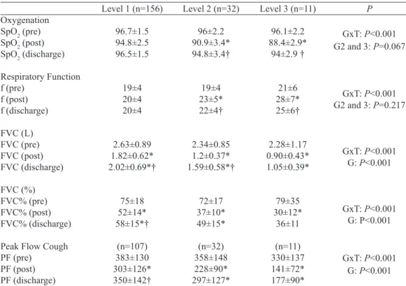
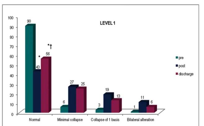
The quantitative parameters of the physical therapy evaluation are shown in Table 4. In the analysis over time, the patients at level 1 did not show signiicant alterations in SpO2 and f. The pulmonary function data revealed sta-tistically signiicant reductions in the postoperative period, with strong improvement at the end of the study, though not returning to the original values. The radiological data shown in Figures 2–4 demonstrate that in this group, the patients had minimum collapse, with collapse in one of the lung bases predominating, which was signiicantly reduced on the ifth day of the study. The behaviour of the patients at levels 2 and 3 were similar. All of the parameters showed signiicant reductions during the postoperative period, with the pulmonary function value not returning to its original value. The relevance of lung collapse was higher in both groups. At level 2, collapse occurred in 41% of the patients, and bilateral alterations occurred in 9% of the patients, with strong reductions in these alterations by the end of the study. In level 3, collapse occurred in 64% of the patients.
In the comparison between the groups, we observed that patients at level 1 showed a significant improvement in pulmonary function by the end of the study, whereas the patients at levels 2 and 3 had the most severe respira-tory impairment. In these groups, only oxygenation and respiratory frequency data showed significant improve-ments by the end of the study when compared with the postoperative period.
Fig. 1 - The lowchart of patients in the study protocol.
Table 2. Anthropometric characteristics and length of hospital stay of the patients classiied according
to level of physical assistance required.
Characteristics N
Sex (M/F) Age (years) Height (m) Body weight (kg)
BMI (kg/m2)
ECC (min) MV (min)
Discharged from ICU (day) Hospital discharge (day)
Level 1 156 (78%)
52/104 46±14 1.61±0.09
63±14 24.1±4.6 78.3±27 495.3±231
4.7±3 14.2±10
Level 2 32 (16%)
10/22 53±15 1.59±0.09
66±13 25.8±4.6 87.5±25 620.4±583
5.1±3 16±10
Level 3 11 (6%) 2/9 58±18 1.55±0.07
60±11 24.9±5.6 78.8±25 1131±1636
6.8±4 18.1±9
P
<0.05(1)
0.073(1)
0.396(1)
0.165(1)
0.074(2)
0.141(2)
0.328(2)
0.099(2)
M=male; F=female; BMI=body mass index; ECC=extracorporeal circulation; MV=mechanical ventilation; ICU=intensive care unit.
(1)Probability descriptive levels of single-factor analysis of variance
Table 3. Qualitative variables of the classiication system in the 3 levels of assistance. TxAbM (pre) Altered TxAbM (post) Altered TxAbM (discharge) Altered PA (pre) Altered PA (post) Altered PA (discharge) Altered Level 1 156 (%) 26 (17) 45 (29) 30 (19) 35 (23) 35(87) 86(55) Level 2 32 (%) 7 (22)
22 (69)#
14 (44)#
8 (25)
30 (94)
24 (75)#
Level 3 11 (%)
3 (27)
7 (64)#
9 (82)#
5 (45)
11 (100)
11 (100)#
P 0.5631 <0.0001 <0.0001 0.2301 0.3748 0.0029
#P< 0.05 versus level 1
TxAbM=thoracoabdominal motion; PA=pulmonary auscultation.
Table 4. Quantitative variables of the classiication system of level 3 assistance.
Oxygenation
SpO2 (pre)
SpO2 (post)
SpO2 (discharge)
Respiratory Function f (pre) f (post) f (discharge) FVC (L) FVC (pre) FVC (post) FVC (discharge) FVC (%) FVC% (pre) FVC% (post) FVC% (discharge)
Peak Flow Cough PF (pre)
PF (post) PF (discharge)
Level 1 (n=156)
96.7±1.5 94.8±2.5 96.5±1.5 19±4 20±4 20±4 2.63±0.89 1.82±0.62* 2.02±0.69*† 75±18 52±14* 58±15*† (n=107) 383±130 303±126* 350±142†
Level 2 (n=32)
96±2.2 90.9±3.4* 94.8±3.4† 19±4 23±5* 22±4† 2.34±0.85 1.2±0.37* 1.59±0.58*† 72±17 37±10* 49±15* (n=32) 358±148 228±90* 297±127*
Level 3 (n=11)
96.1±2.2 88.4±2.9* 94±2.9 † 21±6 28±7* 25±6† 2.28±1.17 0.90±0.43* 1.05±0.39* 79±35 30±12* 36±11 (n=11) 330±137 141±72* 177±90* P
GxT: P<0.001
G2 and 3: P=0.067
GxT: P<0.001
G2 and 3: P=0.217
GxT: P<0.001
G: P<0.001
GxT: P<0.001
G: P<0.001
GxT: P<0.001
G: P<0.001
* P<0.05 versus preoperative period; † P<0.05 versus postoperative period.
Fig. 2 - Percentage of radiological changes of level 1 in the pre, post and discharge of study.
Fig. 4 - Percentage of radiological changes of level 3 in the pre, post and discharge of study.
DISCUSSION
Our results showed that the surgical event altered the pulmonary conditions in the patients who underwent valve surgery. The pulmonary volumes decreased, with smaller diaphragmatic mobility, which increased auscultatory alter-ations and reduced oxygenation. Moreover, the radiological images showed pulmonary collapse. After 5 days of study, pulmonary function improved; however, the preoperative values were not reached.
Our data showed that most of the patients were allocated into level 1 (78%), with a younger mean age. This may be justiied by the fact that the patients with valve disease of rheumatic etiology and a irst surgical intervention was most prevalent among the younger individuals[20]. Advanced age
has been pointed out as a factor associated to a higher in-cidence of postoperative pulmonary complications (CPPO), which has observed in our study, as patients older than 50 years were included at levels 2 and 3.
In this structured evaluation, the respiratory mechanics was conirmed by the TxAbM analysis, palpation of the di -aphragmatic movement, and generation of pulmonary vol-umes, which help the physiotherapist in detecting alterations in the muscle mobility. Our group previously observed that patients with stenosis and mitral regurgitation in the preoper-ative period showed normal TxAbM and breathing patterns,
regardless of the type of valve lesion[21]. This was observed
in our present study again. However, in the postoperative pe-riod, the TxAbM was altered, particularly in the patients at levels 2 and 3. At the end of the study, the patients at level 2 showed a normal TxAbM, and this was partially attribut-ed to the resolution of the pulmonary collapse observattribut-ed on chest radiograph. In the patients at level 3, alterations in the TxAbM (63%) were mainly associated to the diaphragmat-ic dysfunction, whdiaphragmat-ich is a compldiaphragmat-ication of cardiac surgery, occurring at an incidence of 2% to 54%[22,23], depending on
the research method. The lower diaphragmatic mobility in-creases the area of pulmonary collapse and can be triggered through rapid supericial breathing. These patients receive intensive physical therapy support and require more time for recovery, which justiies the small improvements observed at the end of the protocol for level 3.
Pulmonary function was reduced until approximately 25% at level 1, 35% at level 2, and more than 50% at lev-el 3. At the end of the study, the patients in all the groups showed recovery but did not achieve the preoperative val-ues. The lower decline observed at level 1 allowed a fast-er recovfast-ery. In previous studies[10,14,24], FVC and/or FEV
1
slight or very slight and thus did not affect the patients’ clin-ical progress.
Hypoxemia was present in the postoperative period, prob-ably due to the surgical stress and reduction in the pulmonary volumes, with decreased area of gas exchange. The patients at level 1 were those with lower deoxygenation and those who recovered the original values in 5 days. The patients at levels 2 and 3, who showed higher degrees of hypoxemia, had a partial recovery but had persistent gas exchange alter-ations at the end of this study.
The factors that contributed to pulmonary impairment are reportedly multivariate. The presence of median sternotomy, drains, inhibition of deep breaths, hypervolemia, signs of congestive heart failure, lower complacency of the rib cage through manipulation, and diaphragmatic dysfunction may justify these pulmonary alterations[1-8]. All of these factors
were present in our patients. Nevertheless, the presence of pleural changes, with consequent collapse and diaphragmatic dysfunction, was an important element in the reduction of pulmonary function.
For evaluation of pulmonary collapse, we adopted the same classiication system used by Jenkins et al. [11] in patients
with cardiac surgery. The authors observed the presence of collapse in 50% of the patients on the ifth day after coronary artery bypass graft surgery (CABG). In valve surgery, this incidence was 35% [14]. In our country, Vargas et al.[25] found
a collapse incidence of 76% among patients on the seventh day after CABG. Our indings are not different from those
reported in the literature, and we observed pulmonary col-lapse in the postoperative period in all of the patient groups. Among the patients who were discharged from the hospital, 56% of the patients at level 1 achieved normal radiographic data compared with 47% and 27% of the patients at levels 2 and 3, respectively. Thus, the chest radiographic parameter was useful in differentiating patients with a higher degree of impairment, as evident in the lower functional recovery.
The application of physical therapy assessment to clas-sify patients according to lung impairment, patients requir-ing smaller alterations are expected to be allocated into level 1. In fact, in our study, such patients had lower pulmonary function impairment, oxygenation, and pulmonary collapse incidence. In level 2, patients who showed a greater extent of pulmonary changes were included. Meanwhile, in level 3, only 11 patients who presented with lower variation in func-tional gains and had longer hospital stay were included. With the classiication system used in this study, it was possible to characterize the severity of pulmonary alterations and differ-entiate the clinical progress of the patients.
In conclusion, the proposed evaluation method was useful in identifying from among patients who underwent valve sur-gery, those who developed pulmonary impairment and require different levels of physical therapy assistance. The patients at level 1 showed lower decrease in pulmonary function and
had rapid recovery. The patients at level 2 showed signiicant changes in their evolution but had functional improvement due to the treatment applied. The patients at level 3 showed higher levels of impairment, recovered slowly, and required a higher level of physical therapy assistance.
Limitations of the study
Our study has some limitations. The main limitation was the different number of patients in each group, which was due to the random distribution of the clinical cases at the valve disease group Another limitation was that the study was performed in 5 days; thus, improvements achieved by the patients until hospital discharge were not registered.
Our study sample was a convenience sample and included patients indicated for surgery at the valve disease group and those who underwent postoperative follow-up. Most of the patients showed lesions in the mitral valve, with a small num-ber of patients with aortic lesion, which did not allow us to perform a statistical analysis among them. This fact did not allow to evaluate the impact of valve disease on the patients’ progress.
Potential Conlict of Interest
The authors declare no conlict of interest.
Sources of Funding
No external funding was received for the completion of this study.
Academic Level
This study is linked to the postgraduate program of anes-thesiology of the Faculty of Medicine, University of São Paulo.
REFERENCES
1. Sia S, D’Andrea V, Mamone D, Pagnotta L, Verre M. Early postoperative hypoxemia: incidence and effectiveness of oxygen administration. Minerva Anestesiol. 1994;60(11):657-62.
2. Szeles TF, Yoshinaga EM, Alenca W, Brudniewski M, Ferreira FS, Auler JO, et al. Hypoxemia after myocardial revascularization: analysis of risk factors. Rev Bras Anestesiol. 2008;58(2):124-36.
Authors’ roles & responsibilities
SSF Analysis and/or interpretation of data; operations and/or experiments conduct; writing of the manuscript or critical review of its content
LMSM Analysis and/or interpretation of the data MG Conception and design
3. Taggart DP, el-Fiky M, Carter R, Bowman A, Wheatley DJ. Respiratory dysfunction after uncomplicated cardiopulmonary bypass. Ann Thorac Surg. 1993;56(5):1123-8.
4. Matthay MA, Wiener Kronish JP. Respiratory management after cardiac surgery. Chest. 1989;95(2):424-34.
5. Singh NP, Vargas FS, Cukier A, Terra-Filho M, Teixeira LR, Light RW. Arterial blood gases after coronary artery bypass surgery. Chest. 1992;102(5):1337-41.
6. Tenling A, Hachenberg T, Tydén H, Wegenius G, Hedenstierna G. Atelectasis and gas exchange after cardiac surgery. Anesthesiology. 1998;89(2):371-8.
7. Mueller XM, Tinguely F, Tevaearai HT, Revelly JP, Chioléro R, von Segesser LK. Pain location, distribution, and intensity after cardiac surgery. Chest.2000;118(2):391-6.
8. Mueller XM, Tinguely F, Tevaearai HT, Ravussin P, Stumpe F, von Segesser LK. Impact of duration of chest tube drainage on pain after cardiac surgery. Eur J Cardiothorac Surg.2000;18(5):570-4.
9. Pasquina P, Tramèr MR, Walder B. Prophylactic respiratory physiotherapy after cardiac surgery: systematic review. BMJ. 2003;327(7428):1379.
10. Dull JL, Dull WL. Are maximal inspiratory breathing exercises or incentive spirometry better than early mobilization after cardiopulmonary bypass. Phys Ther. 1983;63(5):655-9.
11. Jenkins SC, Soutar SA, Loukota JM, Johnson LC, Moxham J. Physiotherapy after coronary-artery surgery: are breathing exercises necessary? Thorax. 1989;44(8):634-9.
12. Stiller K, Montarello J, Wallace M, Daff M, Grant R, Jenkins S, et al. Eficacy of breathing and coughing exercises in the prevention of pulmonary complications after coronary artery surgery. Chest. 1994;105(3):741-7.
13. Crowe JM, Bradley CA. The effectiveness of incentive spirometry with physical therapy for high-risk patients after coronary artery bypass surgery. Physical Therapy. 1997;77(3):260-8.
14. Johnson D, Kelm C, Thomson D, Burbridge B, Mayers I. The effect of physical therapy on respiratory complications following cardiac valve surgery. Chest. 1996;109(3):638-44.
15. Brasher PA, McClelland KH, Denehy L, Story I. Does removal
of deep breathing exercises from a physiotherapy program including pre-operative education and early mobilisation after cardiac surgery alter patient outcomes? Aust J Physiother. 2003;49(3):165-73.
16. Renault JA , Costa-Val R, Rossetti MB. Respiratory physiotherapy in the pulmonary dysfunction after cardiac surgery. Rev Bras Cir Cardiovasc. 2008;23(4):562-9.
17. Brooks D, Parsons J, Newton J, Dear C, Silaj E, Sinclair L, Quirt J. Discharge criteria from perioperative physical therapy. Chest. 2002;121(2):488-94.
18. Weindler J, Kiefer RT. The eficacy of postoperative incentive spirometry is inluenced by the device-speciic imposed work of breathing. Chest. 2001;119(6):1858-64.
19. Agostini P, Naidu B, Cieslik H, Steyn R, Rajesh PB, Bishay E, et al. Effectiveness of incentive spirometry in patients following thoracotomy and lung resection including those at high risk for developing pulmonary complications. Thorax. 2013;68(6):580-5.
20. Tarasoutchi F, Montera MW, Grinberg M, Barbosa MR, Piñeiro DJ, Sánchez CRM, et al. Diretriz Brasileira de Valvopatias - SBC 2011/I Diretriz Interamericana de Valvopatias - SIAC 2011. Arq Bras Cardiol. 2011;97(5 supl. 3):1-67.
21. Franco SS, Bardi PN, Grinberg M, Feltrim MIZ. Estudo do padrão respiratório e movimento toracoabdominal em valvopatia mitral. Arq Bras Cardiol. 2012;99(5):1049-55.
22. Diehl JL, Lofaso F, Deleuze P, Similowski T, Lemaire F, Brochard L. Clinically relevant diaphragmatic dysfunction after cardiac operations. J Thorac Cardiovasc Surg.1994;107(2):487-98.
23. DeVita MA, Robinson LR, Rehder J, Hattler B, Cohen C. Incidence and natural history of phrenic neuropathy occurring during open heart surgery. Chest.1993; 103:850-956.
24. Dias CM, Vieira R O, Oliveira JF, Lopes AJ, Menezes SL, Guimarães FS. Three physiotherapy protocols: effects on pulmonary volumes after cardiac surgery. J Bras Pneumol. 2011;37(1):54-60