BrazJOtorhinolaryngol.2014;80(6):544---545
Brazilian
Journal
of
OTORHINOLARYNGOLOGY
www.bjorl.org
CASE
REPORT
Mature
teratoma
of
the
nasopharynx
夽
Teratoma
maduro
de
rinofaringe
Claudiney
Candido
Costa
∗,
Valeriana
de
Castro
Guimarães,
Fabiano
Santana
Moura,
Maryana
do
Nascimento
Chediack,
Edson
Júnior
de
Melo
Fernandes
DepartmentofOtorhynolaryngology,HospitaldasClínicas,UniversidadeFederaldeGoiás(UFG),Goiás,GO,Brazil
Received22January2013;accepted8April2013 Availableonline3July2014
Introduction
Teratomas are neoplasms derived from germ cells with componentsofthethreeembryoniclayers(ectoderm, meso-derm, and endoderm), that occur in any age group but are more prevalent in childhood, and have no gender preference.1,2Thelesionscanbebenign(mature,dermoid,
andcysticteratomas)ormalignant(immatureandsolid
ter-atomas),andcanaffectanystructureinthemidline).1---3The
clinicalpresentationvariesaccordingtothelesionsizeand
location.1
Imaging studies are useful to show the location and
extent of the lesion and to aid in clinical management.
Early diagnosis and treatment withexcision of the lesion
arenecessaryforafavorableoutcome.1,4,5
In the present report, the authors describe a case
of nasopharyngeal teratoma, emphasizing diagnosis and
treatment-relatedaspects.
夽 Pleasecitethisarticleas:CostaCC,GuimarãesVD,MouraFS, ChediackMN,FernandesEJ.Matureteratomaofthenasopharynx. BrazJOtorhinolaryngol.2014;80:544---5.
∗Correspondingauthor.
E-mails:orlccp@uol.com.br,claudineyccosta@gmail.com (C.C.Costa).
Case
report
S.A.R.B.,a1year8montholdfemaleinfant,wasseenwith
aclinicalpresentationofnasalobstruction,bilateral
puru-lentnasaldischarge,andsnoring,allpresentsinceshewas
born. At initialexamination, the patienthadnormal vital
signsandexhibitednoabnormalities onoropharyngoscopy
andotoscopy.
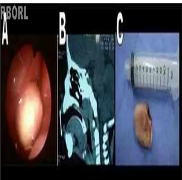
Flexiblenasofibroscopy revealed a whitish
nasopharyn-geal mass (Fig. 1A),completely obstructingthe leftnasal
cavityandpartiallyobstructingtherightnasalcavity.
Computed tomography and magnetic resonance
imag-ingrevealedapoorlyvascularizedobstructivelesioninthe
nasopharynx, with no signs of infiltration or intracranial
extension(Fig.1B).
Utilizing an endoscopic surgical approach, an electric
scalpelwasusedtouneventfullyresectthelesionthatarose
from the left torus tubarius. The lesion macroscopically
appearedsimilartoatongue(Fig.1C).Thepatient’sclinical
courseimprovedintheimmediatepostoperativeperiod.
Thehistopathologicalexaminationidentifiedadipose
tis-sue, mature cartilage tissue, and fibroconnective stroma
consistingofskinandskinappendages,forminganepithelial
inclusioncystconsistentwithamatureteratoma.
Discussion
Thiscaseisrelevantbothbecauseofitsrarityandthe
impor-tance of the differentialdiagnosis of nasalobstruction in
http://dx.doi.org/10.1016/j.bjorl.2014.05.026
Matureteratomaofthenasopharynx 545
Fig. 1 (A)Tumor obstructing therhinopharynx onthe left. (B) Computed tomography showing a lesion limited to the rhinopharynx.(C)Surgicalspecimen.
infants, which should include choanal atresia, intranasal
glioma,encephalocele, rhabdomyosarcoma,dermoidcyst,
lymphangioma,hemangioma,andneurofibromatosis.
Imagingstudiesarehelpfultodeterminethedifferences
between solid and cystic tumors, in addition to showing
thelocationandextentof lesions,thus aidingin the
clin-icalmanagementandthesurgicalapproach.However,they
do not differentiate benign from malignant lesions.2,3 In
thepresentcase,computedtomography(CT)andmagnetic
resonanceimaging (MRI)identifieda massobstructing the
nasopharynx,withnosignsofinfiltrationorcontinuitywith
intracranialstructures.
Knowledgeofthelimitsandsizeofthetumorare
impor-tant aspects to be considered in surgical planning.1,2 In
thereportedcase,thesurgicalexcisionofthetumormass
wasperformed endoscopically,withcompleteresectionof
thelesionwithoutdamagetoadjacentstructures.The
his-tological examination confirmed the diagnosis of mature
teratoma;although the incidence of mature teratomas is
1:4000 livebirths, it is exceedingly rare in the head and
neckandcomprisesonly2%to5%ofcases.2---5
Final
comments
Teratomamustbe consideredin the differentialdiagnosis
oflesionsfoundinthenasopharynxandnasalcavity,mainly
inneonates. Endoscopicandimagingstudies (CTandMRI)
promoteearlydiagnosisandimprovetheoutcome.
Conflicts
of
interest
Theauthorsdeclarenoconflictsofinterest.
References
1.BarksdaleEM,ObokhareI.Teratomasininfantsandchildren.Curr OpinPediatr.2009;21:344---9.
2.IbekweTS, Kokong DD,Ngwu BA,Akinyemi OA, Nwaorgu OG, AkangEE.Nasalseptalteratomainachild.WorldJSurgOncol. 2007;31:58.
3.CukurovaI,GumussoyM,YazA,BayolU,YigitbasiOG.Abenign teratomapresentingasanobstructionofthenasalcavity:acase report.JMedCaseRep.2012;12:147.
4.HuthME,HeimgartnerS,SchnyderI,CaversaccioMD.Teratoma ofthenasal septum inaneonate: anendoscopicapproach.J PediatrSurg.2008;43:2102---5.