Arq Neuropsiquiatr 2004;62(3-B):889-891
Serviço de Neurologia, Departamento de Clínica Médica, Hospital de Clínicas, Universidade Federal do Paraná, Curitiba PR, Brasil: 1Médico Residente, 2Médica Neurologista, 3Professor Assistente, 4Professor Adjunto, 5Professor Titular.
Received 26 December 2003, received in final form 19 April 2004. Accepted 1 June 2004.
Dra. Viviane H.F. Zetola - Rua General Carneiro 181/1103 - 80060-150 Curitiba PR - Brasil. E-mail: viviane@flumignano.com
HEMORRHAGIC STROKE AFTER
NAPHAZOLINE EXPOSITION
Case report
Jorge A.A. Zavala
1, Eduardo R. Pereira
1, Viviane H.F. Zétola
2,
Hélio A.G. Teive
3, Édison M. Nóvak
4, Lineu C. Werneck
5ABSTRACT - Ten percent of all strokes are due to spontaneous cerebral hemorrhages. They are associated to drugs (licit and illicit) in 9.5% of all cases in young adults. This is a case report of a 44-year-old man, without previous morbidities, who presented a sudden onset headache and arterial hypertension 24 hours after use of naphazoline as nasal decongestant. Cranial tomography showed right thalamus hemorrhage. Cerebral angiography showed no aneurisms, vascular malformations or vasculitis. No other risk factors were found during investigation in this patient and the stroke was attributed to naphazoline exposition. KEY WORDS: hemorrhagic stroke, naphazoline, sympathomimetic drug.
Acidente vascular encefálico hemorrágico após exposição à nafazolina: relato de caso
RESUMO - Dez por cento de todos os eventos vasculares encefálicos são devido às hemorragias intracere-brais espontâneas, associados a drogas (lícitas e ilícitas) em 9,5% de todos os casos em adultos jovens. Relatamos o caso de um homem de 44 anos de idade, sem doenças prévias, que apresentou cefaléia súbita e hiperten-são arterial 24 horas após o uso de congestionante nasal contendo nafazolina. A tomografia de crânio eviden-ciou hemorragia talâmica. Durante a investigação não foram encontrados outros fatores de risco e a he-morragia foi atribuída à exposição à nafazolina.
PALAVRAS-CHAVE: doença vascular encefálica hemorrágica, nafazolina, droga simpaticomimética.
Cerebrovascular diseases occur more frequent-ly in elderfrequent-ly people. Peak incidence is between 7th
and 8thdecades1,2. Before age 55, incidence is 10%3,4
and before 45 it falls to 3.9%5.
Ten percent of all cerebrovascular events are due to hemorrhage6. Its estimated incidence in United
States of America is 0.3/100000 in younger than 35 years old7. Main etiologies are vascular
malforma-tions, arterial hypertension and exposition to dru-gs (amphetamines, sympathomimetics and illicit drugs)8,9. In young adults, drugs are associated to
9.5% of ischemic and hemorrhagic stroke10.
We report a case of exposition to sympathomi-metic drug naphazoline followed by thalamic he-morrhage.
CASE
A 44 year-old previously healthy man, with no his-tory of arterial hypertension, was admitted to the emer-gency department of Hospital de Clinicas, Federal
Uni-versity of Parana, complaining of headache and left arm weakness. Five days before he had had some flu-like symptoms and naphazoline nasal decongestant was prescribed. In the following day he had a sudden onset headache associated to nausea and vomiting. He then looked for medical attention. Blood pressure was 190/120 mmHg and captopril 50 mg as a single dose was pre-scribed. A couple of days latter he developed left hemi-paresia that made him seek for our emergency depart-ment. There was no previous history of thrombotic disor-ders and there were no familiar stroke cases.
On physical examination vital signs were normal in-cluding blood pressure (120/70 mmHg), with no positive signs on cardiopulmonary and abdominal examination. Neurological examination revealed an oriented patient, with intact memory and cognition. Cranial nerve exam-ination showed no positive signs. There was left arm and leg hypotonia and slight weakness (4-/5). Babinski sign was not present. He could stand up by himself and there was no gate disorders or cerebellar signs.
x 1.6 cm (Fig 1). The patient was admitted to the Neuro-logic Unit. Chemical constituents of blood and hematolo-gical exams as total cholesterol and fractions, fast gluco-se, triglycerides, complete blood count, coagulation tests (hypercoagulable states from protein C, free protein S, antitrombin III, lupus anticoagulant and anticardiolipin antibody abnormalities) and inflammatory marks were negative (C-reactive protein, ESR). Electrocardiogram and echocardiogram were normal. Cerebral
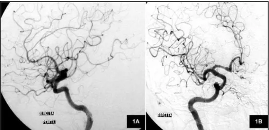
angiogra-890 Arq Neuropsiquiatr 2004;62(3-B)
phy revealed no aneurisms, vascular malformations or vasculitis (Fig 2). No drugs were needed to control blood pressure and the patient was discharged in the 8thday after admission with total recovery of the motor deficit.
DISCUSSION
Cerebrovascular diseases are an example of me-dical conditions with multiple risk factors. Complete investigation of predisposing factors and exhausti-ve laboratory data performance are needed to find their cause. Even these efforts are not always suffi-cient and the diagnostic conclusion is made by ex-clusion. In younger patients it becomes more evi-dent once vascular risk factors can be absent most of the time.
Most common causes of hemorrhagic stroke in young adults are vascular malformations, including cavernous angiomas, and arterial hypertension11.
Risk factors are smoking and hypocholesterolemia. In 1984, Pentel described sympathomimetic drugs exposition as a risk factor for cerebrovascular dise-ase12. Most common drugs associated to stroke
were phenylpropanolamine, ephedrine, pseudo-ephedrine and caffeine leading to hypertension, hypertensive encephalopathy and hemorrhagic stroke. Sloan et al. found a positive previous exposi-tion to drugs in 11% of 116 cases of stroke between 1988 and 198910. There was no difference for
he-morrhagic or ischemic stroke. Related substances were cocaine, heroine, sympathomimetic drugs and phencyclidine. Most recently, Morgenstern et al. showed association between high doses of Ephedra in over-the-counter products for weight loss and energy enhancement and occurrence of hemorrha-gic stroke13.
Fig 1. Cranial tomography showing right thalamus hematoma.
Until 1992, 142 cases of hemorrhagic stroke as-sociated to exposition to phenylpropanolamine as nasal decongestants have been reported14.
Bet-ween 1969 and 1991, there were 22 spontaneous reports of such relation to the FDA.
In order to verify if association of phenylpropa-nolamine to stroke was true, Kernan et al conduc-ted a prospective study between 1994 and 199915.
Association was positive for patients who used it as nasal decongestant for the first time and for wo-man who used it as appetite moderators. A possi-ble mechanism for cerebral infarction is focal arte-rial vasoconstriction and occasionally cerebral vas-culitis. A likely mechanism for intracranial hemor-rhage is acute arterial hypertension. With the ex-ception of endocarditis, management of stroke re-lated to drug abuse is largely supportive, with em-phasis on supportive care to prevent stroke compli-cations, physical and occupational therapy, and ag-gressive addiction rehabilitation.
There are no other risk factors than exposition to naphazoline in the reported case above. No other similar cases were found in medical literature. Our conclusion is that as it is a sympathomimetic drug as phenylpropanolamine, it can lead to the same
Arq Neuropsiquiatr 2004;62(3-B) 891
adverse reactions such as arterial hypertension and stroke.
REFERENCES
1. Sacco RL, Hauser WA, Mohr JP. Hospitalized stroke in black and hispa-nics in Northern Manhattan. Stroke 1991;22:1491-1496.
2. Leno C, Berciano J, Combarros O, et al. A prospective study of stroke in young adults in Cantabrio, Spain. Stroke 1993;24:792-795. 3. Nencini P, Inzitari D, Baruffi MC, et al. Incidence of stroke in young
adults in Florence, Italy. Stroke 1988;19:977-981.
4. Zétola VHF, Nóvak EM, Camargo CH, et al. Stroke in young adults: analysis of 164 patients. Arq Neuropsiquiatr 2001;59:740-745. 5. Kristensen B, Malm J, Carlberg B, et al. Epidemiology and etiology of
stroke patients aged 18 to 44 years in Northern Sweden. Stroke 1997;28:1702-1709.
6. Mohr JP, Caplan LR, Melski JW, et al. The Harvard Cooperative Stroke Registry: a prospective registry. Neurology 1978;28:754-762. 7. Drury I, Whisnant JP, Garraway WM. Primary intracerebral hemorrhage:
impact of CT on incidence. Neurology 1984;34:653-657.
8. Toffol JC, Biller J, Adams HP. Nontraumatic intracerebral hemorrhage in young adults. Arch Neurol 1987;44:483-485.
9. Caplan L, Hier DB, Banks G. Stroke and drug abuse. Stroke 1982;13:869-872. 10. Sloan MA, Kittner SJ, Rigamonti D, Price TR. Occurrence of stroke
as-sociated with use/abuse of drugs. Neurology 1991;41:1358-1364. 11. Ruiz-Sandoval JL, Cantu C, Barinagarrementeria F. Intracerebral
hemor-rhage in young people. Stroke 1999;30:537-541.
12. Pentel P. Toxicity of over-the-counter stimulants. JAMA1984;252:1898-1903. 13. Morgenstern LB, Viscoli CM, Kernam WN, et al. Use of ephedra-containing
products and risk for hemorrhagic stroke. Neurology 2003;60:132-135. 14. Lake CR, Gallant S, Masson E, Miller P. Adverse drug effects attributed
to phenylpropanolamine: a review of 142 case reports. Am J Med 1990;89:195-208.