1 8 2
Case Report
Arq Bras Oftalmol. 2014;77(3):182-4 http://dx.doi.org/10.5935/0004-2749.20140046
Severe scleral dellen as an early complication of pterygium excision with
simple conjunctival closure and review of the literature
“Dellen” escleral grave como complicação precoce de excisão de pterígio com fechamento conjuntival
simples e revisão da literatura
Jose Javier Garcia-Medina1,2,3, Mónicadel-rio-vellosillo4, vicente Zanon-Moreno3,5, aManda ortiZ-GoMariZ1, Manuela Morcillo-Guardiola1, Maria dolores PinaZo-duran3,6
Submitted for publication: August 2, 2013 Accepted for publication: December 17, 2013
Study conducted at Department of Ophthalmology, General University Hospital Reina Sofia, Murcia Spain.
1 Department of Ophthalmology, General University Hospital Reina Sofia, Murcia, Spain. 2 Department of Ophthalmology and Optometry, School of Medicine, University of Murcia, Spain. 3 Ophthalmology Research Unit “Santiago Grisolia”, Valencia, Spain.
4 Department of Anesthesia, University Hospital La Arrixaca, Murcia, Spain.
5 Department of Preventive Medicine & Public Health, School of Medicine, University of Valencia,
Valencia, Spain.
6 Department of Ophthalmology, School of Medicine, University of Valencia, Valencia, Spain.
Funding: No specific financial support was available for this study.
Disclosure of potential conflicts of interest: None of the authors have any potential conflicts of interest to disclose.
Corresponding author: Jose Javier García-Medina. Department of Ophthalmology. General Uni-versity Hospital Reina Sofia. Avenida Intendente Jorge Palacios, 1 - 30003 Murcia, Spain E-mail: [email protected]
INTRODUCTION
Several surgical techniques are available for treating pterygium, such as bare sclera, simple conjunctival closure, sliding conjunctival flaps, and conjunctival autografts. Adjunctive therapies, such as pos toperative beta radiation or thiotepa drops and intraoperative mitomycin C, can be used to prevent the recurrence of pterygium(1).
Severe scleral dellen (SD) is a rare complication during the early postoperative period following pterygium surgery. The few reports describing this condition are all related to the bare sclera technique without adjunctive therapy(2,3) or with intraoperative mitomycin C(4,5) or beta radiation(6). However, we are unaware of any report of SD asso-ciated with pterygium excision followed by minimal cauterization of episcleral vessels and simple conjunctival closure.
ABSTRACT
We describe a patient with acute scleral dellen (SD) after pterygium excision with simple conjunctival closure. In addition, we present a PUBMED review on the medical literature on early SD after pterygium surgery. This case describes a 45-year-old man who presented with severe SD, 7 days after pterygium surgery with minimal cauterization of episcleral vessels and simple conjunctival closure. No other adjunctive therapy was used intraoperatively. The patient refused conjunctival flap coverage of the lesion. Therefore, medical treatment consisted of antibiotic ointment, patching, and daily follow-up. After 7 days, the patching was changed for intensive ocular lubrication. Five weeks later, the surrounding conjunctiva had completely covered the affected sclera. To the best of our knowledge, this is the first report of early SD following pterygium excision and simple conjunctival closure with no other adjunctive therapy. When performing pterygium excision with conjunctival coverage of the sclera, a close follow-up is recommended to rule out wound dehiscence and SD, even when surgical wound closure is considered to prevent SD. If this complication is detected, the treatment can be conservative.
Keywords: Pterygium/surgery; Gentamicins; Scleral diseases/etiology; Case
reports
RESUMO
Descrevemos um paciente com “dellen”escleral agudo (SD) após excisão de pterígio
com fechamento conjuntival simples. Uma revisão adicional da literatura médica sobre SD precoce após a cirurgia de pterígio também é realizada. Este caso descreve um homem de 45 anos de idade, que apresentou SD grave, sete dias após a cirurgia de pterígio com cauterização mínima de vasos episclerais e fechamento conjuntival simples. Nenhuma outra terapia adjuvante foi utilizada no intraoperatório. O pa-ciente recusou-se à cobertura de retalho conjuntival da lesão. Portanto, o tratamento médico consistiu em pomada antibiótica, oclusão e acompanhamento diário. Após sete dias, a oclusão foi mudada para a lubrificação ocular intensiva. Cinco semanas após, a conjuntiva cobriu completamente a esclera afetada. Ao melhor de nosso conhecimento, este é o primeiro relato de SD precoce após a excisão do pterígio e fechamento conjuntival simples com nenhuma outra terapia adjuvante. Ao realizar a excisão do pterígio com cobertura conjuntival da esclera, um acompanhamento frequente é recomendado para descartar a deiscência da ferida e SD. Se esta com-plicação for detectada, o tratamento pode ser conservador.
Descritores: Pterígio/cirurgia; Gentamicinas; Doenças da esclera/etiologia; Relatos de casos
CASE REPORT
Garcia-MedinaJJ, et al.
183
Arq Bras Oftalmol. 2014;77(3):182-4 One week later, the patient arrived at the emergency department
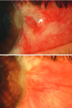
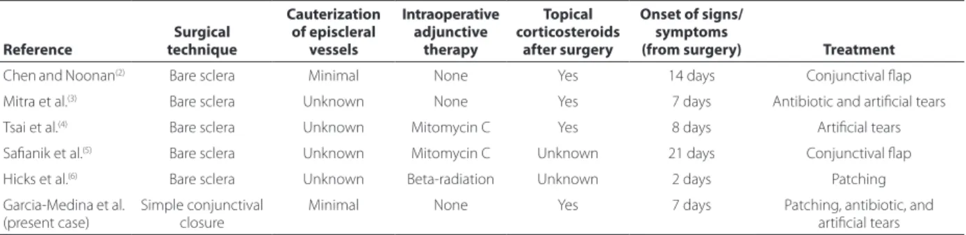
complaining of moderate pain and a black dot in his right eye. His best corrected visual acuity was 10/10 and intraocular pressure was 15 mmHg. Slit lamp examination revealed severe scleral thinning reaching the choroid (Figure 1). The conjunctival sutures were no lon-ger in place. The anterior chamber was deep with no Tyndall effect, whereas the pupils were isocoric and normoreactive. The fundus showed transparent media with no other abnormal findings. The patient refused improper eye handling, therapeutic failure, or going back to work in a dusty environment. Reconstructive surgery through a conjunctival flap was offered, but the patient refused; therefore, a conservative medical treatment was prescribed that included gen-tamicin antibiotic ointment (3 mg/g), patching for 7 days, and daily outpatient monitoring, which was followed by intensive ocular lubri-cation (carmellose 10 mg/mL every hour) for 6 weeks. Within a few weeks, the conjunctival defect was replaced by granulation tissue, and later with a flap of adjacent conjunctiva. The photographic series illustrates the patient’s evolution (Figures 2 and 3).
After solving the case, the patient was referred to internal medici-ne to rule out collagen or infectious disease. Medical history, physical examination, and laboratory test (blood count, biochemistry, rheu-matoid factor, autoantibody screening, infectious serology, and Man-toux) revealed no infectious or autoimmune inflammatory disease.
DISCUSSION
As seen above, several surgical techniques can be used to treat pterygium. The bare sclera technique includes excision of the pte -ry gium, which leaves the conjunctival defect to heal by sclera epi-thelization from the surrounding conjunctiva. Exposing the sclera contributes to drying, and therefore is a risk factor for thinning. All severe scleral thinning cases during the early postoperative period seem to be associated with the bare sclera technique. In our case, however, we used the primary excision technique with simple con-junctival closure (Table 1).
The fact that no stitches were found at about 1 week post-surgery suggests improper eye handling by the patient, excessively lax stitches, or a combination of both. The dehiscence of the wound, possibly resulting in exposure of the underlying sclera and edema, and raised conjunctiva edges, may have caused a discontinuity of the tear film leading to exacerbated local dehydration. In their publica-tion, Chen and Noonan(2) attributed SD after pterygium surgery with the bare sclera technique to raised granulation tissue edges, which developed in the conjunctiva margin of exposed sclera.
Whereas the growth of blood vessels in the wound bed is con-sidered to contribute to pterygium recurrence, some authors have recommended cauterization of episcleral vessels, especially at the limbus. However, it appears that coagulation of the episcleral vessels, which do not bleed, neither avoids recurrences nor contributes to accomplishing better esthetic results during pterygium surgery (personal communication of Lawrence W. Hirst). The cauterization applied to our patient was performed carefully and only with the purpose of avoiding bleeding during surgery. Nonetheless, this factor has to be considered in the etiology of scleral perforation as it may Figure 1. Initial appearance of the scleral dellen reaching the choroid.
Severe scleral dellen as an early complication of pterygium excision with simple conjunctival closure and review of the literature
184 Arq Bras Oftalmol. 2014;77(3):182-4
Table 1. Summary of published cases of early scleral dellen after pterygium surgery
Reference
Surgical technique
Cauterization of episcleral
vessels
Intraoperative adjunctive
therapy
Topical corticosteroids
after surgery
Onset of signs/ symptoms
(from surgery) Treatment
Chen and Noonan(2) Bare sclera Minimal None Yes 14 days Conjunctival flap
Mitra et al.(3) Bare sclera Unknown None Yes 7 days Antibiotic and artificial tears
Tsai et al.(4) Bare sclera Unknown Mitomycin C Yes 8 days Artificial tears Safianik et al.(5) Bare sclera Unknown Mitomycin C Unknown 21 days Conjunctival flap
Hicks et al.(6) Bare sclera Unknown Beta-radiation Unknown 2 days Patching
Garcia-Medina et al. (present case)
Simple conjunctival closure
Minimal None Yes 7 days Patching, antibiotic, and artificial tears
cause local ischemia. In the cases reported in the literature, whether episcleral vessel cauterization was used or its duration is unclear.(2-6) Subconjunctival anesthesia containing a vasoconstrictor agent (epi-nephrine) also may have been a contributory factor for local ischemia in the present case.
Topical corticosteroids enhance collagenases and inhibit colla-gen synthesis, which may have also contributed to SD formation, as pro-posed by Mitra et al.(3). Therefore, they should be removed immediately. The conjunctival flap graft has been used successfully to manage these complications(2,5). As the patient refused this surgical procedu-re, we had the opportunity to assess the evolution of scleral erosion with medical treatment. We found that eye patching, antibiotic co-verage, and intensive lubrication combined are sufficient for healing this com plication. Conservative treatment has also been proved to be successful in the cases that other authors have presented(3,4,6).
In conclusion, we can state that pterygium excision with primary conjunctival closure and cauterization of episcleral vessels may result
in severe SD, even in patients with no history of risk, as was the case presented here. Conservative treatment may be appropriate to ma-nage this rare complication.
REFERENCES
1. Hirst LW. The treatment of pterygium. Surv Ophthalmol. 2003;48(2):145-80. 2. Chen S, Noonan C. Scleral dellen complicating primary pterygium excision. Eye.
2000;14(Pt 1):100-1.
3. Mitra S, Ganesh A, Shenoy R. Scleral dellen complicating primary pterygium excision. Eye. 2000;14 (Pt 6):924-5.
4. Tsai YY, Lin JM, Shy JD. Acute scleral thinning after pterygium excision with intraope-rative mitomycin C: a case report of scleral dellen after bare sclera technique and re view of the literature. Cornea. 2002;21(2):227-9.
5. Safianik B, Ben-Zion I, Garzozi HJ. Serious corneoscleral complications after pterygium excision with mitomycin C. Br J Ophthalmol. 2002;86(3):357-8.