Nasal Septal Perforation closure with bacterial cellulose in rabbits
Abstract
Eulógio Emílio Martinez Neto 1, Jose Eduardo Lutaif Dolci 2
1 MD. MSc student - Otorhinolaryngology - Medical School of Santa Casa - São Paulo.
2 Full Professor of Medicine - Medical School of Santa Casa - São Paulo, Head of the Department of Otorhinolaryngology - Medical School of Santa Casa - São Paulo. Faculdade de Medicina da Santa Casa de São Paulo.
Send correspondence to: Eulógio Marinez - Rua Antonio José Thomaz da Costa 444 Florianópolis SC 88063-610. Paper submited to the BJORL-SGP (Publishing Management System – Brazilian Journal of Otorhinolaryngology) on May 09, 2009;
and accepted on November 03, 2010. cod. 6620
A
lloplastic materials can be used together with tissue and structure to close nasal septal perforation.Aim: to test cellulose use in the closure of septal perforation in rabbits and to compare fibrosis, inflammation, vascular congestion and graft integrity.
Materials and Methods: Fifteen rabbits. The rabbits were divided into two groups: Control: Five rabbits and Bionext® and fibrin glue Tissucol®: Ten rabbits. Septal perforations were done in all of them. In the Bionext® group the closure was performed with the placement of cellulose.
Results: Two rabbits died in the first week. Cellulose group: 2 closures without the cellulose in between the septum membrane and in 4 cases the graft stood in the middle of the perforation locked in place by the edges. No closure in the control group.
Conclusion: There was no closure of the perforation of the nasal septum with the graft between the septum membranes. There was no statistically significant difference concerning acute inflammation, vascular congestion and fibrosis between the 2 groups. In cases in which the graft remained in place, there was no change in its integrity. It may be used as a substructure for reepithelization of the perforation edges.
ORIGINAL ARTICLE Braz J Otorhinolaryngol.
2010;76(4):442-9.
BJORL
Keywords:
adhesive,
fibrin tissue, surgery, cellulose,
nasal septum.
INTRODUCTION
Nasal septum perforation is a disorder of easy diagnosis through the use of a nasal speculum or nasal endoscopy in the office. It can have numerous causes, from very benign ones, to local manifestations of syste-mic diseases. Moreover, nasal septum perforation varies broadly depending on location, size and symptoms; in consequence, many are the treatment options, including conservative treatment and numerous surgical techniques. Treatment must be customized for each patient.
The main cause of septal perforation is iatrogenic
- as a complication of nasal surgery1,2; however, other
iatrogenic causes may happen, such as the use of nasal
steroids3-5, mucosal cauterization to treat epistaxis;
naso-tracheal intubation and turbinate cryosurgery. Moreover, nasal perforations have been described after many types
of trauma surgery. Perforation rarely happens in children6.
Among the diseases associated with nasal perfo-ration, we list: cutaneous mucosal leishmaniasis; nasal abscess; syphilis; tuberculosis; typhoid fever; diphtheria; Wegener granulomatosis; lupus erythematous and sar-coidosis. Neoplasia and carcinomas also can cause nasal
septum perforation7-9. In tropical, developing areas, leprosy
and Leishmaniasis still are among the causes of nasal
septum perforation10,11. When the perforation borders are
covered by mucosa, hardly the perforation is associated to
more severe disorders, such as tuberculosis or neoplasias3,1.
Inhaling irritating products can also cause nasal septum perforation, such as in cases of nasal aspiration
of cocaine, which causes ischemia by vasoconstriction1,
foreign body granuloma caused by substances added to the drug3, besides perforations reported after inhaling chro-mic acid smoke, limo and cement powder, tar, pitch, salt, glass powder, sodium cabonate, calcium nitrate, calcium cyanide, arsenic, mercury and phosphorus.
Often times, the perforation happens before other systemic disease symptoms, and clinical investigation is
ne-cessary for those cases without clear etiological diagnosis12.
Nasal septum perforation can be range from asymp-tomatic to those causing severe epistaxis, nasal cosmetic
deformities, crust forming and nasal obstruction1-3.
The surgeries proposed to close perforations use flaps and grafts. The latter can be autologous (removed from the same being), homologous (from another being of the same species), heterologous (from another being of another species) and alloplastic (synthetic materials). Autologous grafts have the main disadvantage of causing trauma to the donor areas; and homologous and hetero-logous grafts bear risk of contamination for the individual
receiving tissue from another being13.
An inert material which could provide structure to the healing would be very useful in the treatment ar-senal of this disorder. The cellulose film formed by the fermentation from Acetobacter xillinum bacteria is inert,
resistant and insoluble, permeable to liquid and gas and resistant to stretching and traction. It is sterile, non-toxic
and non-pyrogenic14.
Studies in rabbits, replacing the septal cartilage for cellulose showed a partial absorption of the cellulose film
after four weeks15 and when used to cover the open area
after nasal concha resection in rabbits, it proved tolerable to the receiving tissue and there was no difference as to
the healing response concerning the control group16.
We propose an experimental study to close a nasal perforation in rabbits using the cellulose film together with fibrin glue used for fixation, and analyze the tissue response, graft properties and whether or not the perfo-ration would be closed.
LITERATURE REVIEW
Using the LILACS and Pubmed databases and the Internet, we searched the existing literature in relation to the cellulose produced by the Acetobacter xillinum bacte-ria, its development and use; surgical techniques used to close nasal septum perforation and the different materials used for this task.
Nasal septum perforation closure surgery in humans
In 1935, Imperatori et al.17 proposed a surgical
enlargement of the perforation, with the aim of reducing the discomfort brought about by constant hissing caused by smaller perforations, located on the anterior portion of the nasal septum.
The techniques used to close septal perforation can be grouped in: those in which we use flaps from the nasal septum itself to close it with or without grafts, and the techniques which use neighboring flaps.
The papers which suggest flap rotation are unani-mous as to the need for a second surgical procedure to resect the vascular pedicle.
Among the techniques using septal mucosa flaps, we find the description of fascia and perichondrium graf-ting in between the septal mucosa leaflets, by Wright et
al.18. This publication was greatly important, both because
of the promising results, as well as for justifying the use of grafts to close the perforations, instead of simply suturing the borders. The authors also report that, besides the low metabolic requirements, fascia and perichondrium grafts serve as support for the growth of fibroblasts, supporting growth at the margins of the mucosas, one towards the other.
McCollough19 reported a successful closure of the
septal perforation with ear-based graft. In 1980, Fairbanks9
Fairbanks8 also spoke against surgical techniques which propose suturing the borders of the perforation, with or without everting the contralateral mucosa because of the atrophic and fragile characteristic of the tissue of the perforation borders. It also criticizes the techniques which use the oral mucosa, considering its non-transformation in ciliated tissue, thus having its drying as a consequence.
Suggestions concerning the use of artificial material or implants associated with closure techniques were given
by Gyeney and Kerenyi20 who in 1977 reported the closure
of septal perforation through fibrin implants (Bioplast®).
Kridel et al. in 199821 suggested the use of a biosynthetic
substitute (Acellular Human Dermal Allograft®), having reported success in the closure of eleven among 12 per-forations treated. The authors criticized the use of fascias, especially due to the need to use material to dry them, which causes early softening, making them difficult to handle. In cases of failure, there was a diameter reduction
from 3cm to 5mm. In 2006, Lee et al.22 showed the use of
fibrin glue and autologous cartilage grafts used to prevent nasal septum perforation during septoplasty.
Other materials such as gold, ivory, cobalt and cork
have also been used23. Among the most used alloplastic
materials we find those based on silicone, such as Silastic® which are largely used in facial structure surgeries thanks to their possibility of being sculptured and molded accor-ding to need. Although relatively inert from the biological standpoint, after its placement on the receiving site, a fibrous capsule can cover it with time, causing tangible mobility and consequently greater likelihood of migration
and extrusion24.
Stoor et al.25 studied the septal closure and infection
by Haemophilus influenza and Streptococcus pneumoniae with the use of bioactive glass (BAG) in 11 patients with septal perforation not having contamination by these ger-ms in any patient and being able to close the perforation in 10 cases.
The cellulose film
The Bionext® cellulose film arises from the fermen-ting of Acetobacter xylinum bacteria, done through the experiment carried out by microbiologist Luís Fernando
Xavier Farah (1984) from Curitiba (PN).26
The cellulose produced by the bacteria can be in the form of a flexible, semitransparent and yellowish membrane, or as a solid, dense, malleable mantle, of firm and jelly-like consistency, of about 0.5cm thick (Figure 1). The film arising from the biological production of the bacteria, after processing, does not have additives, being pure cellulose, made up of polysaccharides, biodegradable, non-toxic, non-pyrogenic and sterile. It is an inert substan-ce, very resistant and insoluble in all organic solvents, and it has specific physical characteristics, such as: established permeability defined to liquids and gases, resistance to
traction and stretching, also having characteristic weight
and molecular structure.14,27,28
It is efficient for pain relief and to reduce healing time in dermoabrasion lesions, skin donor areas, or in large burns. This composite has been successfully used as bandage in skin sores, burns and skin donor areas. It
was also used as substitute for meninges.29-34
It is seen as a material of numerous possibilities,
from paper to protect documents to bullet proof vests26.
Acetobacter xilinum comercial cellulose, Bionext®, has been approved by ANVISA and the FDA and is used as a transitional skin replacement. Its physical and biocompa-tibility properties, as well as its ease of use and the possi-bility of modeling during insertion makes this substance a possible element in the treatment of bone and cartilage together.
OBJECTIVES
1. To test the use of cellulose produced by Ace-tobacter xylinum bacteria -Bionext® together with the Tissucol® biological fibrin glue in the closing of septal perforation surgically inflicted in rabbits.
2. To histologically compare the degree of fibrosis, vascular congestion in the animals, graft integrity and whether or not the septal perforation was closed.
MATERIALS AND METHODS
Materials
For this study we used fifteen adult New Zealand rabbits weighing approximately 3kg, cellulose mantle (Bionext®) and fibrin biological glue (Tissucol®).
Sample size and selection
The rabbits were randomly assigned (coin flip)
into two groups: Control Group made up of five rabbits; and the Bionext® group associated with the fibrin glue Tissucol®, made up of 10 rabbits.
One rabbit from each group died in the immediate post-op. the rabbits were slaughtered fifty days after the experiment.
Surgical procedure
The surgical procedures were carried out following the ethical principles of experimentation with animals, set forth by the Brazilian Code of Animal Experimentation (COBEA). Protocol 09/07 ICAO.
The surgical procedures were done with the rab-bits under general anesthesia, using Zoletil® (Tiletamine associated with Zolazepan) and Nilperidol® (Fentanyl associated with Droperidol) and kept under spontaneous ventilation, in dorsal decubitus. They were submitted to nasal septum perforation as per described below:
1. 2% lidocaine and noradrenaline soaked cotton in the concentration of 1:50,000 in both nasal cavities.
2. Bilateral subperichondrial septum injection of the same solution.
3. Upper left-side anterior septal incision followed by ipsilateral subperichondrial detachment.
4. Square-shaped incision and removal of appro-ximately 7mm measured with a surgical caliper, on each side of the septal mucosa and septal cartilage.
The experimental group was submitted to the placement of the Bionext® graft in the same procedure when the perforation was made; anchored in between the remaining septal mucosa, which were previously detached and not removed (Fig.2).
septum perforation and underwent histopathology analysis of the surgical specimen.
For the slaughter at the end of the follow up period, the rabbits were again anesthetized and received IV sodium thiopental (dose 40mg/kg). After the slaughter, the facial mesostructure was removed en bloc for histopathology purposes.
The rabbits were evaluated by clinical parameters which could indirectly assess tolerability issues, such as general and breathing discomfort. The parameters evaluated were: amount of food ingested, rabbit weight variation, temperature, respiratory rate and the presence of nasal bleeding. All the rabbits were weighed before the procedure and on a daily basis until their slaughter. The amount of food ingested was controlled daily in grams. Ear temperature, in Celsius was measured twice a day. Respiratory rate was checked twice-a-day. Bleeding was seen daily.
The animals’ facial mesostructures were dissected and fixed in a 10% formaldehyde solution.
Histological evaluation
The animals’ facial mesostructures remained for 6 days in a 5% nitric acid solution for decalcification. Once decalcified, the nose and skull were sliced in 5mm cross-sections. The slices were then dehydrated, clarified and included in paraffin. After inclusion in the paraffin blocs, the specimens were cut in the microtome at an average thickness of 5μm. (Figure 3)
Figure 2. Rabbit positioned and anesthetized for the procedure.
The rabbits were kept under the same daily care conditions as they were prior to treatment, for 50 days, when they were slaughtered for observation of the nasal
Figure 3. Photomicrography of septum histology slides. A - Ruptured septum (macro). HE dye.
pathologist in a blind way as to germ group and species, and classified in degrees by qualitative and semiquantita-tive criteria. Thus, Bionext® (IBT) integrity was classified in: 0=not applicable/ Bionext® absent, 1= intact and 2= fragmented (Figure 4); vascular congestion: 0= absent, 1= mildly congested, 2= dilated and congested, and 3= highly dilated vessels with red blood cell extravasation (Fig. 5); Fibrosis: 0=absent, 1= presence of fibroblasts alone and 2= reparative fibroblastic proliferation with thickness; acute inflammation (AI): we also looked for fibrin and neutrophilic exudate: 0=absent, 1= scattered spots and 2= neutrophil build up involving the structures (Fig. 6).
RESULTS
Two rabbits died in the first week of the experi-ment, one from the Bionext® group and one from the Control Group.
On Table 1 we see the parameters assessed from each rabbit, according to their weight in the beginning of the experiment and after treatment, and the histological characteristics assessed on the specimens analyzed.
On Table 2 we see the data regarding the graft beha-vior and whether or not the septal perforation was closed.
DISCUSSION
Because of the need for a framework for fibroblasts to grow - supporting growth at the margins of the mucosa,
one towards the other through a simple border suturing18,2,
we considered the possibility of using a cellulose film pla-ced between the borders of a surgical perforation done to the nasal septum of rabbits in order to serve as a support for a possible tissue regeneration.
Because the perforation and repair surgery were carried out in the same procedure and with the film already in place since the onset of the healing process, we did not have atrophic margins and we had very little vascularization, which is common in the clinical practice
and does not foster closure8. This is a bias in our study.
Knowing of the ease in intraoperative handling, although of little adhesiveness, we added biological glue to the margins of the perforation with the aim of fixing the film. Thus, the experiment would be similar to the one from Kridel et al., who in 1998, who suggested the use of biosynthetic substitute (Acellular Human Dermal Allograft®) fixed with suture points to one of the sides and the other one is closed with mucosa flap rotation; none-theless, in our case we used cellulose film and biological glue without closing the mucosa in any of the sides of
the perforation.21
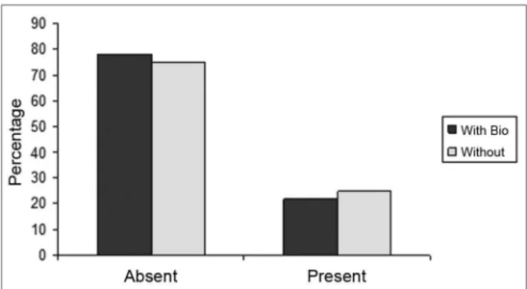
Figure 4. Percentage of animals that presented or did not present biomaterial integrity, in the control group (without) and experimental group (with Bio). (Fisher test: p=0.462).
Figure 5. Percentage of animals who had or did not have congestion, in the control (without) and experimental (with bio) groups. (Fisher test: p=0.308).
Figure 6. Percentage of animals who had or did not have an acute inflammatory process, in the control (without) and experimental (with bio) groups.
(Fisher Test: p=0.706)
Statistical Assessment
The film incorporation and absorption were more
significant after four weeks in the studies by Oliveira15
studying the replacement of the septal cartilage for cellu-lose film. For this reason we chose to assess our sample after 50 days.
All the studies carried out evaluating the cellulose film had it in contact with some tissue, we found it inte-resting to assess its behavior when it is attached only to the borders of the perforation.
The perforation closure with the cartilage repla-cement for the cellulose did not happen. In the cases in which the perforation closed, either the cellulose film was kept attached to the perforation’s epithelized bor-ders like a “cork” (Fig. 7), or it was lost very likely due to graft shifting. In the cases in which the closure happened
without the graft (Fig. 8), the question remains whether or not the graft helps close the perforation, and at what time the shifting happened. Despite the lack of statistical significance, we can suspect the film helped in the closure since the control group did not show any closure of the perforated septa; and such significance was due to the small number of rabbits in the sample.
The graft shifting may have happened because of the smooth surface of the cellulose film, and due to the lack of efficacy of the fibrin glue in contact with the cellulose or
Table 1. Distribution of the rabbits as to the treatment given, weight (g) and histopathology parameters analyzed. Rabbit With
Bionext W/out Weight pre Weight post bti ai Cong fibrosis Septum
1 x 5100 6200 0 0 2 0 Ruptured
2 X 4080 4237 0 0 3 0 Ruptured
3 x 3517 3691 0 0 2 0 Closed
4 x 4167 4180 0 0 1 0 Closed
5 X 4157 4238 0 0 2 0 Ruptured
6 X 3438 3751 0 0 0 0 Ruptured
7 X 3570 3693 0 2 3 0 Ruptured
8 x 3150 3774 0 0 3 0 Ruptured
9 x 3305 3508 1 1 1 0 Bt
10 x 3210 3109 0 0 2 0 Ruptured
11 x 3200 3422 0 0 2 0 Closed
12 x 3230 3247 1 1 2 0 Bt
13 x 3185 3392 0 0 2 0 Closed
bti =biotissue integrity (0=not applicable/ Bionext® absent, 1= intact and 2= fragmented); ai= acute inflammation (fibrin and neutrophilic exu-date: 0=absent, 1= scattered spots and 2= neutrophil build up involving the structures); cong= vascular congestion:( 0= absent, 1= mildly congested, 2= dilated and congested and 3= vessels highly dilated with red blood cell spill over); fibrosis: (0=absent, 1= scattered fibroblasts and 2= reparative fibroblastic proliferation with thickening) and septum (Bt= Bionext® between the perforation borders)
Table 2. Histological changes seen in relation to the integrity of the biomaterial and characteristics of the septum, according to the group studied.
Groups n BTI SEPTUM
0 1 Ruptured Bionext® Closed
With Bio 9 7 2 3 2 4
W/out 4 4 0 4 0 0
Bionext®: Cellulose present between the borders of the perforation We did not notice statistically significant difference between the groups as to biomaterial integrity (BTI) (p=0.462) and septum closure (p=0.176).
by the mechanical force of sneezes. Postoperative packing for a long period of time, such as the 12 days proposed
by Kratz in 197335 or for shorter periods such as the two
days proposed by Lee in 200836 is not feasible in rabbits
with a delicate graft on the nasal septum.
The lack of statistically significant difference as far as acute inflammation, vascular congestion and fibrosis are concerned, prove that the implant is inert and well accepted by the nasal mucosa.
The weight gain of the group submitted to the graft shows it is well tolerated by the animals and also shows little suffering with the experiment.
CONCLUSIONS
a) There was no statistically significant difference concerning acute inflammation, vascular congestion and fibrosis between the two groups.
b) In the cases in which the graft was kept positio-ned, there was no change to its integrity.
c) It can be useful in the therapeutic weaponry as a basis for reepithelization of the perforation borders.
REFERENCES
1. Judson MAJ, Belmont R. An Approach to Large Nasoseptal Perfora-tions and Attendent Deformity. Arch Otolaryngol. 1985;111(7):450-5. 2. Teichgraeber JF, Russo RC. The Management Of Septal Perforations.
Plast Reconstr Surg. 1993;91(2):229-35.
3. Miller FF. Occurrence Of Nasal Septal Perforation With Use Of Intranasal Dexamethasone Aerosol. Ann Allergy. 1975;34:107-9. 4. Ferguson, B. J. Nasal Steroid Sprays And Septal Perforations. ENT
J. 1997;76(2):75-6.
5. Soderberg-Warner ML. Nasal Septal Perforation Associated With Topical C orticosteroid Therapy. J Pediatr. 1984;105(5):840-1. 6. Bridger GP. Surgical Closure Of Septal Perforations. Arch Otolaryngol
Head Neck Surg. 1986;112(12):1283-5.
7. Fairbanks DNE, Chen SCA. Closure Of large Nasal Septum Perfora-tions. Arch Otolaryngol. 1970;91:403-6.
8. Fairbanks DNE, Fairbanks GR. Nasal Septal Perforation:Prevention And Management. Ann Plast Surg. 1980;5(6):452-9.
9. Fairbanks DNF. Closure Of Nasal Septum Perforation. Arch Otola-ryngol. 1980;106:509-13.
10. Goulart IMB, Patrocínio LG, Nishioka SA, Patrocínio JA, Ferreira MS, Fleury RN. Concurrent leprosy and leishmaniasis with mucosal involvement. Lepr Rev. 2002;73:283-4.
11. Dolci JE, Bussolotti F, Liquidato BM. Etiologia das perfurações septais. Revista Portuguesa de Otorrinolaringologia. 2001;39(4):369-72. 12. Diamantopoulos II JNS. The investigation of nasal septal perforations
and ulcers. Laryngol Otol. 2001;115(7):541-4.
13. Sofia OB. O uso de cartilagem costocondral em reconstrução de dorso nasal. Acta ORL. 2005;23 (4):176-81.
14. Costa HO, Souza FC. Avaliação da regeneração tecidual da pele de porco submetida à lesão térmica seguida de colocação de Biotissue. ACTA ORL. 2005;23(3):23-7.
15. Oliveira RCB. Avaliação da resposta tecidual quando da substituição da cartilagem do septo nasal de coelhos por manta de celulose bacteriana. Estudo experimental. Tese de mestrado. São Paulo: Faculdade de Ciências Médicas da Santa Casa de São Paulo, 2007. 16. Osman Salah Ali. Estudo experimental sobre a aplicação de película
de celulose (Bionext®) em área cruenta de ressecção de concha nasal de coelhos. Tese de mestrado. São Paulo: Faculdade de Ciências Médicas da Santa Casa de São Paulo, 2007.
17. Imperatori CJ, Burman HJ. Diseases of the Nose and Throat. Ha-gerstown, Md, Harper & Row Publishers Inc, 1935.p. 122-124. 18. Wright WK. Tissues For Tympanic Grafting. Arch Otolaryngol.
1963;78:291.
19. McCollough EG. An Approach To Repair Of Septal Perforations. ORL Digest. 1976;38:13.
20. Gyeney L, Kerenyi G. Bioplast fibrin implants in nasoseptal perfo-ration. Arch Otorhinolaryngol. 1977;20;218(1-2):143-5.
21. Kridel RW, Foda H, Lunde KC. Septal perforation repair with acellu-lar human dermal allograft. Arch Otoacellu-laryngol Head Neck Surg. 1998;124(1):73-8.
22. Lee JY, Lee SH, Kim SC, Koh YW, Lee SW. Usefulness of Autolo-gous Cartilage and Fibrin Glue for the Prevention of Septal Perfo-ration during Septal Surgery: A Preliminary Report. Laryngoscope. 2009;116:934-7.
23. Juraha LZ. Experience with alternative material for nasal augmenta-tion. Aesthetic Plast Surg. 1992;16(2):133-40.
24. Lindsey WH, Ogle RC, Morgan RF, Cantrell RW, Sweeney TM. Nasal reconstruction using an osteoconduetive collagen gel matrix. Arch Otolaryngol Head Neck Surg. 1996;122(1):37-40.
25. Stoor P, Sóderling E, Grénman R. Bioactive glass S53P4 in repair of septal perforations and its interactions with the respiratory infection-associated microorganisms Haemophilus influenzae and Streptococcus pneumoniae. J Biomed Mater Res. 2001;58(1):113-20. 26. Abrantes ACS. Do avião à urna eletrônica. Fonte:Ministério da Ciência
& Tecnologia (jovem). Disponível em <http://ctiovem.mct.qov.br>. Acesso em 03/11/2006.
27. Fontana JD, De Souza AM, Fontana CK, Torriani IL, Moreschi JC, Gallotti BJ et al. Acetobacter cellulose pellicle as a temporary skin substitute. Appl Biochem Biotechnol. 1990;24(25):253-64.
28. Hart J, Silcock D, Gunnigle S, Cullen B, Light ND, Watt PW. The role of oxidized regenerated cellulose/collagen in wound repair: effects in vitro on fibroblast biology and in vivo in a model of compromised healing. Int J Biochem Cell Biol. 2002;34(12):1557-70.
29. Cabral LM, Gattaz MD, Factore LAP, Mattar JA, Diament D, Oliveira AM. Curativo biológico no tratamento do grande queimado:apresentação de caso. Rev Bras Cir. 1987;77(6):383-9. 30. De Paola DQ, Souza MGPP. Película celulósica - Novo curativo
biológico para melhoria do leito receptor de enxertia cutânea. Rev Bras Cir. 1987;77(3):135-8.
31. Hilário AH, Vasquez LAM. Utilização de um substituto temporário de pele nas perdas cutâneas de pacientes ambulatoriais. Rev Bras Cir. 1988;78(6):393-8.
32. Peixoto R, Santos DLN. Biofil. Uso e avaliação clínico de uma película celulósica em lesões cutâneas. Rev Bras Cir. 1988;78(2):141-5.
33. Pitanguy I, Salgado F, Maracajá PF. Utilização de película de celulose (Biofill®) como curativo biológico. Rev Bras Cir. 1988;78(5):317-26. 34. Rebello C, Almeida DA, Lima Jr. EM, Dornelas MP. Biofill, um novo
substituto de pele:nossa experiência. Rev. Bras Cir. 1987;77(6): 407-14.
35. Kratz RC. Repair of Septal Perforations With Composite Grafts. Arch Otolaryngol. 1973;98:380-2.