ABSTRACT
Histomorphometric analysis of the repair process
Jônatas Caldeira ESTEVES1, Albanir Gabriel BORRASCA2, Alessandra Marcondes ARANEGA3, Idelmo Rangel GARCIA JUNIOR3, Osvaldo MAGRO FILHO3
1- DDS, MSc, Master in Oral and Maxillofacial Surgery, PhD student in Implantology, Araraquara Dental School, UNESP - Univ. Estadual Paulista, Araraquara, SP, Brazil.
2- DDs, MSc in Oral and Maxillofacial Surgery, PhD student in Oral and Maxillofacial Surgery, Araraquara Dental School, UNESP - Univ. Estadual Paulista, Araraquara, SP, Brazil.
3- DDS, MSc, PhD, Assistant Professor, Division of Oral and Maxillofacial Surgery, Department of Diagnostics and Surgery, Araraquara Dental School, UNESP - Univ. Estadual Paulista, Araraquara, SP, Brazil.
Corresponding address: Jônatas Caldeira Esteves - Rua Humaitá, 1680, 20 andar, Departamento de Periodontia - Centro - 14801-903 - Araraquara-SP - Phone: 55 16 8839-0803 - Fax: 55 16 3301-6369 - e-mail: [email protected]
O
!"#$%&'()'((* + right parietal bone for graft obtainment using a 4-mm-diameter trephine drill. Then, the * #/0/&1 2 3 * 5 #/ *0/& & the presence of adhesive at the recipient-site/graft interface. Graft incorporation to the & 2 #/0/)* & '(( & 2 & 5
Key words: Tissue adhesives. Cyanoacrylates. Bone transplantation.
INTRODUCTION
Autogenous grafting is the most commonly used surgical procedure for bone reconstruction due to its & such as biocompatibility and bone regeneration potential4. Several possibilities are reported by & among these solutions it is possible to cite intra and extra-oral autogenous grafts, allografts, alloplastic & membranes for guided bone regeneration19,20.
Autogenous bone grafts are considered the most suitable for the reconstruction of defects at oral and maxillofacial regions, mainly due
to their characteristics of osteoinduction and osteoconduction. It is the only type of graft that provides live immunocompatible bone cells, essential to the stage I of osteogenesis. This makes this type of graft more advantageous, since the higher the amount of transplanted living cells, the
4,5,20.
position1,27. On the other hand, disadvantages such 5 & researchers to the search for an alternative method && & are a resource potentially capable of providing stability for the healing process12.
Tissue adhesives based on cyanoacrylate (CA) are substances that have been successfully employed for skin laceration synthesis and surgical incisions6,10,13,22,25& 2 of thin bone fragments in orbital fractures13 and osteochondral fractures26. The most interesting properties of the adhesives are their rapid polymerization, strong adhesion to the surfaces
& 2,8,12,
bacteriostatic15,22 and hemostatic actions18, besides
EF 3,10,21.
J)*& )*& & presenting excellent adhesive strength11K & its apparent toxicity to soft tissues conducted the researches for adhesives of longer chain, )*& biocompatibility9.
( & of ethyl-CA has still been tested for biological use in
& 3,14,16,24.
' & )* behavior on mineralized tissues fixation. The and histometric analyses of the repair process of ethyl-CA adhesive
MATERIAL AND METHODS
3 J ) !L MN//1 //PP1Q% (Rattus norvegicus albinus, Wistar)) NP/& N of 16 animals each: Group I – Control and Group (( U * V & F X Department of Surgery and Integrated Clinics and
!'Z®, Mogiana
* & \] L& \L& ^2% ad libitum, except in the fasting period (14-16 hours) &
* 2 hydrochloride (0.2 mL/250 g) (Xilazin®, Syntec do Brasil Ltda, Cotia, SP, Brazil) and ketamine hydrochloride (Cetamin®, Syntec do Brasil Ltda, Cotia, SP, Brazil) (0.1 mL/250 g), shaving and #/q LXL( the frontoparietal region. The animals received
N/ incision in the scalp over the sagittal calvarial suture, & parietal bones. In each animal, a rounded osteotomy w saline irrigation to obtain a rounded bone fragment, preserving the integrity of the dura mater and brain. F ( & & 2 M1 ( ' (& & ( ' ((& ethyl-CA adhesive (Super Bonder®; Loctite-Henkel, ( &\L&^2% 5 spiraled suture using Mononylon 5-0 (Mononylon®, J&J Ethicon, São José dos Campos, SP, Brazil).
2 overdose at 10 and 30 postoperative days (8 % & #/q laboratorial processing. Six-micrometer-thick cross and eosin for histological and histometric analyses.
In the histological (qualitative) analysis, the graft/recipient site interface, the presence of the & 5 & & deep tissues.
In the histometric (quantitative) analysis, the (Aristoplan-Leitz®, Leica, Wetzlar, Hesse, Germany) NP& !*) MRc5®& &\*&\L&^2% *XwP & ( & 2 & pixels per inch. The bone graft size at 10 and 30 E & ( N/// !V ^ ( & Vargem Grande do Sul. SP, Brazil). The evaluation & # F & statistical analysis.
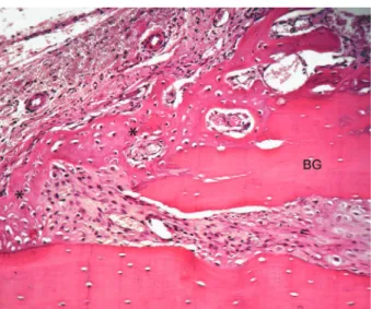
Figure 1- Group I – 10 days: Peripheral region of the graft. New bone formation, with primary bone tissue (asterisks), from the graft fragment (bone graft - BG) and parietal bone (hematoxylin-eosin – Original 160x)
Figure 2- Group II – 10 days: Bone graft (BG) with osteocyte lacunae containing basophilic nucleus. Fragments of the bone graft and the recipient site and discrete areas of new bone formation (BN) can be observed in the recipient site (hematoxylin-eosin - Original 63x)
#/ 0/ individual comparisons (p<0.05).
RESULTS
* & #/ postoperatively, the soft tissues handled during the &
Histological results 10 days – Group I
Superficially, the periosteum presented 5 & & * & remodeling both at the graft and the parietal bone. * & ! #%
10 days – Group II
\ & ( specimens, a predominance of lymphocytes together * containing basophilic nucleus. Several bone lacunae cells, and, in most of the specimens, a large number of neutrophils. Deeply and adjacent to the graft, )* * & osteoclastic-type multinucleate cells. A large number & V ( bone graft and the recipient site (Figure 2).
30 days – Group I
At 30 days, the periosteum layer over the graft & 5 ^ bone tissue at the interface region. In several &
at the central third of the graft, and their margins remained joined to the donor area by a compact connective tissue (Figure 3). The graft had an aspect & 2 K
30 days – Group II
Figure 3- Group I – 30 days: Bone graft (BG) incorporated to the recipient site. New bone formation at the interface observed only at the graft central region, where the (hematoxylin-eosin – Original 100x)
Figure 4- Group II – 30 days: Underneath, bone graft is linked to the recipient site only in the graft margin. Areas of new bone formation (BN) are observed in the internal by a high amount of cyanoacrylate (CA) remains between !!"#$
Figure 5- Group II – 30 days: New bone formation observed between the bone graft and the recipient site, in contact with the cyanoacrylate (CA) (hematoxylin-eosin - Original 160x)
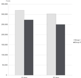
Figure 6- Area of the bone graft fragment in Groups I and II at 10 and 30 days
in most of the animals, this union could not be & 5 & V & )* & & )* ( & & the regions adjacent to the graft proximal surface ! w% observed in this space until the time period studied. Regarding the recipient site, resorption areas and some of the specimens (Figure 5).
Histometric and statistical results
2 ! % 0#&1$P !N#&$##% 0/N&N$1 !#1&/%
Group I at 10 and 30 days, respectively, and NQ0&#w !#&#Q1% NP/&0# !NP&#NQ% Group II at 10 and 30 days, respectively (Figure $% ( & #/0/ & ' I and II (p<0.05).
DISCUSSION
makes impracticable its use. Thus, bone fragments recipient site.
The partial graft resorption is a natural process & of inflammatory reaction or micromovements tends to accelerate and intensify this process17. It is important to emphasize that the bone graft at rat calvaria, as proposed in the experimental model of the present study, is not subjected to many movements; it is maintained in position and becomes properly incorporated to the recipient site, as observed in the control group at 10 and 30 days. Differently from the calvaria, in the mouth, the constant movements of the muscles and food, F blocks. Graft stability is essential for the occurrence of revascularization and graft incorporation17,20.
According to clinical observations, the technique & & mechanically, the adhesive promoted a good demonstrated by the fact that the bone fragment & )*&
11,23,27.
It has been reported that the smaller the ester chain, the higher its histotoxicity10 K & 5 & #/ * 0/ & 5 exhibited foreign body-type multinucleate cells in areas adjacent to the CA fragments. These 5 reaction, produced by the material, has a short duration. According to Celik, et al.7 (1991), the 5 on tissue oxygen, explained by the transformation of the cellular membrane polyunsaturated fatty & metabolism of the local arachidonic acid, unleashing the synthesis of tromboxane and prostaglandin.
in vivo on CA behavior in the bone tissue. Clinical and experimental produce a stable union of bone segments1,8,12,14,16,27, & Saska, et al.23 (2006), in a histomorphometric study utilizing rabbits as experimental models, )* 5 #P & the posterior periods. In the present study, in fact, 5
K & of multinucleated cells, especially at the graft & & &
Similarly to the results of Saska, et al.23 (2006), )* )* & & 5 & formation of osseous trabeculae occurred after this & an osteoconductive or osteoinductive behavior, formation and graft incorporation to the recipient & 5 higher graft resorption observed at both 10 and 30 postoperative days.
L complete repair process, providing results similar to those provided by the conventional internal rigid K & )* still observed in the graft/recipient site interface, the area and the maintenance or not of the graft bone size.
CONCLUSION
According to the methodology employed in this & & fragment at 30 days, the permanence of adhesive & 5
REFERENCES
1- Ahn DK, Sims CD, Randolph MA, O'Connor D, Butler PE, Amarante MTJ, et al. Craniofacial skeletal fixation using biodegradable plates and cyanoacrylate glue. Plast Reconstr Surg. 1997;99:1508-15.
N*F\&V^&) FV&)F^&2&^F ZN to the anterior nasal spine in rabbits: experimental study. Eur Arch Otorhinolaryngol. 2007;264:1425-30
5- Boyne PJ, Lynch SE, Genco RJ, Marx RE. Studies of the surgical application of osteoconductive and osteoinductive materials. In: Lynch SE, Genco R, Marx R. Tissue engineering: applications in maxillofacial surgery and periodontics. Chicago: Quitessence Publishing; 1999.
6- Bruns TB, Simon HK, McLario DJ, Sullivan KM, Wood RJ, Anand KJ. Laceration repair using a tissue adhesive in a children's emergency department. Pediatrics. 1996;98:673-5.
7- Celik H, Caner H, Tahta K, Ozcan OE, Erbengi A, Onol B. Nonsuture closure of arterial defect by vein graft using isobutyl-2-cyanoacrylate as a tissue adhesive. J Neurosurg Sci. 1991;35:83-7.
1V^&*F\&)&^F 2 ZN & effects on the tissues. Eur Arch Otorhinolaryngol. 2007;264:539-44.
9- Eaglstein WH, Sullivan T. Cyanoacrylates for skin closure. Dermatol Clin. 2005;23:193-8
10- Farion KJ, Osmond MH, Hartling L, Russell KF, Klassen TP, Crumley E, et al. Tissue adhesives for traumatic lacerations: a 2 * J Med. 2003;10:110-8.
11- García-Páez JM, Jorger-Herrero E, Rocha A, Maestro M, Castillo-Olivares JL, Millan I, et al. Comparative study of the mechanical behavior of a cyanoacrylate and a bioadhesive. J Mater Sci Mater Med. 2004;15:109-15.
12- Gonzalez E, Orta J, Quero C, Niemshik L, Galera R, Onay D, JN 5 craniotomy. Surg Neurol. 2000;53:288-9.
13- Gosain AK, Lyon VB, Plastic Surgery Educational Foundation DATA Committee. The current status of tissue glues: part II. For adhesion of soft tissues. Plast Reconstr Surg. 2002;110:1581-4. 14- Gosain AK, Plastic Surgery Educational Foundation DATA ) ( Plast Reconstr Surg. 2002;109:2581-3.
#PK &^ *&\+&VK\&J^* Comparison of effects of suture and cyanoacrylate tissue adhesive on bacterial counts in contaminated lacerations. Antimicrob Agents Chemother. 1995;39:559-60.
16- Kim YO. Use of cyanoacrylate in facial bone fractures. J Craniofac Surg. 1997;8:229-34.
17- Lin KY, Bartlett SP, Yaremchuk MD, Fallon M, Grossman 3& F * onlay bone graft: an experimental study. Plast Reconst Surg. 1990;86:449-56.
18- Lumsden AB, Heyman ER, Closure Medical Surgical Sealant Study Group. Prospective randomized study evaluating an absorbable cyanoacrylate for use in vascular reconstructions. J Vasc Surg. 2006;44:1002-9.
19- Macedo NL, Macedo LG, Matuda FS, Ouchi SM, Monteiro *\& ) 3 ' implants of PTFE and hydroxyapatite physical barriers in rats. Braz Dent J. 2003;14:119-24.
20- Misch CE. Aumento do osso para inserção do implante: soluções para o enxerto ósseo. In: Misch CE. Implantes dentários contemporâneos. São Paulo: Ed. Santos; 2000. p. 451-65. 21- Pérez M, Fernández I, Marquez D, Bretaña RMG. Use of n-butyl-2-cyanoacrylate in oral surgery: biological and clinical evaluation. Artif Organs. 2000;24:241-3.
NN & '& \ & F & & \ (& et al. A randomized trial comparing octylcyanoacrylate tissue adhesive and sutures in the management of lacerations. JAMA. 1997;277:1527-30.
23- Saska S, Hochuli-Vieira E, Minarelli-Gaspar AM, Gabrielli MF, ) X&' * rabbits. Int J Oral Maxillofac Surg. 2009;38:180-6.
24- Shermak MA, Wong L, Inque N, Crain BJ, Im MJ. Chao EYS, et F N and its effect on histotoxicity and healing. Plast Reconst Surg. 1998;102:309-18.
NP \ K& V& ^ ^& F & 3& Sullivan KM. Long-term appearance of lacerations repaired using a tissue adhesive. Pediatrics. 1997;99:193-5.