Materials Research, Vol. 10, No. 1, 1-4, 2007 © 2007
*e-mail: [email protected]
Article presented at the IV Congresso Latino Americano de Órgãos Artificiais e Biomateriais (COLAOB 2006), August 8 and 11, 2006, Caxambu, MG, Brazil
Evaluation of Shrinkage Temperature of Bovine Pericardium Tissue
for Bioprosthetic Heart Valve Application by Differential Scanning Calorimetry
and Freeze-drying Microscopy
Virgilio Tattini Jr.a, Jivaldo do Rosario Matosb, Bronislaw Polakiewicza,
Ronaldo Nogueira de Moraes Pitomboa*
a
Department of Biochemical and Pharmaceutical Technology, Pharmaceutical Sciences School,
University of Sao Paulo, Av. Prof. Lineu Prestes, 580, Bloco 16, 05508-900 São Paulo - SP, Brazil
Prof. Lineu Prestes, 580, Bloco 16, 05508-900 São Paulo - SP, Brazil
b
Chemestry Institute, University of São Paulo, Av. Prof. Lineu Prestes, 748, Bloco 8,
Prof. Lineu Prestes, 748, Bloco 8,
05508-900 São Paulo - SP, Brazil
Received: July 12, 2006; Revised: November 30, 2006
Bovine pericardium bioprosthesis has become a commonly accepted device for heart valve replacement. Present practice relies on the measurement of shrinkage temperature, observed as a dramatic shortening of tissue length. Several reports in the last decade have utilized differential scanning calorimetry (DSC) as an alternative method to determine the shrinkage temperature, which is accompanied by the absorption of heat, giving rise to an endothermic peak over the shrinkage temperature range of biological tissues. Usually, freeze-drying microscope is used to determine collapse temperature during the lyophilization of solutions. On this experiment we used this technique to study the shrinkage event. The aim of this work was to compare the results of shrinkage temperature obtained by DSC with the results obtained by freeze-drying microscopy. The results showed that both techniques provided excellent sensitivity and reproducibility, and gave information on the thermal shrinkage transition via the thermodynamical parameters inherent of each method.
Keywords: Freeze-drying, DSC, Freeze-drying microscope, bovine pericardium
1. Introduction
Almost 30 years after the introduction of valvular prostheses, patients worldwide are receiving cardiac valve substitutes each year. Among these, the bovine pericardium has become a commonly ac-cepted device for heart valve replacement. The use of glutaraldehyde as a cross-linking agent has become a standard practice for most manufacturers of bioprosthetic valves, each of them having developed their own proprietary method of processing. Glutaraldehyde stabi-lizes the collagen structure, prevents tissue digestion by enzymes or bacteria and reduces the antigenicity of the material1.
Biological tissues used in the construction of bioprostheses are mainly composed of collagen. The structure of water surrounding collagen has been studied by a wide variety of techniques2. Water plays a very important role in maintaining the conformation of col-lagen molecules and the mechanical properties of colcol-lagen fibrils3. It is commonly accepted that in the simplest scheme, water associated with collagen can be divided into three types: structural, bound and free or bulk water. Structural water is believed to stabilize the triple helix by participating in the H-bond backbone. The second fraction corresponds to hydrogen-bonded water between triple helices and between the microfibrils. The third fraction consists of free water between the microfibrils and fibrils4.
Present practice relies on the measurement of hydrothermal shrinkage temperature, observed as a dramatic tissue length short-ening as much as 80%, within a narrow temperature range of only 2-3 °C when a sample is subjected to a temperature programme. This effect is essentially a heat-induced denaturation of tropocollagen5. The temperature at which denaturation - and hence shrinkage under con-stant load begins is termed the “shrinkage temperature”: a term often applied to the collagen denaturation temperature, even if measured by other means. A typical procedure involves clamping the sample
to the load cell of a tensometer followed by heating of the sample in a water or saline bath at a slow rate of 0.5-1 °C.min-1. When the tissue contracts, the tension increases rapidly and the temperature at the onset of this contraction is defined as the shrinkage temperature6. The limitations of the hydrothermal method are that it measures only dimensional changes on a macroscopic scale, has a maximum work-able temperature of 100 °C and, if the samples are preloaded with weights to keep them straight, the load may retard contraction of the specimen and give an erroneous shrinkage temperature.
Several reports in the last decade have utilized differential scanning calorimetry (DSC) as an alternative method to determine the shrinkage temperature of tanned biological tissues7-12. DSC, by contrast, is a well-developed analytic tool used for measurement of transitions and associated enthalpies in polymers and other chemi-cal systems. For collagenous materials, one pan contains a small sample of tissue in a bathing solution while the other pan contains-as a blank- an equivalent volume of solution only. The two pans are increased in temperature, and a difference in heat flow between the two pans indicates a transition dependent enthalpy in the tissue. At the denaturation temperature, at least one peak is observed and the heat flow/temperature curve is recorded as well as the enthalpy of the transition calculated from the area under the peak13.
2 Tattini Jr. et al. Materials Research
event. To ensure that the measured denaturation temperatures were accurate, we have also performed DSC and freeze-drying microscopy measurements on each of two sample materials: one natural (fresh bovine pericardium) and the other one of interest for cardiovascular devices (bovine pericardium crosslinked with glutaraldehyde).
The aim of this work was to compare the results of shrinkage temperature obtained by DSC with the results obtained by freeze-drying microscopy.
2. Materials and Methods
Pericardium tissues from bovine’s hearts were obtained fresh from slaughter. Fatty tissues and sections with heavy vasculature or attached ligaments together with a serosal layer were gently removed from each pericardium. The contribution of collagen type I can be estimated as 71.8 ± 2.1% (dry weight). Three 15 x 10 mm rectangles were excised from each pericardium.
2.1. Chemical treatment of pericardium tissue
The glutaraldehyde cross-linking process was carried out with a 0.2% glutaraldehyde (GA) solution in 0.2 M phosphate buffer (pH 7.4) at 4 °C for 2 hours. Tissue samples were immersed in an excess of GA solution, 2 mL.mg-1 dry tissue. After chemical treat-ment, pericardium tissue samples were rinsed three times with saline solution.
2.2. DSC measurement
DSC curves were obtained in a DSC-50 cell (Shimadzu) using aluminum crucibles with about 2 mg of samples, under dynamic nitrogen atmosphere (50 mL.min-1) and heating rate of 10 °C.min-1 in temperature range from 25 to 100 °C. DSC cell was calibrated with indium (m.p. 156.6 °C; ∆Hfus. = 28.54 J.g-1) and zinc (m.p. 419.6 °C). A heating rate of 10 °C.min-1 was used to evaluate various instru-ment parameters that influence the profile of DSC curve of biological tissues. All tissue samples submitted to DSC experiments were cut into 4 x 4 mm 2 pieces, rinsed in saline solution with stirring for 3 times x 15 minutes, blot-dried with filter paper for 1 minute, im-mediately sealed in aluminum crucibles and placed in the DSC cell at the programmed heating rate.
2.3. Visual observation of shrinkage temperature
The denaturation temperature was also determined for the bo-vine pericardium by a freeze-drying microscope (Linkam, England) equipped by a video camera and a computer to capture the shrinkage image. The equipment consists of a small freeze-drying chamber containing a temperature-controlled stage, a vacuum pump to ensure the evacuation and an optical window through which the drying sample can be observed by a microscope. A heating rate of 2, 5 and 10 °C.min-1 were used to evaluate various instrument parameters that influence the denaturation profile of freeze-drying microscope of biological tissue. Direct observation of the shrinkage during heating the tissue was done by using a Olympus BX-51 microscope equipped with objectives plan of 10 x magnification, equipped with a Nikon Elipse E600 (Nikon, Japan) polarized microscope with a condenser extension lens (Linkam). Video images were obtained with a JAI M50 CCD monochrome video camera (Imasys, Suresnes, France).
It is important to stand out the freeze-drying microscope was used just to heat the sample up to 100 °C under atmospheric pressure. The vacuum unit remained off during the experiment.
3. Results and Discussion
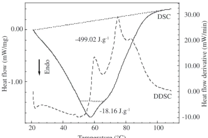
Figures 1 and 2 show the DSC and DDSC curves from bovine pericardium untreated and treated with glutaraldehyde cross-linked
respectively. The shrinkage of bovine pericardium is accompanied by the absorption of heat, giving rise to an endothermic peak on the DSC curve. The area under the peak is directly proportional to the enthalpy change ∆H (Enthalpy energy) while its height is a measure of the heat capacity (i.e. dH/dT) of the transition. The temperature of the transition can be defined as either the onset or the peak maximum.
We also observed that the peak temperature values used to report the shrinkage temperature generally displayed less deviation than the extrapolated onset value. According to Loke and Khor14, we recommend that the peak temperature value should be used as the shrinkage temperature.
The untreated bovine pericardium tissue presented an endother-mic peak that is directly related to the shrinkage of the material at 57 °C (Figure 1). The same thermal event was detected for the GA cross-linked bovine pericardium (Figure 2) but at a higher temperature (3 °C) when compared to the untreated tissue.
The ∆H involved during the denaturation event were different between the samples too. The GA cross-linked bovine pericardium showed a 5-fold increase on the ∆H value when compared to un-treated bovine pericardium. This difference could be explained due to the strong net structure originated between collagen fibers after the GA treatment.
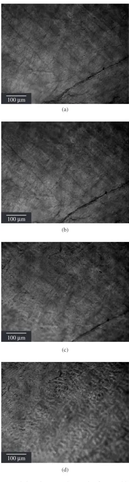
On Figures 3 and 4 we could observe the shrinkage of bovine pericardium tissue (treated and untreated) at approximately 60 °C through freeze-drying microscopy.
20 40 60 80 100
Temperature (°C) -1.00
0.00
Heat flo
w (mW/mg)
-18.16 J.g-1
-499.02 J.g-1
Endo Heat flo w deri v ati v e (mW/min) DSC DDSC 30.00 20.00 10.00 0.00 -10.00
Figure 1. DSC/DDSC curves obtained in dynamic N2 atmosphere
(50 mL.min-1) and heating rate 10 °C.min-1 of the untreated bovine
pericar-dium sample.
20 40 60 80 100
-1.11 J.g-1
-98.02 J.g-1
Temperature (°C) Heat flo w deri v ati v e (mW/min) Heat flo w (mW/mg) Endo DSC DDSC 30.00 20.00 10.00 0.00 - 10.00 1.00 0.00 -1.00 -2.00 -3.00
Figure 2. DSC/DDSC curves obtained in dynamic N2 atmosphere
(50 mL.min-1) and heating rate 10 °C.min-1 of GA cross-linked bovine
Vol. 10, No. 1, 2007 Evaluation of Shrinkage Temperature of Bovine Pericardium Tissue for Bioprosthetic Heart Valve Application by Differential Scanning Calorimetry and Freeze-drying Microscopy 3
Figure 3. Freeze-drying microscope photographs of GA bovine pericardium
at different temperatures: a) 30.5 °C; b) 40.8 °C; c) 49.9 °C; and d) 59.9 °C. Heating rate of 10 °C.min-1.
(a) 100Mm
(b) 100Mm
(c) 100Mm
(d) 100Mm
100Mm
(a)
100Mm
(b)
100Mm
(c)
Figure 4. Freeze-drying microscope photographs of untreated bovine
peri-cardium at different temperatures: a) 30.9 °C; b) 40.4 °C; c) 50.5 °C; and d) 60.5 °C. Heating rate of 10 °C.min-1.
4 Tattini Jr. et al. Materials Research
These temperatures are closely related to DSC results (n = 5, S.D. = 0.61). On both cases we could observe the visual shrinkage of bovine pericardium tissue when the temperature reached 60 °C (n = 5, S.D. = 0.87).
The results showed that shrinkage temperature of bovine pericar-dium tissue obtained from freeze-drying microscopy is quite similar to that obtained from DSC curves. The freeze-drying microscopy can be used as a very useful tool to determine the shrinkage temperature from biological tissues composed mainly of collagen fibers.
Acknowledgments
The authors acknowledge the financial assistance of CNPq, CAPES and FAPESP.
References
1. Nimni ME, Cheung D, Strates B, Kodama M, Sheikh K. Chemically modified collagen: A natural biomaterial for tissue replacement. Journal of Biomedical Materials Research. 1987; 21(6):741-771.
2. Renou JP, Bonnet M, Bielicki G, Rochdi A, Gatellier P. NMR study of collagen-water interactions. Biopolymers. 1994; 34(12):1615. 3. Bella J, Brodsky B, Berman HM. Crystal and Molecular
Struc-ture of a Collagen-like Peptide at 1.9Å Resolution. Science. 1994; 266(5182):75-81.
4. Jastrzebska M, Wrzalik R, Kocot A, Zalewska-Rejdak J, Cwalina BJ. Hy-dration of glutaraldehyde-fixed pericardium tissue: Raman spectroscopic study. Journal of Raman Spectroscopy. 2003; 34(6):424-431.
5. Flory PJ, Garrett RR. Phase transitions in collagen and gelatin systems. Journal of the American Chemical Society. 1958; 80:4836-4845.
6. Simionescu D, Simionescu A, Radu D. Mapping of glutaraldehyde-treated bovine pericardium and tissue selection for bioprosthetic heart valves. Journal of Biomedical Materials Research. 1993; 27(6):697-704. 7. Fisher J, Gorham SD, Howie AM, Wheatley DJ. Examination of fixative
penetration in glutaraldehydetreated bovine pericardium by stratigraphic analysis of shrinkage temperature measurements using differential scan-ning calorimetry. Life Support Systems. 1987; 5(3):189-193.
8. Bigi A, Cojazzi G, Roveri N, Koch MHJ. Differential scanning calorimetry and X-ray diffraction study of tendon collagen thermal denaturation. Inter-national Journal of Biological Macromolecules. 1987; 9(6):363-367. 9. Fortune F, Chantal B, Daniel H. A differential scanning calorimetry
analysis of the age-related changes in the thermal stability of rat skin collagen. Biochimica et Biophysica Acta. 1984; 791(2):205-211. 10. Ruijgrok JM, de Wijn JR, Boon ME, de Groot K. Shrinkage
tempera-ture profiles of glutaraldehyde tanned collagen in a multilayer diffusion model. Advanced Biomaterials. 1992; 10(Biomaterial Tissue inter-face):453-457.
11. Ignatieva NY, Lunin W, Averkiev SV, Maiorova AF, Bagratashvili VN, Sobol EN. DSC investigation of connective tissues treated by IR-laser radiation. Thermochimica Acta. 2004; 442(1-2):43-48.
12. Siapi E, Mavromoustakos T, Trandafir V, Albu B, Budrugeac P. The Use of Differential Scanning Calorimetry to Study the Effects of Gentamy-cin on Fibrous Collageneous Membranes. Thermochimica Acta. 2005; 425(1-2):165-171.
13. Lee JM, Pereira CA, Abdulla D, Naimark WA, Crawford I. A multi-sample denaturation temperature tester for collagenous biomaterials. Medical Engineering and Physics. 1995; 17(2):115-121.