Discovery of Mosquito Saliva MicroRNAs
during CHIKV Infection
Payal D. Maharaj1, Steven G. Widen2, Jing Huang1, Thomas G. Wood2, Saravanan Thangamani1,3,4*
1Department of Pathology, University of Texas Medical Branch, Galveston, Texas, United States of America,2Department of Biochemistry and Molecular Biology, University of Texas Medical Branch, Galveston, Texas, United States of America,3Institute for Human Infections and Immunity, University of Texas Medical Branch, Galveston, Texas, United States of America,4Center for Tropical Diseases, University of Texas Medical Branch, Galveston, Texas, United States of America
*sathanga@utmb.edu
Abstract
Mosquito borne pathogens are transmitted to humans via saliva during blood feeding. Mosquito saliva is a complex concoction of many secretory factors that modulate the feed-ing foci to enhance pathogen infection and establishment. Multiple salivary proteins/factors have been identified/characterized that enhance pathogen infection. Here, we describe, for the first time, the identification of exogenous microRNAs from mosquito saliva. MicroRNAs are short, 18–24 nucleotide, non-coding RNAs that regulate gene expression, and are generally intracellular. However, circulating miRNAs have been described from serum and saliva of humans. Exogenous miRNAs have not been reported from hematophagous arthro-pod saliva. We sought to identify miRNAs in the mosquito saliva and their role in Chikungu-nya virus (CHIKV) infection. Next generation sequencing was utilized to identify 103 exogenous miRNAs in mosquito saliva of which 31 miRNAs were previously unidentified and were designated novel. Several miRNAs that we have identified are expressed only in the CHIKV infected mosquitoes. Five of the saliva miRNAs were tested for their potential to regulated CHIKV infection, and our results demonstrate their functional role in the transmis-sion and establishment of infection during blood feeding on the host.
Author Summary
Mosquito saliva contains a complex repertoire of bioactive factors that are secreted into blood feeding site, the skin. Infected mosquitoes transmit pathogens to the host during feeding via saliva. The bioactive factors in mosquito saliva are responsible for modulating host hemostasis, immune defenses and pain/itch responses, and have been implicated to enhance pathogen infection and establishment in the host. In our efforts to identify and characterize salivary immunomodulators that enhance Chikungunya virus (CHIKV) transmission, we have discovered, for the first time, exogenous microRNA in mosquito saliva. MicroRNAs (miRNAs) are short, 18–24 nucleotide, non-coding RNAs that regulate gene expression. Short non-coding RNAs were extracted from the saliva of Chikungunya
OPEN ACCESS
Citation:Maharaj PD, Widen SG, Huang J, Wood TG, Thangamani S (2015) Discovery of Mosquito Saliva MicroRNAs during CHIKV Infection. PLoS Negl Trop Dis 9(1): e0003386. doi:10.1371/journal. pntd.0003386
Editor:Ken E. Olson, Colorado State University, UNITED STATES
Received:August 4, 2014
Accepted:October 30, 2014
Published:January 22, 2015
Copyright:© 2015 Maharaj et al. This is an open access article distributed under the terms of the Creative Commons Attribution License, which permits unrestricted use, distribution, and reproduction in any medium, provided the original author and source are credited.
Data Availability Statement:All relevant data are within the paper and its Supporting Information files.
Funding:This work is supported by the departmental start-up fund, and funds provided by the Institute for Human Infections and Immunity to ST. ST is partially supported by NIAID/NIH # AI097675. PDM is supported by NIAID/NIH T32 fellowship (#5T32AI007536-15). The funders had no role in study design, data collection and analysis, decision to publish, or preparation of the manuscript.
virus (CHIKV) infected and uninfectedAedes aegyptiandAedes albopictussaliva, and subjected to Illumina next generation sequencing. Bioinformatic analysis revealed the presence of miRNAs in the mosquito saliva. We have also identified several novel miRNAs that are expressed only during CHIKV infection. Though the functional roles of these miRNAs are yet to be established, ourin-vitrodata from testing 5 miRNAs demonstrate their role in the regulation of CHIKV infection. These miRNAs may play an important role in regulating the establishment of CHIKV infection in the mammalian host during blood feeding.
Introduction
Mosquitoes are a significant public health concern due to their ability to transmit a variety of emerging and reemerging arboviruses [1,2]. Chikungunya virus (CHIKV) is an excellent example of globalization of a mosquito borne disease, as evident from the CHIKV epidemics in the past seven years [3,4]. Chikungunya virus is anAlphavirusbelonging to the Togaviridae family and is transmitted predominantly byAedes aegyptiandAedes albopticus(www.cdc.gov/ ncidod/dvbid/Chikungunya).Aedes aegyptiandAe. albopictustransmit CHIKV during blood meal acquisition, along with the saliva the mosquitoes inject into the skin. The complex reper-toire of secretory proteins/factors in the mosquito saliva creates an immunologically compro-mised micro-environment that can have a profound effect on the transmission efficiency, pathogen establishment, and disease development [5–7]. The presence ofAe.aegyptisaliva causes a differential host immune response to CHIKV infections in mice [6], suppresses re-cruitment of T cells to the initial bite site thus enhancing West Nile virus dissemination [8], suppresses antimicrobial peptides and IFNs thus enhancing Dengue virus (DENV) infection in human keratinocytes [9] and modulates Rift Valley Fever virus pathogenicity in mice [10]. To that end, several saliva proteins have been isolated that are facilitators of mosquito feeding, modulators of skin immunity and regulators of virus transmission and dissemination in the vertebrate host [11]. For example, the aegyptin protein isolated fromAe.aegyptisaliva aids in blood feeding [12]. Another isolated putative 34 kDa protein modulates DENV infection in human keratinocytes via immunomodulation [13] and serine proteases inAe.aegyptisaliva fa-cilitate DENV dissemination in mice [11]. These studies provide important information about the complex roles of salivary proteins in virus-host interactions however, other components of saliva and their functions have not been identified or characterized.
MicroRNAs (miRNAs) are short 18–24 nucleotide non-coding RNAs that regulate gene ex-pression post-transcriptionally by binding to complementary regions mainly in the 30UTRs of targeted messenger RNAs. MicroRNA expression patterns have been profiled in mosquitoes of medical importance such asAnopheles gambiae[14],Anopheles stephensi [15], Ae.aegypti[16],
Ae. albopictus[17],Culex quinquefasciatus[18] andAnopheles anthropophagus[19]. Function-al studies of these mosquito miRNAs have demonstrated their role in blood digestion and egg development inAe. aegypti[20], blood-meal induced miRNA expression for regulation of im-mune genes inAe. aegypti[21] andAe. albopictus[22], altered patterns of expression inAn. ste-phensipost-blood feeding [23] and growth-stage specific expression inAn. anthropophagus
[19]. These miRNA expression profiles are altered in mosquitoes infected with parasites. For instance, the obligate endosymbiont,Wolbachia pipientis, regulates specific miRNA levels for maintenance of its life cycle inAe.aegyptimosquitoes [24,25]. MicroRNA levels were also manipulated inAn.stephensi[23] andAn. gambiae[14] infected withPlasmodiumand in
While miRNAs have been detected and profiled from mosquito cell lines and mosquitoes, miRNA profiles in mosquito saliva have not been investigated. In the present study, we sought to detect and identify miRNAs in the saliva ofAe.aegyptiandAe. albopictusmosquitoes via deep se-quencing. Furthermore, to investigate the effect of CHIKV infection on saliva miRNA expression profiles, deep sequencing was also performed on CHIKV-infectedAe.aegyptiandAe.albopictus
saliva. A total of 103 mature miRNAs were discovered inAe.aegyptiandAe.albopictussaliva. Sev-enty-two of the detected miRNAs aligned with previously identified miRNAs while 31 were po-tential novel miRNAs. Furthermore, 59 and 30 known miRNAs were upregulated inAe.aegypti
andAe. albopictusCHIKV-infected saliva respectively indicating the possible functional impor-tance of these miRNAs in CHIKV dissemination and transmission in the host.
Methods
Cells and viruses
African green monkey kidney (Vero) cells were maintained with Dulbecco’s Modified Eagle Medium (DMEM; Gibco, Carlsbad, CA) and baby hamster kidney (BHK-21) cells were main-tained with Modified Eagle’s Medium (MEM; Gibco, Carlsbad, CA) supplemented with 10% fetal bovine serum (FBS; Gibco, Carlsbad, CA) and 5% penicillin/streptomycin (P/S; 100U/mL/ 100μg/mL, Gibco, Carlsbad, CA) at 37°C with 5% CO2. TheAedes albopictus(C6/ 36) cell line
was maintained in Leibowitz’s media (Invitrogen) supplemented with 10% FBS and 5% P/S at 28°C without CO2. TheAedes Ae.aegypti(AAG-2) cell line was maintained in Schneider’s
In-sect Cell Media (Invitrogen) supplemented with 10% FBS and 5% P/S at 28°C without CO2.
The infectious clone, CHIKV-LR 50GFP (CHIKV), used in this study has been described and characterized previously [27], and was provided by Dr. Stephen Higgs.
Mosquitoes
TheAedes aegypti(Higgs White eye) strain and theAedes albopictus(La Reunion) strain are well characterized and competent vectors for CHIKV and CHIK-LR 50GFP viruses [27]. Mos-quitoes were reared as previously described [16] within the UTMB insectary services core facil-ity. Both species of mosquitoes were maintained at 28°C at a 14:10 hour (L: D) photoperiod with 10% sucrose solution providedad libitum.Three to five day old females were used for all intrathoracic inoculations.
Intrathoracic inoculations
Three to 5 day-oldAe.aegyptiandAe. albopictusmosquitoes were cold-anesthetized and intra-thoracically inoculated with an approximately 0.1μL inoculum of CHIKV-LR 50GFP: 4.6
TCID50/ mL. One hundred mosquitoes were inoculated per species after which inoculated
mosquitoes were placed in 1 pint cartons in a 28°C incubator with 10% sucrose suppliedad li-bitumand a 14:10 hour (L:D) photoperiod. After 10 days post-infection (d.p.i), 50 infected and 50 uninfected mosquitoes were collected for each species, cold-anesthetized and saliva was col-lected. Briefly, saliva was collected by inserting each mosquito proboscis in a capillary tube with approximately 10μL of immersion oil and letting each mosquito salivate for 30 minutes at
room temperature. Saliva were pooled according to infection status and species of mosquito, mixed with 250μL of DMEM and stored at−80°C until further processing.
RNA extractions
mosquito saliva samples for virus inactivation and incubated overnight at−20°C. After 24 hours post-inactivation, RNA samples were thawed and 150μL of chloroform was added to
each tube and shaken vigorously for 30 seconds. The samples were centrifuged for 15 minutes at 10000 × g at 4°C after which the clear, top layer was transferred to a new tube for total RNA and miRNA extraction using the Qiagen RNeasy extraction kit and Qiagen microRNA extrac-tion kit respectively.
Next generation sequencing
The Illumina TruSeq SmallRNA kit was used to prepare libraries of the microRNA samples. Briefly, short unique adapters were ligated to the 50and 30ends of short RNAs. Reverse-tran-scriptase and PCR were used to add the full length adapters required for Illumina sequencing, followed by gel purification of the correct size templates. The samples were tracked using
“index tags”incorporated into the adapters. Library quality was evaluated using an Agilent DNA-1000 chip on an Agilent 2100 Bioanalyzer. Quantification of library DNA templates was performed using qPCR and a known-size reference standard.
Sequence analysis
Cluster formation of the library DNA templates was performed using the TruSeq PE Cluster Kit v3 (Illumina) and the Illumina cBot workstation using conditions recommended by the manufacturer. Template input was adjusted to obtain a cluster density of 700–850 K/mm2. 50 base sequencing by synthesis was performed using TruSeq SBS kit v3 (Illumina) on an Illumina HiSeq 1000 using protocols defined by the manufacturer.
Data analysis
The miRDeep2 software package [28] identified potential miRNA precursors by scanning for pileups of short reads in the genome alignment data. The region surrounding the pileup was excised computationally and analyzed for miRNA features. The structure of the potential pre-cursor RNA was analyzed by RNAfold to determine the predicted secondary structure of the region and that structure was compared to typical miRNA precursor structures. If a likely structure was found, reads mapped to the precursor were counted and analyzed for the pres-ence of mature and star miRNA sequpres-ences and then compared to the level of background se-quences. The miRDeep2 algorithm used these results to score the likelihood that the predicted miRNA was real. The number of reads for each unique sequence was tracked. Following the miRDeep2 workflow the microRNAs were then compared against known microRNAs from the miRBase database (Version 20) withAedes aegypti(AaegL1) as the reference species and
Anopheles gambiae(AgamP3) as a related species. As theAe.albopictusgenome sequence was unavailable and miRNAs are highly conserved between species, reads fromAe.albopictussaliva were compared to knownAe. aegyptiandAn. gambiaemiRNAs from miRBase database (Ver-sion 20). Novel microRNAs were identified by mapping the reads to theAe. aegyptigenome (AaegL2 from VectorBase VB-2014-02). Finally a table of known and potentially novel miR-NAs was output with mapped read counts for each. Relative abundance of miRmiR-NAs in CHIKV infected samples were calculated by normalizing the data by tags per million (TPM) reads of total RNA as described previously [29].
MicroRNA inhibition assay
MicroRNA inhibitors were designed based on the sequences of the following select
inhibitor with random sequence, Scramble, that was designed based on a previous study [30]. All miRNA inhibitors (MIR-12, MIR-125, MIR-184, MIR375 and MIR-2490) were synthesized by Integrated DNA Technologies©. The microRNAs that were chosen for this miRNA inhibi-tion study were selected based on relative abundance levels of CHIKV- infected saliva, as well as, previous reports indicating their importance in modulating DENV andWolbachia replica-tion [21,24,31,32]. However, they have not been studied in the context of CHIKV replication. Additionally, these miRNAs have been identified and characterized in both AAG-2 and C6/36 mosquito cell lines [21,24,25,32,33]. Baby hamster kidney cells were used for this study as they are a fibroblast cell line and CHIKV targets and replicates in fibroblast cells in a natural infec-tion [34,35]. The cell lines, AAG-2, BHK-21 and C6/36 cells, were grown to confluency and transfected in triplicate with 100 nanograms of each miRNA inhibitor via Cellfectin transfec-tion reagent. As a control, cells were mock transfected without template. Cells were re-trans-fected at 48 hours post-transfection and were inre-trans-fected with CHIKV at a multiplicity of infection of 0.01 at 72 hours after initial transfection. As a control, mock transfected cells were also infected with CHIKV. Daily timepoints of 50μL were collected from each replicate until
72 hours post-infection, added to 450μL of diluent and stored at−80°C until further process-ing. A standard plaque assay on Vero cells was used to determine CHIKV titer at each time-point as previously described [36].
Statistical analysis
A 2-tailed student’s T-test (α0.05) was used to analyze the significance of viral titer differences
in the miRNA inhibition assay at each time point.
Results
Small RNA sequencing of
Aedes
spp. saliva
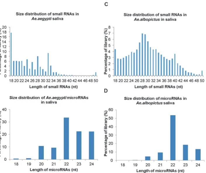
Small RNAs were extracted from the saliva of uninfectedAe.aegyptiandAe.albopictus mosqui-toes andAe.aegyptiandAe. albopictusmosquitoes infected with CHIKV. These small RNAs were then sequenced via Illumina-based high-throughput sequencing in order to identify small non-coding RNAs. A total of 14 × 106small RNAs were detected inAe.aegyptisaliva with a predominant size distribution of 18–33 nucleotides (nt) (Fig. 1A). Out of these, 18–24mers represented 56% of the library where 18mers represented a higher percentage of the library at 19% (Fig. 1A). After these RNAs were aligned with theAe.aegyptigenome, 43% of theAe. aegyptilibrary was composed of 18–24mers with 22mers exhibiting the highest frequency of reads (Fig. 1B). In comparison, small RNA sequencing ofAe.albopictussaliva, detected 3 × 10⁶ small RNAs and demonstrated a larger size range of 18–40 nts out of which 21% were repre-sented by 18–24mers (Fig. 1C).Ae.albopictussaliva small miRNAs were matched to knownAe. aegyptiandAn.gambiaemiRNAs and demonstrated a 48.8% representation of 18–24mers with 22mers having the highest frequency of reads (Fig. 1D).
these data provide strong evidence for the presence of mature insect miRNAs inAe.aegypti and Ae.albopictussaliva.
Identification of
Ae.aegypti
saliva miRNAs
After aligning the sequencing reads fromAe.aegyptisaliva to theAe.aegyptimiRNA database, 72 distinct known miRNAs were identified in both uninfected and CHIKV-infected mosquito saliva (Table 1). In uninfectedAe.aegyptisaliva, a total of 298283 reads were obtained with 283197 reads aligning with known miRNAs and 15086 reads that were novel miRNAs. In com-parison, the total read count in CHIKV-infectedAe.aegyptisaliva was 305894 reads with 251277 known miRNA reads and 54617 novel miRNA reads). The highest expressing miRNA in uninfectedAe.aegyptisaliva was aae-mir-281-2-5p at 80151 reads. The other highly express-ed miRNAs in uninfectexpress-edAe.aegyptisaliva were aae-mir-281 (56394), aae-mir-2940 (25307),
Figure 1. Identification of microRNAs inAedesspp. saliva.Aedes aegyptiandAedes albopictuswere intra-thoracically infected with Chikungunya virus. At 10 days post infection, saliva was collected from both infected and uninfected mosquitoes. Small RNAs were extracted from the saliva and subjected to deep sequencing, small RNA libraries were created and mapped toAe.aegyptiandAn.gambiaemiRNA databases. Figure a) size distribution and percentages of small RNAs inAe.aegyptisaliva, b) percentages of 18–24 nucleotide microRNAs inAe.aegyptisaliva library, c) size distribution and percentages of small RNAs inAe. albopictussaliva, d) percentages of 18–24 nucleotide microRNAs inAe. albopictussaliva library.
Table 1.Read counts of individual microRNAs detected in uninfected and CHIKV-infectedAe.aegyptisaliva.
MicroRNA Read counts Difference between infected and uninfected
Infected Uninfected Difference Fold Difference
aae-mir-8 50004 47613 2391 1.2
aae-mir-2940 21514 25307 −3793 1.0
aae-mir-263a 20584 9084 11500 2.6
aae-bantam 18002 9969 8033 2.0
aae-mir-125 15735 5863 9872 3.0
aae-mir-281 5833 56394 −50561 0.1
aae-mir-281-2-5p 9818 80151 −70333 0.1
aae-mir-100 13160 4309 8851 3.4
aae-mir-14 12958 5389 7569 2.7
aae-mir-285 10006 31 9975 363.8
aae-mir-276-1 9384 2584 6800 4.1
aae-mir-276-2 9291 2570 6721 4.1
aae-mir-317-1 6263 3367 2896 2.1
aae-mir-317-2 6263 3367 2896 2.1
aae-mir-184 5080 10105 −5025 0.6
aae-mir-12 4288 537 3751 9.0
aae-mir-277 3210 798 2412 4.5
aae-mir-10 2815 2035 780 1.6
aae-mir-279 2315 587 1728 4.4
aae-mir-2a 1695 580 1115 3.3
aae-mir-11 1650 2309 −659 0.8
aae-mir-1891-2 1576 1078 498 1.6
aae-mir-1891-1 1576 1078 498 1.6
aae-mir-1889 1195 298 897 4.5
aae-mir-2c 1133 313 820 4.1
aae-mir-210 1080 28 1052 43.5
aae-mir-34 1036 790 246 1.5
aae-mir-2b 905 219 686 4.7
aae-mir-92a 891 227 664 4.4
aae-mir-306 866 419 447 2.3
aae-mir-927 771 116 655 7.5
aae-mir-71 764 352 412 2.4
aae-mir-275 735 271 464 3.1
aae-mir-92b 700 430 270 1.8
aae-mir-996 693 169 524 4.6
aae-let-7 665 616 49 1.22
aae-mir-305 575 111 464 5.8
aae-mir-970 543 259 284 2.4
aae-mir-957 533 43 490 14.0
aae-mir-999 478 144 334 3.7
aae-mir-252 410 340 70 1.4
aae-mir-13 409 67 342 6.9
aae-mir-9c 332 299 33 1.3
aae-mir-980 330 18 312 20.7
aae-mir-133 277 11 266 28.4
mir-8 (47613), mir-184 (10105) mir-bantam (9969), mir-263a (9084) and aae-mir125 (5863) (Table 1). Similarly, the highest expressing miRNAs in CHIKV-infectedAe. aegyptisaliva were 8 (50004), 2940 (21514), 263a (20584), bantam (18002), 125 (15735), 100 (13160), 14 (12958) and aae-mir-285 (10006) (Table 1).
Detection of novel
Ae.aegypti
saliva miRNAs
Thirty-one novel mature miRNAs were detected fromAe.aegyptisaliva after the predicted miRNAs were compared to theAe.aegyptimiRNA database and AaegL2 (Table 2). The highest ex-pressed novel miRNA was aae-mir-143 with a count of 4275 and with a seed sequence match to aga-mir-14 (Table 2). Aae-mir-249, aae-mir-80 and aae-mir-5 were also highly expressed novel miRNAs in uninfectedAe.aegyptisaliva with counts of 5385, 3773 and 2566 respectively (Table 2).
Identification of
Ae.albopictus
saliva miRNAs
A total of 43 miRNAs were identified inAe.albopictussaliva. In uninfectedAe.albopictussaliva, a total of 12075 reads were obtained with 9180 reads aligning with known miRNAs and 4741
Table 1. (Continued)
MicroRNA Read counts Difference between infected and uninfected
Infected Uninfected Difference Fold Difference
aae-mir-1000-2 226 2 224 127.4
aae-mir-1000-1 226 2 224 127.4
aae-mir-998 211 140 71 1.7
aae-mir-190 190 36 154 5.9
aae-mir-308 172 44 128 4.4
aae-mir-307 161 0 161 161.0
aae-mir-315 146 10 136 16.5
aae-mir-263b 133 7 126 21.4
aae-mir-9a-2 112 67 45 1.9
aae-mir-9a-1 112 67 45 1.9
aae-mir-1890 109 33 76 3.7
aae-mir-932 109 50 59 2.5
aae-mir-2941-2 104 177 −73 0.7
aae-mir-2941-1 100 168 −68 0.7
aae-mir-87 98 34 64 3.2
aae-mir-2945 96 21 75 5.2
aae-mir-278 91 80 11 1.3
aae-mir-33 86 11 75 8.8
aae-mir-31 74 11 63 7.6
aae-mir-981 73 10 63 8.2
aae-mir-989 72 616 −544 0.1
aae-mir-2946 68 234 −166 0.3
aae-mir-375 54 189 −135 0.3
aae-mir-283 49 225 −176 0.2
aae-mir-9b 34 64 −30 0.6
aae-mir-1174 18 158 −140 0.1
aae-mir-1175 12 96 −84 0.1
reads that were novel miRNAs (Table 3). In contrast, the total read count was 2-fold higher in CHIKV-infectedAe.albopictussaliva with a total count of 32593 reads with 16050 known miRNA reads and 16543 novel miRNA reads. Twenty-eight known miRNAs were identified in
Ae.albopictussaliva (Table 3). The highest expressed miRNA in uninfectedAe.albopictussaliva was aae-mir-8 with a count of 12874 followed by aae-mir-2940 (2574), aae-mir-bantam (2127) and aae-mir-125 (2132) (Table 3). Highest read counts in CHIKV-infectedAe.albopictussaliva were from aae-mir-125 (4333), aae-mir-263a (4293), aae-mir-8 (2609), aae-mir-184 (2332) and aae-mir-100 (2255) (Table 3). With the exception of aae-mir-8, these miRNAs were upre-gulated at least 1.3-fold or higher in comparison with uninfected saliva (Table 3).
Identification of novel miRNAs in
Ae.albopictus
saliva
Twenty-four novel, mature miRNAs were detected inAe.albopictussaliva (Table 4). The high-est expressing miRNA in uninfectedAe.albopictussaliva was aal-mir-43b which had a read
Table 2.Read counts of individual novel microRNAs detected in uninfected and CHIKV-infectedAe.aegyptisaliva.
Assigned Name Consensus sequence Read Counts Difference between infected and
uninfected
Infected Uninfected Difference Fold Difference
aae-mir-143 aacccguagauccgaacuugug 13129 4275 8854 0.8
aae-mir-249 ucagucuuuuucucucuccu 12951 5385 7566 0.7
aae-mir-5 uaggaacuucauaccgugcucu 9219 2566 6653 1.0
aae-mir-229 ucauaagacacacgcggcuau 544 259 285 0.6
aae-mir-778 uugguccccuucaaccagcugu 278 11 267 7.0
aae-mir-620 auuagaauguggaaucuguuuu 51 3 48 4.7
aae-mir-3069 uuuguucguuuggcucgagu 54 188 −134 0.1
aae-mir744 caucacagucugaguucuugcu 1451 1783 −332 0.2
aae-mir-115 ugugaugugacguagugguac 71 616 −545 0.0
aae-mir-23 uagcaccauucgaaaucaguac 10021 0 10021 10021.0
aae-mir-576 ggggauguagcucagugguagag 2033 0 2033 2033.0
aae-mir-320 uuucggauauguuuuagaaauuc 1262 0 1262 1262.0
aae-mir-214 uucccggacgagccccca 606 433 173 0.4
aae-mir-402 uucccggacgagccccca 606 433 173 0.4
aae-mir-65 ugcacacgacucgaugggauagac 397 0 397 397.0
aae-mir-341 gcaggaucguaggaggcu 294 0 294 294.0
aae-mir-3 agggucggagguucgaauccc 250 0 250 250.0
aae-mir-3798 auauuguccugucacagcag 226 0 226 226.0
aae-mir-187 auauuguccugucacagcag 226 0 226 160.0
aae-mir-242 caucgaucgcgcaccuga 160 0 160 139.0
aae-mir-309 guaggccggcggaaacuacuugc 139 21 118 1.8
aae-mir-210 uuuaccauuucaagaugacc 129 0 129 129.0
aae-mir-1571 gagaggccuguguaaucu 88 0 88 88.0
aae-mir-1247 guucgacucccagucggu 81 12 69 1.9
aae-mir-40 uugcguugauuaaguccc 81 0 81 81.0
aae-mir-843a guccugucacggucgcca 74 0 74 81.0
aae-mir-117 uagcagaauccugaguaggac 73 0 73 74.0
aae-mir-360 gugagcaaauuuucaggugugu 67 0 67 73.0
aae-mir-359 guaacugacgcugaggag 56 0 56 67.0
aae-mir-80 auucucuguucguccacca 0 3773 −3773 0.0
aae-mir-109 aucacgucggggucacca 0 227 −227 0.0
Table 3.Read counts of individual microRNAs detected in uninfected and CHIKV-infectedAe.albopictussaliva.
MicroRNAs Read Counts Difference between Infected and Uninfected
Infected Uninfected Difference Fold difference
aae-mir-125 4333 2132 2201 2.2
aae-mir-263a 4293 1343 2950 3.4
aae-mir-8 2609 12874 −10265 0.2
aae-mir-184 2332 1885 447 1.3
aae-mir-100 2255 1204 1051 2.0
aae-mir-2940 1923 2574 −651 0.8
aae-mir-281 377 210 167 1.9
aae-mir-281-2-5p 752 292 460 2.7
aae-bantam 1116 2127 −1011 0.6
aae-mir-276-1 1046 482 564 2.3
aae-mir-276-2 1038 482 556 2.3
aae-mir-14 1032 731 301 1.5
aae-mir-10 887 153 734 6.2
aae-mir-927 795 151 644 5.6
aae-mir-317-1 632 844 −212 0.8
aae-mir-317-2 632 844 −212 0.8
aae-mir-277 430 212 218 2.2
aae-let-7 423 195 228 2.3
aae-mir-999 409 146 263 3.0
aae-mir-11 385 381 4 1.1
aae-mir-957 361 107 254 3.6
aae-mir-34 339 619 −280 0.6
aae-mir-92b 304 45 259 7.2
aae-mir-275 276 109 167 2.7
aae-mir-315 273 16 257 18.1
aae-mir-2a 212 171 41 1.3
aae-mir-2c 182 143 39 1.4
aae-mir-2b 148 147 1 1.069
aae-mir-12 145 132 13 1.2
aae-mir-1891-2 120 41 79 3.1
aae-mir-1891-1 120 41 79 3.1
aae-mir-133 109 25 84 4.6
aae-mir-252 105 202 −97 0.6
aae-mir-970 98 95 3 1.1
aae-mir-306 88 79 9 1.2
aae-mir-71 85 119 −34 0.8
aae-mir-279 46 131 −85 0.4
aae-mir-190 40 54 −14 0.8
aae-mir-305 35 55 −20 0.7
aae-mir-996 24 77 −53 0.3
aae-mir-210 18 808 −790 0.0
aae-mir-932 10 141 −131 0.1
aae-mir-285 1 114 −113 0.0
count of 2134 followed by aal-mir-13 and aal-mir-43a at 1874 and 1200 reads respectively (Table 4). Highly expressed miRNAs in CHIKV-infectedAe.albopictussaliva were aal-mir-43b, aal-mir-43a, aal-mir-413a, aal-mir-5 and aal-mir-249 with read counts of 4339, 2253, 1643, 1035 and 1032 respectively (Table 4).
Relative abundance of miRNAs in CHIKV-infected saliva
In comparison with uninfectedAe.aegyptisaliva, CHIKV-infected saliva miRNA reads were slightly lower out of which 251277 reads corresponded with previously identifiedAe. aegypti
miRNAs (Table 1) and 54617 reads were novel miRNAs (Table 2). The highly expressed miRNAs, aae-mir-bantam, aae-mir-263a, aae-mir-125 and aae-mir-285 were upregulated in CHIKV-infectedAe.aegyptisaliva with counts of 18002 (2.0-fold), 20584 (2.6-fold), 15735 (3.0-fold) and 10006 (>100-fold) when compared with uninfected read counts (Table 1). The novel miRNAs also did not demonstrate a significant total fold difference between the unin-fected and inunin-fected saliva total read counts but individual miRNAs demonstrated differential expression (Table 2). In comparison with uninfected reads, highly expressed mir-23, aae-mir-576 and aae-mir-320 were upregulated in CHIKV-infectedAe.aegyptisaliva (Table 2) however aae-mir-80 was highly expressed in uninfected saliva (3773) but undetected in infected saliva.
Table 4.Read counts of individual novel microRNAs detected in uninfected and CHIKV-infectedAe.albopictussaliva.
Assigned Name Consensus sequence Read Counts Difference between infected and
uninfected
Infected Uninfected Difference Fold Difference
aal-mir-43a aacccguagauccgaacuugug 2253 1200 1053 0.88
aal-mir-5 uaggaacuucauaccgugcucu 1035 482 553 1.00
aal-mir-249 ucagucuuuuucucucuccu 1032 729 303 0.66
aal-mir-778 uugguccccuucaaccagcugu 108 25 83 2.02
aal-mir-774 caucacagucugaguucuugcu 378 336 42 0.53
aal-mir-229 ucauaagacacacgcggcuau 98 95 3 0.48
aal-mir-43b ucccugagacccuaacuuguga 4339 2134 2205 0.95
aal-mir-413a guucgaauccuguucugg 1643 0 1643 1643.00
aal-mir-305 guucgauucccguucgag 984 0 984 984.00
aal-mir-6 guucgaauccugguaaga 892 0 892 892.00
aal-mir-47 ggggauguagcucagugguagag 751 0 751 751.00
aal-mir-157 uguggcguaguugguaac 398 0 398 398.00
aal-mir-28 guggagcaguauggaagc 373 0 373 373.00
aal-mir-69 guggcguaauugguagac 267 0 267 267.00
aal-mir-137 aggucguggguucgaacccc 232 0 232 232.00
aal-mir-2308 ggucggugguucgaaucc 119 0 119 119.00
aal-mir-214 uucccggacgagccccca 117 201 −84 0.27
aal-mir-309 guaggccggcggaaacuacuugc 82 36 46 1.07
aal-mir-62 acgucaaaucaucauguc 80 103 −23 0.36
aal-mir-143 accccugaaggaguuuucggag 71 0 71 71.00
aal-mir-127 guagccagaggaagagaaa 61 0 61 61.00
aal-mir-446 ucaaaucuugucgcgccg 53 0 53 53.00
aal-mir-408 guucgaauccuagucggga 52 0 52 52.00
aal-mir-13 uggacggagaacugauaagggc 0 1874 −1874 0.00
Similar toAe.aegypti, aae-mir-8 was also highly expressed at 12874 reads in uninfectedAe. albopictussaliva but in contrast toAe.aegypti,was detectable in CHIKV-infectedAe.albopictus
saliva (Table 3). Aae-mir-2940 was also downregulated (0.8-fold) in CHIKV-infected
Ae.albopictussaliva whereas aae-mir-125 (2.2-fold), aae-mir-263a (3.4-fold), aae-mir-184 (1.3-fold) and aae-mir-100 (2.0-(1.3-fold) were all upregulated in comparison with uninfected Ae.albo-pictussaliva. The highly expressed novel miRNAs, aal-mir-43b (1-fold), aal-mir-43a (0.9-fold), aal-mir-413a (>100-fold), aal-mir-5 (1-fold) and aal-mir-249 (0.7-fold) were upregulated in CHIKV-infectedAe.albopictussaliva in comparison with uninfectedAe.albopictussaliva with the exception of aal-mir-413a, which was not detected in uninfected saliva at all (Table 4). MicroRNA aal-mir-13 was highly expressed in uninfectedAe.albopictussaliva but was unde-tected in CHIKV-infected saliva.
Aedes
spp. saliva miRNAs modulate viral replication in mosquito and
mammalian cells
In order to investigate the role of saliva miRNAs in the CHIKV replication, miRNA inhibitors were designed and transfected into mosquito (AAG-2 and C3/36) and mammalian (BHK-21) cells to profile CHIKV replication over time. In all three cell lines, there were no significant dif-ferences in CHIKV replication in non-transfected cells (CHIKV only), mock transfected cells (Transfected +CHIKV) and Scramble transfected cells. CHIKV replication in Scramble control cells peaked at 6.62 ± 0.03 log10PFU/mL at 48 hours post infection (h.p.i.) in AAG-2 cells. In
Scramble BHK-21 cells and C6/36 cells, CHIKV peaked at 48 h.p.i. with a titer of 7.41 ± 0.15 and 8.88 ± 0.21 log10PFU/ mL, respectively.AAG-2 cells:At 24- 48 h.p.i., CHIKV titers were
significantly lower (p<0.05) in cells transfected with MIR-12 (Fig. 2A), MIR-125 (Fig. 2B) and mir-2490 (Fig. 2E) than in Scramble cells. At 48 h.p.i., CHIKV titers were significantly lower (p<0.05) in cells transfected with MIR-184 (Fig. 2C) and MIR-375 (Fig. 2D). CHIKV titers peaked at 72 h.p.i. in AAG-2 cells transfected with miRNA inhibitors demonstrating an attenuated growth pattern compared to Scramble control cells where CHIKV titers peaked at 48 h.p.i.BHK-21 cells:Cells transfected with MIR-12 and MIR-125 did not exhibit any significant differences in CHIKV titers at any timepoint when compared with Scramble control cells. At 24 h.p.i., MIR-184 inhibited cells showed a significantly lower CHIKV titer of 7.16 ± 0.12 log10PFU/mL in comparison to 7.5 ± 1.2 log10PFU/mL (p<0.05). CHIKV titers
were significantly lower (p<0.05) in MIR-375 and MIR-2940 inhibited cells at both 24 and 48 h.p.i. No significant viral titer differences were observed at 72 h.p.i. for any miRNA inhibitor.C6/36 cells:No significant differences were observed in titers for any miRNA inhibitor with the exception of MIR-184. At 24 and 48 h.p.i., CHIKV titers were 7.49 ± 0.29 and 8.40 ± 0.20 log10PFU/ mL respectively, which was significantly lower in comparison with
Scramble control cells at those timepoints (p<0.05).
Discussion
MicroRNAs are generally considered to be intra-cellular. However circulating microRNAs have also been identified from human serum, saliva and other biofluids [37–40] but have not been described before in mosquito saliva. In the present study, mature microRNAs were dis-covered in the saliva of two species ofAedesspp. mosquitoes,Ae.aegyptiandAe.albopictus. To our knowledge, this is the first documentation of the presence of exogenous miRNAs in mos-quito saliva where at least 70% of these miRNAs were found within theAe.aegyptiand related
Similar miRNAs were identified in both species of mosquitoes which corresponds with previ-ous studies withAe.albopictusandAe.aegyptimosquito miRNAs [18] thus indicating the evo-lutionary pressure for miRNA sequence conservation and also potential multiple functions of each miRNA. Interestingly, the same miRNAs were highly expressed in bothAe.albopictusand
Ae.aegyptisaliva and these include aae-mir-8, aae-mir-2940, aae-mir-263a, aae-mir-bantam, aae-mir-125, aae-mir-184, aae-mir-281and aae-mir-100 all of which have been identified in
Aedesspp. before [18].
Recent studies have shown exosomes to be the extracellular vesicles that transport miRNAs in biofluids like saliva and serum [37,41,42]. Microvesicles, such as exosomes, play a major role in intercellular communication and has been shown to transfer functional and intact proteins, lipids and nuclei acids between cells. The argonaute family of proteins has also been shown to transport miRNAs via serum [43]. Studies with Epstein-Barr virus (EBV) have demonstrated infected B cells releasing exosomes that contain EBV-miRNAs [44]. Therefore it is possible that exosomes or argonaute proteins are transporting miRNAs from the mosquito salivary glands to the bite site via saliva to potentially modulate viral replication.
The miR-184 was highly expressed in both species. High expression of miR-184 has been reported in other insects as well [18,45] where miR-184 is ubiquitously expressed in varying levels at all stages ofDrosophiladevelopment [31]. In comparison with uninfected saliva, aae-mir-184 was highly expressed but downregulated in CHIKV infectedAe.aegyptisaliva and upregulated in infectedAe.albopictussaliva. In our miRNA inhibition assays, CHIKV replica-tion was inhibited in AAG-2 and BHK-21 cells at 48 and 24 h.p.i but not at 72 h.p.i. This corresponds with a previous study, where upregulation of miR-184 was observed inS.
frugiperdacells after baculovirus infection at 24 h.p.i. but downregulated by 72 h.p.i. and could potentially explain the lack of CHIKV inhibition in our study at 72 h.p.i. [31]. Significant inhi-bition of CHIKV replication in both AAG-2 and BHK-21 cells also indicates the important role of miR-184 in arboviral infections in both mosquito and mammalian host. MicroRNA-184 has also been shown to increase in response to interleukin-22 (IL-22), a proinflammatory cytokine associated with inflammatory skin disorders, thereby reducing expression of Argo-naute-2 (AGO 2) protein in human keratinocytes [46]. The AGO 2 protein recognizes and cleaves targeted dsRNA as part of the RNA-induced silencing complex (RISC) in the RNA in-terference (RNAi) pathway. As the RNAi pathway is an important defense pathway against viral infections in several mosquito species [47–50] differential expression of aae-miR-184 post-infection in mosquitoes could modulate AGO 2 levels thereby regulating viral replication at the initial site of infection. The C6/36 cell line has a dysfunctional RNAi pathway where Dicer-2, part of the RISC that associates with AGO 2, is lacking [51,52]. In the present study, CHIKV replication was inhibited at 24 and 48 h.p.i. in C6/36 cells suggesting a potentially more complex role of miR-184 in the RISC.
The highly expressed aae-miRNA-125 and aae-miR-100 were both upregulated in CHIKV-infectedAe.aegyptiandAe.albopictussaliva. MicroRNA-125, a homolog ofDrosophila miR-let-7, is expressed in specific developmental stages of Drosophila [53]. MicroRNA-125, miR-100 and miR-let-7 are part of the same primary transcript and originate from a common genomic locus inDrosophila[54]. Additionally, clustering of the paralogs of these miRNAs also exists in the mouse genome suggesting multiple roles of these miRNAs across different species [55,56]. Target sites for mir-125a and mir-125b have been predicted to be within the 30UTR of both
Figure 2. Saliva microRNAs regulate CHIKV replication in mosquito and mammalian cells.Mosquito (AAG-2 and C6/36) and mammalian (BHK-21) cells were transfected with miRNA inhibitors, a) MIR-12, b) MIR-125, c) MIR-184, d) MIR-375 and e) MIR-2940, and then infected with CHIKV at 72 hours post-transfection. Supernatant was collected daily for 72 hours and viral titers for each timepoint were determined via standard plaque assay on Vero cells.
mouse and human TNF-αtranscripts [57] and miR-125b levels either increase or decrease in
response to TNF-αstimulated macrophages bothin vitroandin vivo[57]. Additionally,
down-regulation of TNFAIP results in increased levels of NF-κB which contributes to increased
im-mune cell activity [58]. Therefore, both aae-mir-125 and aae-mir-100 could be contributing to regulating immune cell activity at the bite site in order to influence CHIKV replication.
The aae-miR-375 has been shown to be important in DENV replication [21] and was down-regulated at least 34-fold inAe.aegyptiand undetected inAe.albopictusin the present study. Predicted target sites for miR-375 include theREL1andprohibitin, the 50UTR ofcactus, the 30UTR ofDEAD box ATP-dependent RNA helicase, a hypothetical protein and the coding re-gion ofkinesinall of which showed significant modulation in response toAe.aegypti mosqui-toes injected with aae-miR-375 mimics [21].CactusandREL1regulate the Toll immune pathway and were differentially expressed in response to aae-miR-375 mimics inAe.aegypti
mosquitoes and AAG-2 cells [21]. Furthermore, presence of aae-miR-375 mimics increased DENV-2 levels in AAG-2 cells which corresponded with our miRNA inhibition assay where a decrease in CHIKV replication was observed in AAG-2 and BHK-21 cells after exposure to aae-miR-375 inhibitors. As thecactusgene inhibits NF-κB transcription factor activation, it
seems that aae-miR-375 allows for enhanced virus infection in AAG-2 cells via downregulation ofcactus. Indeed, DENV infection was attenuated when thecactusgene was silencedAe.aegypti
mosquitoes [59]. In another study, miR-375 function was enhanced by increased expression of AGO2 in mice suggesting a potential interaction of aae-miR-375 and AGO2 [60].
In AAG-2 cells and BHK-21 cells, aae-miR-2490 inhibitors significantly reduced CHIKV replication at 24 and 48 h.p.i. which corresponds with reducedWolbachiareplication in AAG-2 cells exposed to aae-miR-AAG-2490 inhibitors [25]. Additionally, the aae-miR-2940 has been shown to target and upregulate metalloprotease m41 ftsh expression in AAG-2 cells andAe. aegyptimosquitoes afterWolbachiainfection which enhances its replication [25]. In another study, aae-miR-2490-5p was shown to enhance West Nile virus replication in C6/36 cells but not miR-2490-3p. In contrast, in our study, CHIKV replication was unaffected by miR-2490-3p inhibition in C6/36 cells as the miR-2490 inhibitor was designed against aae-miR-2490-3p due to the predominant number of read counts in the saliva (aae-miR-2490).
The aae-miR-12 was highly upregulated in CHIKV-infectedAe.aegyptisaliva but was unaf-fected inAe.albopictussaliva. In cells transfected with aae-miR-12 inhibitors, reduced CHIKV replication was observed in AAG-2 cells but not BHK-21 or C6/36 cells. While aae-miR-12 has not been characterized with viruses, a similar pattern was observed in AAG-2 cells inhibition of aae-miR-12 greatly reducedWolbachiadensity [25]. Potential targets of aae-miR-12 were predicted to beMCM6(DNA replication licensing factor),MCT1(monocarboxylate transport-er) and theExonucleasegene however onlyMCM6andMCT1were down-regulated when ex-posed to aae-miR-12 mimics in AAG-2 cells [24].
Out of the 5 miRNAs inhibited, all demonstrated lower CHIKV titers in AAG-2 cells how-ever, only miR-184, miR-375 and miR-2490, demonstrated decreased CHIKV titers in both mosquito (AAG-2) and mammalian (BHK-21) cells. This suggests a multiple roles and multi-ple target sites of these miRNAs across various species. It further suggests that these 5 miRNAs, along with the other highly expressed discovered miRNAs, could be acting in concert at the bite site to regulate viral replication, viral dissemination and immune cell activity in the host. Because inhibiting the miRNAs decreased viral replication, the presence of these miRNAs and upregulation of these miRNAs in mosquito saliva most likely enhances CHIKV replication and dissemination in the host and at the site of infection.
miRNAs only in the CHIKV infected saliva suggests a possible importance in CHIKV trans-mission and establishment of infection in the host. Though the functional roles of these miR-NAs are yet to be established, ourin-vitrodata from testing 5 miRNAs demonstrate their role in the regulation of CHIKV infection. These miRNAs may play an important role in regulating the establishment of CHIKV infection in the mammalian host during blood feeding, and are a subject of our future study.
Supporting Information
S1 Fig. Predicted stem-loop structures of novel microRNAs.The miRDeep2 software was used to confirm the presence of miRNAs based on nucleotide length, star sequence, stem-loop folding and homology to AaegL1 and AgamP3 genomes. Figures a) 249 b) aae-miR-23 c) aal-miR-43b and d) aal-miR-5 show the predicted stem-loop structures, star and mature sequences of highly expressed novel microRNAs inAe.aegyptiandAe.albopictussaliva. As the
Ae.albopictusgenome has not been described, aal-miR, was used to designate novel miRNAs identified inAe.albopictus.
(TIFF)
Author Contributions
Performed the experiments: PDM JH SGW ST. Analyzed the data: PDM SGW TGW ST. Con-tributed reagents/materials/analysis tools: PDM SGW TGW ST. Wrote the paper: PDM SGW ST. Conceived the idea: ST. Designed experiments: ST PDM.
References
1. Gratz NG (1999) Emerging and resurging vector-borne diseases. Annu Rev Entomol 44: 51–75.
2. Gubler DJ (2002) The global emergence/resurgence of arboviral diseases as public health problems. Arch Med Res 33: 330–342. PMID:12234522
3. Weaver SC (2014) Arrival of Chikungunya Virus in the New World: Prospects for Spread and Impact on Public Health. PLoS Negl Trop Dis 8: e2921. doi:10.1371/journal.pntd.0002921PMID:24967777 4. Higgs S (2014) Chikungunya Virus: A Major Emerging Threa. Vector-Borne and Zoonotic Diseases 14.
5. Schneider BS, Higgs S (2008) The enhancement of arbovirus transmission and disease by mosquito saliva is associated with modulation of the host immune response. Transactions of The Royal Society of Tropical Medicine and Hygiene 102: 400–408. doi:10.1016/j.trstmh.2008.01.024PMID:18342898 6. Thangamani S, Higgs S, Ziegler S, Vanlandingham D, Tesh R, et al. (2010) Host Immune Response to
Mosquito-Transmitted Chikungunya Virus Differs from That Elicited by Needle Inoculated Virus. PLoS ONE 5: e12137. doi:10.1371/journal.pone.0012137PMID:20711354
7. Styer LM, Lim P-Y, Louie KL, Albright RG, Kramer LD, et al. (2011) Mosquito Saliva Causes Enhance-ment of West Nile Virus Infection in Mice. Journal of Virology 85: 1517–1527. PMID:21147918 8. Schneider BS, Soong L, Coffey LL, Stevenson HL, McGee CE, et al. (2010)Aedes aegyptiSaliva Alters
Leukocyte Recruitment and Cytokine Signaling by Antigen-Presenting Cells during West Nile Virus In-fection. PLoS ONE 5: e11704. doi:10.1371/journal.pone.0011704PMID:20661470
9. Surasombatpattana P, Patramool S, Luplertlop N, Yssel H, Misse D (2012)Aedes aegyptiSaliva En-hances Dengue Virus Infection of Human Keratinocytes by Suppressing Innate Immune Responses. J Invest Dermatol 132: 2103–2105. doi:10.1038/jid.2012.76PMID:22475758
10. Le Coupanec A, Babin D, Fiette L, Jouvion G, Ave P, et al. (2013)AedesMosquito Saliva Modulates Rift Valley Fever Virus Pathogenicity. PLoS Negl Trop Dis 7: e2237. doi:10.1371/journal.pntd. 0002237PMID:23785528
11. Conway MJ, Watson AM, Colpitts TM, Dragovic SM, Li Z, et al. (2014) Mosquito Saliva Serine Protease Enhances Dissemination of Dengue Virus into the Mammalian Host. Journal of Virology 88: 164–175. doi:10.1128/JVI.02235-13PMID:24131723
Glycoprotein VI, Integrinα2β1, and von Willebrand Factor. Journal of Biological Chemistry 282: 26928–26938. PMID:17650501
13. Surasombatpattana P, Ekchariyawat P, Hamel R, Patramool S, Thongrungkiat S, et al. (2014)Aedes aegyptiSaliva Contains a Prominent 34-kDa Protein that Strongly Enhances Dengue Virus Replication in Human Keratinocytes. J Invest Dermatol 134: 281–284. doi:10.1038/jid.2013.251PMID:23752041 14. Winter F, Edaye S, Huttenhofer A, Brunel C (2007)Anopheles gambiaemiRNAs as actors of defence
reaction againstPlasmodiuminvasion. Nucl Acids Res 35: 6953–6962. PMID:17933784
15. Mead E, Tu Z (2008) Cloning, characterization, and expression of microRNAs from the Asian malaria mosquito,Anopheles stephensi. BMC Genomics 9: 244. doi:10.1186/1471-2164-9-244PMID:
18500992
16. Li S, Mead E, Liang S, Tu Z (2009) Direct sequencing and expression analysis of a large number of miRNAs inAedes aegyptiand a multi-species survey of novel mosquito miRNAs. BMC Genomics 10: 581. doi:10.1186/1471-2164-10-581PMID:19961592
17. Gu J, Hu W, Wu J, Zheng P, Chen M, et al. (2013) miRNA Genes of an Invasive Vector Mosquito,
Aedes albopictus. PLoS ONE 8: e67638. doi:10.1371/journal.pone.0067638PMID:23840875 18. Skalsky R, Vanlandingham D, Scholle F, Higgs S, Cullen B (2010) Identification of microRNAs
express-ed in two mosquito vectors,Aedes albopictusandCulex quinquefasciatus. BMC Genomics 11: 119. doi:10.1186/1471-2164-11-119PMID:20167119
19. Liu W, Huang H, Xing C, Li C, Tan F, et al. (2014) Identification and characterization of the expression profile of microRNAs inAnopheles anthropophagus. Parasites & Vectors 7: 159. doi: 10.1186/1756-3305-7-159PMID:24690438
20. Bryant B, Macdonald W, Raikhel AS (2010) microRNA miR-275 is indispensable for blood digestion and egg development in the mosquitoAedes aegypti. Proceedings of the National Academy of Sci-ences 107: 22391–22398. doi:10.1073/pnas.1016230107PMID:21115818
21. Hussain M, Walker T, O’Neill SL, Asgari S (2013) Blood meal induced microRNA regulates develop-ment and immune associated genes in the Dengue mosquito vector,Aedes aegypti. Insect Biochemis-try and Molecular Biology 43: 146–152. doi:10.1016/j.ibmb.2012.11.005PMID:23202267
22. Puthiyakunnon S, Yao Y, Li Y, Gu J, Peng H, et al. (2013) Functional characterization of three Micro-RNAs of the Asian Tiger Mosquito,Aedes albopictus. Parasites & Vectors 6: 230. doi: 10.1186/1756-3305-6-230PMID:23924583
23. Jain S, Rana V, Shrinet J, Sharma A, Tridibes A, et al. (2014) Blood Feeding and<Plasmodium
Infec-tion Alters the miRNome ofAnopheles stephensi. PLoS ONE 9: e98402.
24. Osei-Amo S, Hussain M, O’Neill SL, Asgari S (2012)Wolbachia-Induced aae-miR-12 miRNA Negative-ly Regulates the Expression ofMCT1andMCM6Genes inWolbachia-Infected Mosquito Cell Line. PLoS ONE 7: e50049. doi:10.1371/journal.pone.0050049PMID:23166816
25. Hussain M, Frentiu FD, Moreira LA, O’Neill SL, Asgari S (2011) Wolbachia uses host microRNAs to ma-nipulate host gene expression and facilitate colonization of the dengue vectorAedes aegypti. Proceed-ings of the National Academy of Sciences 108: 9250–9255.
26. Campbell CL, Harrison T, Hess AM, Ebel GD (2014) MicroRNA levels are modulated inAedes aegypti
after exposure to Dengue-2. Insect Molecular Biology 23: 132–139. doi:10.1111/imb.12070PMID:
24237456
27. Tsetsarkin K, Higgs S, McGee CE, Lamballerie XD, Charrel RN, et al. (2006) Infectious Clones of Chi-kungunya Virus (La Réunion Isolate) for Vector Competence Studies. Vector-Borne and Zoonotic Dis-eases 6: 325–337. PMID:17187566
28. Friedlander M, Chen W, Adamidi C, Maaskola J, Einspanier R, et al. (2008) Discovering microRNAs from deep sequencing data using miRDeep. Nat Biotechnol 26: 407–415. doi:10.1038/nbt1394PMID:
18392026
29. Shrinet J, Jain S, Jain J, Bhatnagar RK, Sunil S (2014) Next Generation Sequencing Reveals Regula-tion of DistinctAedesmicroRNAs during Chikungunya Virus Development. PLoS Negl Trop Dis 8: e2616. doi:10.1371/journal.pntd.0002616PMID:24421911
30. Hussain M, Torres S, Schnettler E, Funk A, Grundhoff A, et al. (2012) West Nile virus encodes a micro-RNA-like small RNA in the 30untranslated region which up-regulates GATA4 mRNA and facilitates
virus replication in mosquito cells. Nucleic Acids Research 40: 2210–2223. doi:10.1093/nar/gkr848
PMID:22080551
32. Slonchak A, Hussain M, Morales ST, Asgari S, Khromykh A (2014) Expression of mosquito microRNA aae-miR-2940-5p is down-regulated in response to West Nile Virus infection to restrict viral replication. Journal of Virology.
33. Zhang G, Hussain M, O’Neill SL, Asgari S (2013)Wolbachiauses a host microRNA to regulate tran-scripts of a methyltransferase, contributing to dengue virus inhibition inAedes aegypti. Proceedings of the National Academy of Sciences 110: 10276–10281. doi:10.1073/pnas.1303603110PMID:
23733960
34. Tang BL (2012) The cell biology of Chikungunya virus infection. Cellular Microbiology 14: 1354–1363. PMID:22686853
35. Selvamani SP, Mishra R, Singh SK (2014) Chikungunya Virus Exploits miR-146a to Regulate NF-κB Pathway in Human Synovial Fibroblasts. PLoS ONE 9: e103624. doi:10.1371/journal.pone.0103624
PMID:25083878
36. Beaty BJ, Calisher CH, Shope RE (1995) Diagnostic procedures for viral, rickettsial, and chlamydial in-fections. In: Lennette EH, Lennette DA, Lennette ET, eds, editors. Arboviruses. Washinton D.C: Amer-ican Public Health Association. pp. 189–212.
37. Gallo A, Alevizos I (2013) Isolation of Circulating MicroRNA in Saliva. In: Kosaka N, editor. Circulating MicroRNAs: Humana Press. pp. 183–190.
38. Hanson EK, Lubenow H, Ballantyne J (2009) Identification of forensically relevant body fluids using a panel of differentially expressed microRNAs. Analytical Biochemistry 387: 303–314. doi:10.1016/j.ab. 2009.01.037PMID:19454234
39. Park NJ, Zhou H, Elashoff D, Henson BS, Kastratovic DA, et al. (2009) Salivary microRNA: Discovery, Characterization, and Clinical Utility for Oral Cancer Detection. Clinical Cancer Research 15: 5473–
5477. doi:10.1158/1078-0432.CCR-09-0736PMID:19706812
40. Weber JA, Baxter DH, Zhang S, Huang DY, How Huang K, et al. (2010) The MicroRNA Spectrum in 12 Body Fluids. Clinical Chemistry 56: 1733–1741. doi:10.1373/clinchem.2010.147405PMID:20847327 41. Gallo A, Tandon M, Alevizos I, Illei GG (2012) The Majority of MicroRNAs Detectable in Serum and
Sali-va Is Concentrated in Exosomes. PLoS ONE 7: e30679. doi:10.1371/journal.pone.0030679PMID:
22427800
42. Valadi H, Ekstrom K, Bossios A, Sjostrand M, Lee JJ, et al. (2007) Exosome-mediated transfer of mRNAs and microRNAs is a novel mechanism of genetic exchange between cells. Nat Cell Biol 9: 654–659. PMID:17486113
43. Arroyo JD, Chevillet JR, Kroh EM, Ruf IK, Pritchard CC, et al. (2011) Argonaute2 complexes carry a population of circulating microRNAs independent of vesicles in human plasma. Proceedings of the Na-tional Academy of Sciences 108: 5003–5008. doi:10.1073/pnas.1019055108PMID:21383194 44. Pegtel DM, van de Garde MDB, Middeldorp JM (2011) Viral miRNAs exploiting the endosomal—
exoso-mal pathway for intercellular cross-talk and immune evasion. Biochimica et Biophysica Acta (BBA)—
Gene Regulatory Mechanisms 1809: 715–721. doi:10.1016/j.bbagrm.2011.08.002PMID:21855666 45. Rao Z, He W, Liu L, Zheng S, Huang L, et al. (2012) Identification, Expression and Target Gene
Analy-ses of MicroRNAs inSpodoptera litura. PLoS ONE 7: e37730. doi:10.1371/journal.pone.0037730
PMID:22662202
46. Roberts JC, Warren RB, Griffiths CEM, Ross K (2013) Expression of microRNA-184 in keratinocytes represses argonaute 2. Journal of Cellular Physiology 228: 2314–2323. doi:10.1002/jcp.24401PMID:
23696368
47. Blair CD (2011) Mosquito RNAi is the major innate immune pathway controlling arbovirus infection and transmission. Future Microbiology 6: 265–277. doi:10.2217/fmb.11.11PMID:21449839
48. Brackney DE, Beane JE, Ebel GD (2009) RNAi Targeting of West Nile Virus in Mosquito Midguts Pro-motes Virus Diversification. PLoS Pathog 5: e1000502. doi:10.1371/journal.ppat.1000502PMID:
19578437
49. Khoo C, Piper J, Sanchez-Vargas I, Olson K, Franz A (2010) The RNA interference pathway affects midgut infection- and escape barriers for Sindbis virus inAedes aegypti. BMC Microbiology 10: 130. doi:10.1186/1471-2180-10-130PMID:20426860
50. Sánchez-Vargas I, Scott JC, Poole-Smith BK, Franz AWE, Barbosa-Solomieu V, et al. (2009) Dengue Virus Type 2 Infections ofAedes aegyptiAre Modulated by the Mosquito’s RNA Interference Pathway. PLoS Pathog 5: e1000299. doi:10.1371/journal.ppat.1000299PMID:19214215
51. Brackney DE, Scott JC, Sagawa F, Woodward JE, Miller NA, et al. (2010) C6/36Aedes albopictus
52. Scott JC, Brackney DE, Campbell CL, Bondu-Hawkins V, Hjelle B, et al. (2010) Comparison of Dengue Virus Type 2-Specific Small RNAs from RNA Interference-Competent and -Incompetent Mosquito Cells. PLoS Negl Trop Dis 4: e848. doi:10.1371/journal.pntd.0000848PMID:21049014
53. Caygill EE, Johnston LA (2008) Temporal Regulation of Metamorphic Processes inDrosophilaby the let-7 and miR-125 Heterochronic MicroRNAs. Current Biology 18: 943–950. doi:10.1016/j.cub.2008. 06.020PMID:18571409
54. Bashirullah A, Pasquinelli AE, Kiger AA, Perrimon N, Ruvkun G, et al. (2003) Coordinate regulation of small temporal RNAs at the onset of Drosophila metamorphosis. Developmental Biology 259: 1–8. PMID:12812783
55. Schulman BRM, Esquela-Kerscher A, Slack FJ (2005) Reciprocal expression of lin-41 and the micro-RNAs let-7 and mir-125 during mouse embryogenesis. Developmental Dynamics 234: 1046–1054. PMID:16247770
56. Griffiths-Jones S, Grocock RJ, van Dongen S, Bateman A, Enright AJ (2006) miRBase: microRNA se-quences, targets and gene nomenclature. Nucleic Acids Research 34: D140–D144. PMID:16381832 57. Tili E, Michaille J-J, Cimino A, Costinean S, Dumitru CD, et al. (2007) Modulation of 155 and miR-125b Levels following Lipopolysaccharide/TNF-αStimulation and Their Possible Roles in Regulating the Response to Endotoxin Shock. The Journal of Immunology 179: 5082–5089. PMID:17911593 58. Kim S-W, Ramasamy K, Bouamar H, Lin A-P, Jiang D, et al. (2012) MicroRNAs 125a and
miR-125b constitutively activate the NF-κB pathway by targeting the tumor necrosis factor alpha-induced protein 3 (TNFAIP3, A20). Proceedings of the National Academy of Sciences 109: 7865–7870. doi:10. 1073/pnas.1200081109PMID:22550173
59. Xi Z, Ramirez JL, Dimopoulos G (2008) TheAedes aegyptiToll Pathway Controls Dengue Virus Infec-tion. PLoS Pathog 4: e1000098. doi:10.1371/journal.ppat.1000098PMID:18604274