Identification of microRNAs in
Macaca
fascicularis
(Cynomolgus Monkey) by
Homology Search and Experimental
Validation by Small RNA-Seq and RT-qPCR
Using Kidney Cortex Tissues
Yaligara Veeranagouda1*, Pierrick Rival2, Catherine Prades2, Claire Mariet1, Jean-François Léonard1, Jean-Charles Gautier1, Xiaobing Zhou3, Jufeng Wang3, Bo Li3, Marie-Laure Ozoux1, Eric Boitier1
1Sanofi R&D, Disposition Safety and animal Research, Vitry-sur-Seine, France,2Sanofi R&D, Global Biotherapeutics, Vitry-sur-Seine, France,3National Center for Safety Evaluation of Drugs (NCSED), National Institutes for Food and Drug Control, Beijing, China
*veereshagy@gmail.com
Abstract
MicroRNAs (miRNAs) present in tissues and biofluids are emerging as sensitive and specific safety biomarkers. MiRNAs have not been thoroughly described inM.fascicularis, an animal model used in pharmaceutical industry especially in drug safety evaluation. Here we investi-gated the miRNAs inM.fascicularis. ForMacaca mulatta, a closely related species ofM. fas-cicularis, 619 stem-loop precursor miRNAs (pre-miRNAs) and 914 mature miRNAs are
available in miRBase version 21. UsingM.mulattamiRNAs as a reference list and homology search tools, we identified 604 pre-miRNAs and 913 mature miRNAs in the genome ofM. fascicularis. In order to validate the miRNAs identified by homology search we attempted to
sequence miRNAs expressed in kidney cortex fromM.fascicularis. MiRNAs expressed in kidney cortex may indeed be released in urine upon kidney cortex damage and be potentially used to monitor drug induced kidney injury. Hence small RNA sequencing libraries were pre-pared using kidney cortex tissues obtained from three naiveM.fascicularisand sequenced. Analysis of sequencing data indicated that 432 out of 913 mature miRNAs were expressed in kidney cortex tissues. Assigning these 432 miRNAs to pre-miRNAs revealed that 273 were expressed from both the -5p and -3p arms of 150 pre-miRNAs and 159 miRNAs expressed from either the -5p or -3p arm of 176 pre-miRNAs. Mapping sequencing reads to pre-miRNAs also facilitated the detection of twenty-two new miRNAs. To substantiate miRNAs identified by small RNA sequencing, 313 miRNAs were examined by RT-qPCR. Expression of 262 miRNAs in kidney cortex tissues ware confirmed by TaqMan microRNA RT-qPCR assays. Analysis of kidney cortex miRNA targeted genes suggested that they play important role in kidney development and function. Data presented in this study may serve as a valuable resource to assess the renal safety biomarker potential of miRNAs in Cynomolgus monkeys.
a11111
OPEN ACCESS
Citation:Veeranagouda Y, Rival P, Prades C, Mariet C, Léonard J-F, Gautier J-C, et al. (2015)
Identification of microRNAs inMacaca fascicularis
(Cynomolgus Monkey) by Homology Search and Experimental Validation by Small RNA-Seq and RT-qPCR Using Kidney Cortex Tissues. PLoS ONE 10 (11): e0142708. doi:10.1371/journal.pone.0142708
Editor:Yu Xue, Huazhong University of Science and Technology, CHINA
Received:August 27, 2015
Accepted:October 26, 2015
Published:November 12, 2015
Copyright:© 2015 Veeranagouda et al. This is an open access article distributed under the terms of the Creative Commons Attribution License, which permits unrestricted use, distribution, and reproduction in any medium, provided the original author and source are credited.
Data Availability Statement:All relevant data are within the paper and its Supporting Information files.
Funding:This work was partially supported by the National Major Scientific and Technological Special Project for“Significant New Drugs Development”(No. 2012ZX09302001) and“Key Technological Study”
Introduction
MicroRNAs (miRNAs) are a class of small (18–24 nucleotide long) RNAs that are involved in regulation of gene expression by targeting messenger RNAs (mRNA). Unlike other regulators, miRNAs exert highly complex combinatorial gene regulations by targeting hundreds of mRNA transcripts [1,2]. Among all miRNAs identified so far, those exhibiting high expression levels have been shown to be conserved across mammalian species [3]. Extensive research in the past decade indicates the involvement of miRNAs in various biological processes such cell develop-ment, proliferation, cell differentiation and apoptosis [1]. Dysregulation of miRNAs has been reported in many human diseases such as cancer, cardiovascular disease and autoimmune dis-orders [4,2]. Since miRNAs can be efficiently inhibited by modified/synthetic antisense oligo-nucleotides, there is great interest in developing anti-miRNA therapies for several diseases. For example, anti-miRNA therapies for treatment of liver cancer and hepatitis C infections are in clinical Phase I and Phase II respectively [5]. In addition to tissues/cells, miRNAs were also detected in various biofluids such as serum/plasma, urine, saliva, cerebrospinal fluid and amni-otic fluid [6,7]. Usually miRNAs present in biofluids are packed in exosomes or associated to proteins or lipoproteins and hence protected from enzymatic degradation. Because of their sta-bility and specificity, several studies demonstrated the utility of circulating miRNAs as diagnos-tic and prognosdiagnos-tic biomarkers in disease such as cancer, cardiac disease and autoimmune disease [8,9and10]. More recently, microRNAs have been investigated as potential safety bio-markers [11]. Thus miRNAs received much attention not only as global intracellular and inter-cellular regulators but also as therapeutic targets and disease or safety biomarkers [11]. Hence there is great interest in miRNA identification and profiling.
Macaca fascicularis(also known by different names: Cynomolgus monkey, long-tailed macaque, crab-eating macaque) belongs to Cercopithecinae subfamily of the old world mon-keys and native to Southeast Asian countries such as Indonesia and Philippines [12]. It emerged as an alternative model animal whenMacaca mulatta(Rhesus macaque) was banned from India. As a non-human primate species,M.fascicularishas been used in pharmaceutical research, especially for the safety evaluation of biopharmaceuticals. Use ofM.fascicularisin preclinical drug safety evaluation promoted the sequencing of its genome. As a result, firstM.
fascicularisof Mauritius origin was sequenced by a shotgun sequencing approach [13]. Com-parative genomic analysis indicated that the genome ofM.fascicularisexhibited 99.2% and 92.8% identity toM.mulattaandH.sapiens, respectively. Comparison of 10,919 mRNAs betweenM.fascicularisand humans indicated higher sequence conservation in the coding DNA sequences and higher divergence in 50
and 30
untranslated regions (UTRs) [13]. This finding supported an earlier hypothesis of King and Wilson who proposed that organismal dif-ferences between human and chimpanzee resulted from changes in gene expression rather than changes (mutations) in protein coding sequences [14]. Since UTRs are involved in epige-netic imprints and harbor binding sites for transcription factors and miRNAs, they contribute to phenotypic and physiological differences and hence play critical role in evolution. In fact, a recent study involving humans and non-human primates (chimpanzee and Rhesus macaque) showed that miRNAs influence large proportions of genes especially by targeting transcription factor genes [15]. Although whole-genome assemblies are available for more than nine strains ofM.fascicularis, relatively little is known about miRNAs in this species [13,16–18]. So far less than hundred miRNAs were proposed forM.fascicularisand many of them are not supported by systematic experimental data [19,20]. Studying miRNAs inM.fasciculariswould not only help to understand evolutionary adaptations and disease models, but would also provide an opportunity for biomarker development, especially to monitor drug-induced toxicity. In view of drug development still there is a need for sensitive and specific nephrotoxicity biomarkers
miRNA Investigation inM.fascicularis
MLO and EB ], but did not have any additional role in the study design, data collection and analysis, decision to publish, or preparation of the manuscript.
which can reliably indicate kidney injury in experimental animals and humans. Recently miR-NAs have been identified as potential biomarkers of nephrotoxicity in rat [21,22]. Exploring urinary miRNAs as potential biomarkers of nephrotoxicity inM.fascicularismay have transla-tional applications in humans. Thorough evaluation of kidney and urinary miRNAs inM. fas-cicularismay provide a basis for the development of novel nephrotoxicity biomarkers which could be used in safety evaluation of biopharmaceuticals and new drugs.
In this study, we investigated the miRNAs inM.fascicularisgenome by homology search and attempted to validate miRNAs by small RNA sequencing and RT-qPCR using kidney cor-tex tissues. We focused on miRNAs in kidney corcor-tex as miRNAs expressed in this tissue could be released into urine upon tissue damage and thus be used as potential non-invasive biomark-ers of drug induced kidney injury in this species.
Results
Identification of miRNAs in
M
.
fascicularis
genome by homology search
When compared to the human genome,M.fascicularisshares a higher degree of sequence homology withM.mulatta(99.2% versus 92.8%). Hence we used stem-loop pre-miRNA and mature miRNAs fromM.mulattaas a reference list to search homolog candidates inM. fascicu-larisgenome. Previously Yue et al. identified 454 miRNAs inM.mulattagenome using human miRNAs as a reference [23]. In its recent release, miRBase (version 21) contains 619 stem-loop pre-miRNAs and 914 mature miRNAs fromM.mulatta[24]. The workflow used for the identi-fication ofM.fascicularismiRNAs is shown inFig 1. For stem-loop pre-miRNA identification, Macaca_fascicularis_5.0 (Washington University) and CE_1.0 (Beijing Genomics Institute) genomes were used as first and second choice respectively as the former genome was better annotated than the latter. To avoid too many false positive hits with mature miRNAs (due to short length),M.mulattastem-loop sequences were used as queries for the BLASTN search againstM.fascicularisgenome. The criteria used for searching stem-loop pre-miRNAs inM. fas-cicularisgenome were the following ones: 1) should match (90%) to full length stem-loop miRNA ofM.mulatta, 2) no multiple genomic locations with same number of mismatches, 3) no same genomic location for same family members, and 4) manual curation of BLASTN results if the first 3 criteria failed. With these criteria, 604 stem-loop pre-miRNA genes were identified inM.fascicularisgenome. Out of these 604 genes, 528 had 0 mismatch, 58 had one mismatch and the remaining 18 had 2–5 mismatches with respect toM.mulattastem-loop miRNA genes. Details of identifiedM.fascicularisstem-loop pre-miRNA gene sequences, proposed names, chromosomal location and strand information are provided inS1 Table.
In order to inferM.fascicularismature miRNAs,M.mulattamature miRNA sequences were compared against correspondingM.fascicularisstem-loop pre-miRNA genes using fuzz-nucalgorithm. With this approach, out of the 914 matureM.mulattamiRNAs, 913 were iden-tified inM.fascicularis. When compared toM.mulattamature miRNA sequences, majority of
M.fascicularismiRNAs (881) had identical sequences (0 mismatch). The rRemaining 28, 2 and 2M.fascicularismiRNAs exhibited 1, 2 and 3 mismatches, respectively. The identified miRNA sequences, proposed names, and length of miRNAs are shown inS2 Table. Thus, usingM.
mulattamiRNA reference sequences, we identified 604 stem-loop and 913 mature miRNAs in
M.fascicularisgenome.
Comparison of
M
.
fascicularis
miRNAs against human miRNAs
In order to determine the identity between miRNAs fromM.fascicularisandH.sapiens, theM.
fascicularismature miRNAs were aligned to their human counterparts (i.e. same miRBase name/family) to highlight the identity and potential overlapping between these two species. Interestingly 763 of the 913M.fascicularismature miRNAs were found in human miRNAs: 417 had perfect match across the full length, 150 had 0 mismatch across>15 internal nucleo-tides, and except mfa-miR-607 which had 10 mismatches, the remaining 195 had 1 to 5 mis-matches with same length (S4 Table). Thirty-fiveM.fascicularismature miRNAs could be mapped to human stem-loop pre-miRNAs with less than 2 mismatches (S5 Table). The remaining 115 mature miRNA sequences, corresponding to 67M.fascicularisstem-loop miRNA sequences were screened against the human genome (GRCh38.p2) to determine if human homologues could be observed (S6 Table). At least 46 of them were present in the human genome. These results indicate that the majority ofM.fascicularismiRNAs are con-served in the human genome.
Confirmation of
M
.
fascicularis
miRNAs by small RNA-sequencing
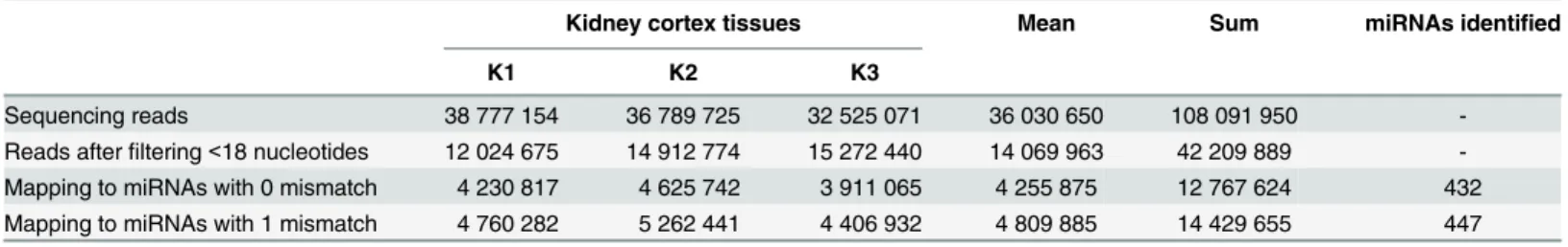
Next, we attempted to experimentally validate the newly-discoveredM.fascicularismiRNAs by small RNA sequencing. For this purpose, small RNA sequencing libraries were prepared from three naiveM.fasciculariskidney cortex tissues and sequenced as described in Methods sec-tion. On average, we obtained 36 million reads for each kidney cortex tissue sample (Table 1). RNA sequencing reads with a length below 18 nucleotides were removed and remaining reads were mapped to the 913M.fascicularismiRNAs. We considered miRNAs as genuine only when they had a minimum of 15 combined sequencing reads from the three kidney cortex tis-sues analyzed. With this criterion we identified 432 and 447 miRNAs with 0 and 1 mismatch, respectively; 432 miRNAs were present in both groups and 15 were unique to 1 mismatch list (Table 1). Although mapping with 1 mismatch increased the total number of mapped reads
Fig 1. Workflow used for the identification ofM.fascicularismiRNA genes.
doi:10.1371/journal.pone.0142708.g001
(from 12.7 to 14.2 million), it only yielded 15 additional unique miRNAs; 14 of them had 15–
32 reads. In fact, all 15 miRNAs identified with 1 mismatch were also present in the 0 mis-match list but had 7–14 reads; hence they did not pass the set criteria. From this result it is clear that allowing 1 mismatch only contributed to the increase in sequencing reads to the existing miRNAs rather than discovery of new miRNAs. Thus in subsequent studies, we retained miRNAs with 0 mismatch (432 miRNAs—S7 Table).
Assigning 432 miRNAs to pre-miRNAs
Generally, miRNAs are represented with suffix -5p or -3p only when a stem-loop pre-miRNA produces mature sequences from both -5p and -3p arms. On the other hand, if a pre-miRNA produces only one mature microRNA, arm is not usually specified [24]. In ourM.fascicularis
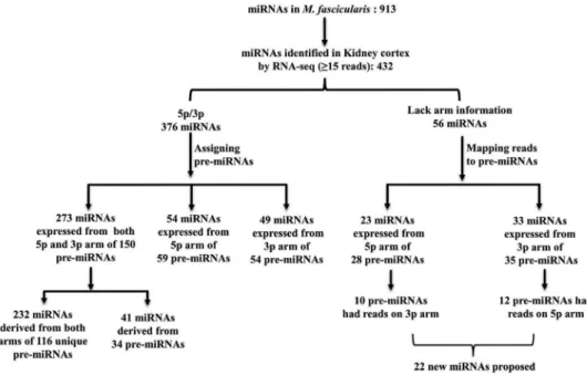
list resulting from the initial analysis, 687 of 913 miRNAs were indicated with a -5p or -3p suf-fix. The remaining 226 miRNAs lacked the arm information. In order to understand whether miRNAs identified in kidney cortex by RNA sequencing resulted from single or both arms of a given precursor, we sorted the 432 mature miRNAs based on stem-loop pre-miRNA informa-tion. 376 out of the 432 miRNAs presented either a -5p or -3p in their nomenclature and the remaining 56 miRNAs lacked the arm information (Fig 2). Then we assigned the 376 miRNAs to respective pre-miRNAs usingS1 Tableand examined for the presence/absence of mature sequence/reads on each arm. As shown inFig 2, 273 out of 376 miRNAs were expressed from both arms of 150 pre-miRNAs: 232 were derived from both arms of 116 unique pre-miRNAs (-5p or -3p miRNAs derived from unique/ single pre-miRNA) and 41 miRNAs derived from both arms of 34 miRNAs (-5p or -3p mature miRNAs derived from more than one pre-miRNA). In addition, 54 miRNAs were expressed from only the -5p arm of 59 pre-miRNAs and 49 miRNAs resulted from only the -3p arm of 54 pre-miRNAs. Hence these 113 mature miRNAs (54 from -5p arm and 49 from -3p arm) derived from only one arm of pre-miRNAs and the corresponding mature miRNA counterparts were not expressed in the present dataset.
For the remaining 56 miRNAs which lacked arm information, instead of assigning them to pre-miRNAs (S1 Table), FASTQ files were mapped against pre-miRNA sequences. This not only helped to assign arm information, but also allowed to visualize reads on both arms of pre-miRNAs. As shown inFig 2, 23 of 56 miRNAs were expressed from the -5p arm of 28 pre-miR-NAs. Interestingly, 10 of the 28 pre-miRNAs also had reads (>15) on the -3p arm and these miRNAs were not described inM.mulatta. Thirty-three of 56 miRNAs were expressed from the -3p arm of 35 pre-miRNAs. Here also 12 of the 35 pre-miRNAs had reads (>15) on the -5p arm (which have also not been described before inM.mulatta). We retrieved conserved miRNA sequences from 22 newly identified mature miRNAs (10 miRNAs from -5p arm and 12 miRNA from -3p arm) and searched in miRBase (S8 Table). Interestingly all 22 miRNAs were present in human and other species with little variation in nucleotide sequence (data not shown).
Table 1. Mapping RNA sequencing reads toM.fascicularismiRNAs.
Kidney cortex tissues Mean Sum miRNAs identified
K1 K2 K3
Sequencing reads 38 777 154 36 789 725 32 525 071 36 030 650 108 091 950
-Reads afterfiltering<18 nucleotides 12 024 675 14 912 774 15 272 440 14 069 963 42 209 889
-Mapping to miRNAs with 0 mismatch 4 230 817 4 625 742 3 911 065 4 255 875 12 767 624 432 Mapping to miRNAs with 1 mismatch 4 760 282 5 262 441 4 406 932 4 809 885 14 429 655 447
From the above results it is clear that manyM.fascicularismiRNAs identified by homology search were present / expressed in kidney cortex tissue. Altogether 432 mature miRNAs expressed in kidney cortex were derived from 326 pre-miRNAs and many of them were expressed from both arms of pre-miRNA (S8 Table). In addition, mapping sequencing reads to the pre-miRNAs allowed us to propose 22 new miRNAs which were not previously described inM.mulatta.
RT-qPCR validation of
M
.
fascicularis
miRNAs
In order to confirm the miRNAs identified by RNA sequencing, we performed RT-qPCR anal-ysis on some of the miRNAs. TaqMan Low Density Array (TLDA) cards have 754 RT-qPCR assays specific to human miRNAs (TLDA-A and B cards). Comparison of the 754 human mature miRNA sequences to the 913M.fascicularismiRNA sequences indicated that at least 294 miRNAs had exact match and 19 had 1 mismatch (total 313 miRNAs). Hence we used TLDA cards to examine 313M.fascicularismiRNAs. Total RNA prepared from 3 kidney cor-tex tissues was subjected to RT-qPCR using human TLDA A and B cards as described in Meth-ods section. The Ct (cycle threshold) values obtained from the 3 tissues were averaged and miRNAs with Ct32 were considered as expressed. Using this criterion, 262 of 313M. fasci-cularismiRNAs were detected by RT-qPCR (S9 Table).
Next we compared miRNAs identified by sequencing and qRT-PCR. As shown inFig 3, 432 and 262 miRNAs were detected inM.fasciculariskidney cortex tissues by RNA sequencing and RT-qPCR, respectively; 222 of 913 miRNAs were validated by both RNA sequencing and RT-qPCR (S10 Table). Interestingly 11 miRNAs identified by RNA sequencing were not detected by RT-qPCR. Also 40 miRNAs identified by RT-qPCR were not detected by RNA sequencing. The observed discrepancy may be due to the differences in sample processing and sensitivity of each platform.
Fig 2. Mapping 432 expressed miRNAs to respective pre-miRNAs.
doi:10.1371/journal.pone.0142708.g002
Expression analysis and target prediction
For the expression analysis, read counts obtained from the three kidney cortex tissues were normalized with edgeR package and examined for kidney specific miRNAs (S11 Table). Several of the 432 miRNAs had been previously reported to be kidney-specific miRNAs: miR-10b-5p, miR-30c-5p, miR-204-5p, miR-10a-5p, miR-30d-5p, miR-200a-3p, miR-196a-5p, miR-196b-5p and miR-146a-miR-196b-5p [25]. Many of those miRNAs were among the top 10 highly expressed miRNAs in kidney cortex samples (Fig 4A). Enriched expression of a cluster of miRNAs such as miR-192, miR-194, miR-204 and miR-215 in human kidney tissue was previously confirmed by Northern blot analysis [26]. Expression of miR-23, miR-24, miR-26a, miR-10a and miR-30c in glomerulus and renal tubules has also been previously reported in mice [27]. Also involve-ment of miR-30 family in kidney developinvolve-ment is docuinvolve-mented [28].
Next, to understand the functional relevance of miRNAs expressed in kidney cortex, we attempted to link the miRNAs to target genes. Since most of the target prediction tools are designed for human miRNAs, expressedM.fasciculariskidney cortex miRNAs which had human orthologs (392 miRNAs) were used (S4 Table) in further analysis. Using Ingenuity Pathway Analysis solftware, only experimentally observed miRNA-target genes interactions in Human were considered. With these setting 392 miRNAs yielded 1084 target genes. Kidney cortex miRNA targeted genes associated with molecular and cell functions are shown inFig 4B. Especially, miRNA targeted genes with lowest p-value are involved in basic cellular pro-cesses such as cell growth and proliferation, cellular development, tissue morphology and cell death. Overall results of target analysis indicate that miRNAs expressed in kidney cortex are involved in regulation of various key cellular processes in kidney cortex cells fromM.
fascicularis.
Discussion
In this study we systematically investigated the miRNAs inM.fascicularis, an animal model with high translational potential to human. By comparing metazoan miRNAs sequence to expressed sequence tags (EST) ofM.fascicularis, Yang et al. (2012) identified eight miRNAs in
M.fascicularis[19]. With the exception of one (rno-miR-3591), all miRNAs described in this previous study are present in our current list of 913M.fascicularismature miRNAs. More
Fig 3. Comparison ofM.fascicularismiRNAs identified by RNA sequencing and RT-qPCR.
recently, Yang et al. (2014) identified 86 putative miRNAs inM.fascicularisby transcriptome analysis [20]. Interestingly only 39 out of 86 miRNAs were present in our list of 913M. fascicu-larismature miRNAs and 47 were absent (S3 Table). This discrepancy may be mainly due to the difference in workflows used in each study. Indeed, Yang et al. (2014) used all existing metazoan miRNA sequences to map their transcriptome data against, whereas we followed a more focused and specific approach usingM.mulattastem-loop pre-miRNA sequences against
M.fascicularisgenome.
Results presented in this investigation indicate thatM.mulattamiRNAs are found to be highly conserved inM.fascicularisas the vast majority ofM.mulattamiRNA sequences are identical to miRNA sequences identified inM.fascicularisgenome (881 out of 913 miRNAs). This result is consistent with the fact that both macaque species exhibit high degree of genome identity (99.2% identical) [13]. Also the majority ofM.fascicularismiRNAs are conserved in
Homo sapiens. A recent miRNA expression study in human and non-human primates showed positive correlations between miRNA sequence conservation and expression levels. Highly conserved miRNAs exhibited higher levels of expression in human, chimpanzee and rhesus macaque [15]. Conversely negative correlation was observed between miRNA hairpin SNP number and miRNA expression. Similar results were also observed in humans: less expressed miRNA showed weaker selection and hence had less constraint evolution than highly expressed miRNAs [29]. High conservation of miRNAs amongM.fascicularis,M.mulattaand human may reflect similar regulatory networks across these three primate species.
Recently, miRBase (version 21) set several criteria to distinguish authentic miRNAs from false annotations. One of the criteria for high confidence miRNA is that pre-miRNA should have at least 10 reads corresponding to each of the two possible mature miRNA sequences
Fig 4. A. Top 10 highly expressed miRNAs in kidney cortex fromM.fascicularis. B. Functions of
kidney cortex miRNAs targeted genes.
doi:10.1371/journal.pone.0142708.g004
[24]. Sequencing of kidney cortex miRNAs at high depth (total 12.7 million miRNA reads/3 animals) not only facilitated the sequencing of a large number of miRNAs but also aided the detection of both -5p and -3p mature sequences of pre-miRNAs. Using stringent mapping cri-teria, 432 miRNAs were detected in kidney cortex tissue; 273 out of 432 miRNAs derived from both -5p and -3p arms of 150 pre-miRNAs and in most cases only one arm showed dominant expression. Thus our results confirm the expression of both -5p and -3p arm of 150 pre-miR-NAs (S8 Table). Recent progress in miRNA research indicated that mature miRpre-miR-NAs produced from both arms (-5p and -3p) of pre-miRNAs are biologically active and expression of a domi-nant mature sequence varies with developmental stages, tissue type and even with species [30].
Tissue-specific miRNAs and their target genes play vital role in organ or tissue differentia-tion, development and function. MiRNAs expressed in kidney cortex ofM.fascicularisdescribed in this study are in good agreement with previously reported kidney specific miRNAs [25]. Many of those kidney specific miRNAs were expressed at very high level and their target genes were involved in various basic cellular functions indicating that they play vital role in kidney. Interestingly, in our preliminary investigation on urinary miRNAs inM.fascicularisconducted in the context of the Health and Environmental Sciences Institute (HESI) Biomarkers of Neph-rotoxicity Committee we observed 9 out of top 10 kidney cortex miRNAs (Fig 4A) in urine sam-ples ofM.fascicularisand they were also among the top 10 highly represented urinary miRNAs (data not shown). Similar miRNAs were also reported for human urine: miR-30a-5p, hsa-miR-10b-5p, hsa-miR-10a-5p, hsa-miR-26a-5p and hsa-miR-30c-5p are among the 10 highly represented miRNAs in exosomal preparation of human urine [31]. Thus kidney (cortex, medulla and papilla) miRNA profiles may help to understand miRNAs present in urine.
In conclusion we identified 913 miRNAs inM.fascicularisand experimentally validated 432 miRNAs in kidney cortex tissues by RNA-sequencing. In addition expression of 222 miRNAs was confirmed by both RNA sequencing and RT-qPCR. The data presented in this investiga-tion will serve as valuable resource especially for exploring biomarker potential of miRNAs in
M.fascicularis. Urinary miRNAs indeed hold great promise as kidney safety biomarkers [32]. Following this line of thought, we are currently exploring biomarker potential of urinary miR-NAs inM.fascicularistreated with various nephrotoxicants.
Materials and Methods
Animals and Ethics Statement
All animal experiment protocols were reviewed and approved by the Institutional Animal Care and Use Committee (IACUC) of National Center for Safety Evaluation of Drugs (NCSED). Cynomolgus monkeys (ages 2–3 years old) were maintained in stainless steel cages (L×W×H: 800×700×750 mm) under condition of 16–26°C, 40–70% relative humidity, a 12-h light-dark cycle and a room air exchange of 8–10 times per hour. Each monkey was provided with 300 g of standard monkey keeping diet and fruits per day, and sterilized water was offered ad libitum. The monkey was individually housed with toys (such as mirror) and monitored daily by the animal care staff. At the end of this experiment, monkeys were exsanguinated following the deep anesthesia by IV injection of pentobarbital sodium, and kidney cortex samples were col-lected, transferred to cryotubes containing RNALater reagent and stored at -80°C.
RNA extraction, qualitative and quantitative analysis
Agilent RNA 6000 Nano kit and 2100 Bioanalyzer (Agilent). All RNA samples had a RNA integrity number between 7 to 8. The RNA quantity was determined using RediPlate™96 Ribo-Green1RNA Quantitation kit (Thermo Fisher).
Small RNA enrichment and sequencing library preparation
Five micrograms of total RNA were used for small RNA enrichment. Small RNA enrichment was performed using Total RNA-Seq kit v2 (Life Technologies) as per manufacturer’s recom-mendations. The sequencing library was prepared from 12 ng (quantified using High Sensitiv-ity DNA analysis kit and 2100 Bioanalyzer) of enriched small RNAs using Total RNA-Seq kit v2 with custom barcode primers.
Sequencing template preparation, Ion PI Chip loading and sequencing
Seventyμl (75 pM) of the sequencing library were used for template preparation. Sequencing templates were prepared using ION PI IC 200 kit (Life Technologies) and Ion Chef System (Life Technologies). One Ion PI Chip kit v2 BC (Life Technologies) was used for each sequenc-ing library. Sequencsequenc-ing templates loaded on Ion PI Chip were sequenced ussequenc-ing ION PI IC 200 kit and Ion Proton System (Ion Proton Sequencer and Ion Proton Torrent Server, Life Tech-nologies), according to manufacturer’s protocol. The Ion Proton Torrent Server trims (removes sequencing key, barcode and adaptor sequences, lower-quality -3p ends with low quality scores) and filters (reads with<8 nucleotides, adaptor dimers, reads lacking key, polyclonal reads, reads with low signal) the sequencing reads and generates FASTQ files.
Analysis of sequencing data
FASTQ files were analyzed using Array studio V.8.0.3.67 (OmicSoft Corp). In the preprocess-ing step, reads with a length below 18 nucleotides and average quality score below 20 were removed from FASTQ files. Then FASTQ files were mapped againstM.fascicularismiRNA reference sequences (generated in this study) and resulting BAM files were used for read quan-titation. Generated miRNA reads were normalized by edgeR method.
RT-qPCR using TaqMan MicroRNA Arrays
Complementary DNA was synthesized from kidney cortex total RNA (200 ng) using TaqMan MicroRNA Reverse Transcription kit (Life Technologies) and Megaplex™TLDA A v2.1 and TLDA B v3.0 RT Primers. Resulting cDNA was used for qPCR. Quantitative real-time PCR was performed using TaqMan Universal Master Mix II (Life Technologies) and TaqMan TLDA Human MicroRNA A version 2.0 and TLDA Human MicroRNA B version 3.0 cards. Sample loaded TLDA cards were run on QuantStudio 12K Flex real-time PCR system (Life Technologies). Generated raw data files were analyzed using Expression Suite Software v1.0.3 (Life Technologies).
miRNA target prediction
Only Human orthologs ofM.fascicularismiRNAs expressed in kidney cortex were used for tar-get prediction. A total of 392 miRNAs were used for tartar-get prediction using Ingenuity Pathway Analysis software version 23814503 (QIAGEN). Initial analysis yielded 17 000 predicted target messenger RNAs. Filters were applied to focus on a reduced but more relevant target gene set. The relationships column was refined with the selection of Ingenuity Expert, Expert Assist find-ings and miRecords, including experimentally observed and / or high or moderate prediction. With these settings a finalized list of 1084 target genes was obtained. Core analysis was carried
out on this list using the following filters: direct and indirect relationships, microRNA-mRNA interactions including miRecords, TargetScan human and Tarbase. Only the human species was taken into account and the confidence level selected was“experimentally observed”. The chart corresponding to Molecular and Cell Function was customized to display biofunctions only.
Bioinformatics analysis
For the homology search, BLASTN package was used [33].M.fascicularismature miRNAs were extracted with fuzznuc, a string-search algorithm from EMBOSS package by comparing eachM.mulattamature miRNA against each correspondingM.fascicularispredicted stem-loop [34]. Venn diagram was constructed using VENNY 2.0 [35].
Supporting Information
S1 Table. Identification ofM.fascicularisstem-loop miRNAs by BLASTN searching of M. mulatta stem-loop miRNA reference sequences inM.fascicularisgenome.
(XLSX)
S2 Table. Identification ofM.fascicularismiRNAs by comparingM.mulattamiRNA refer-ence sequrefer-ence againstM.fascicularisstem-loop miRNA sequences.
(XLSX)
S3 Table. Comparison ofM.fascicularismiRNAs with previously reported 86 M. fascicu-laris miRNAs.
(XLSX)
S4 Table. Comparison ofM.fascicularismiRNAs versusH.sapiensmiRNAs. (XLSX)
S5 Table. Comparison ofM.fascicularisstem-loop miRNA sequences versusH.sapiens stem-loop miRNAs sequences.
(XLSX)
S6 Table. Comparison ofM.fascicularisstem-loop miRNAs versusH.sapiensgenome. (XLSX)
S7 Table. Mapping reads from 3 kidney cortex samples (K1, K2 and K3) toM.fascicularis miRNAs with 0 mismatch.MiRNA reads were counted from 3 kidney cortex tissues. MiRNA having reads15 were considered as genuine—With this criterion, a total of 432 mature miR-NAs were identified.
(XLSX)
S8 Table. Assigning reads toM.fascicularisstem-loop pre-miRNAs and mature miRNAs. (XLSX)
S9 Table. Detection of miRNAs by RT-qPCR.TaqMan1
MicroRNA Arrays (Human Arrays A and B) were used for detection of miRNAs expressed in kidney cortex tissues. MiRNAs spe-cific toM.fascicularisare shown in table. For each miRNA, average Ct is calculated from three kidney cortex samples (K1, K2 and K3). MiRNAs with Ct32 were considered as expressed. With this criterion, a total of 262 miRNAs were considered as expressed.
(XLSX)
detected by RT-qPCR (262). (XLSX)
S11 Table. EdgR normalized miRNAs values from three kidney cortex samples.M. fascicu-larismiRNAs ortholog to human miRNAs were used for target identification.
(XLSX)
Acknowledgments
This work was supported by the National Major Scientific and Technological Special Project for“Significant New Drugs Development”(No. 2012ZX09302001) and“Key Technological Study”(No. 2012ZX09505001-004) during the Twelfth Five-year Plan Period from the Minis-try of Science and Technology, People's Republic of China.
Author Contributions
Conceived and designed the experiments: YV XZ JW BL JFL JCG MLO EB. Performed the experiments: YV. Analyzed the data: JFL JCG MLO EB. Wrote the paper: YV JFL JCG MLO EB. Performed NGS and analyzed the NGS data: YV. Performed the bioinformatics analysis: PR CP. Performed the RT-qPCR experiments: CM. Contributed all the animal tissue and bio-fluid samples used in the study: XZ JW BL. All authors read and approved the final
manuscript.
References
1. Ha M, Kim VN. Regulation of microRNA biogenesis. Nat Rev Mol Cell Biol. 2014; 15: 509–524. doi:10. 1038/nrm3838PMID:25027649
2. He L, Hannon GJ. MicroRNAs: small RNAs with a big role in gene regulation. Nat Rev Genet. 2004; 5: 522–531. doi:10.1038/nrg1415PMID:15211354
3. Meunier J, Lemoine F, Soumillon M, Liechti A, Weier M, Guschanski K, et al. Birth and expression evo-lution of mammalian microRNA genes. Genome Res. 2013; 23: 34–45. doi:10.1101/gr.140269.112
PMID:23034410
4. Iorio M V, Croce CM. MicroRNA dysregulation in cancer: Diagnostics, monitoring and therapeutics. A comprehensive review. EMBO Mol Med. 2012. pp. 143–159. doi:10.1002/emmm.201100209PMID:
22351564
5. Li Z, Rana TM. Therapeutic targeting of microRNAs: current status and future challenges. Nat Rev Drug Discov. 2014; 13: 622–638. doi:10.1038/nrd4359PMID:25011539
6. Weber JA, Baxter DH, Zhang S, Huang DY, Huang KH, Lee MJ, et al. The microRNA spectrum in 12 body fluids. Clin Chem. 2010; 56: 1733–1741. doi:10.1373/clinchem.2010.147405PMID:20847327
7. Cortez MA, Bueso-Ramos C, Ferdin J, Lopez-Berestein G, Sood AK, Calin GA. MicroRNAs in body flu-ids—the mix of hormones and biomarkers. Nat Rev Clin Oncol. 2011; 8: 467–477. doi:10.1038/ nrclinonc.2011.76PMID:21647195
8. Hayes J, Peruzzi PP, Lawler S. MicroRNAs in cancer: biomarkers, functions and therapy. Trends Mol Med. 2014; 20: 460–469. doi:10.1016/j.molmed.2014.06.005PMID:25027972
9. D’Alessandra Y, Devanna P, Limana F, Straino S, Di Carlo A, Brambilla PG, et al. Circulating micro-RNAs are new and sensitive biomarkers of myocardial infarction. Eur Heart J. 2010; 31: 2765–2773. doi:10.1093/eurheartj/ehq167PMID:20534597
10. Zeng L, Cui J, Wu H, Lu Q. The emerging role of circulating microRNAs as biomarkers in autoimmune diseases. Autoimmunity. 2014; 47: 419–429. doi:10.3109/08916934.2014.929667PMID:24953646
11. Mikaelian I, Scicchitano M, Mendes O, Thomas RA, LeRoy BE. Frontiers in preclinical safety biomark-ers: microRNAs and messenger RNAs. Toxicol Pathol. 2012. doi:10.1177/0192623312448939
12. Rogers J, Gibbs RA. Comparative primate genomics: emerging patterns of genome content and dynamics. Nat Rev Genet. 2014; 15: 347–359. doi:10.1038/nrg3707PMID:24709753
13. Ebeling M, Küng E, See A, Broger C, Steiner G, Berrera M, et al. Genome-based analysis of the nonhu-man primateMacaca fascicularisas a model for drug safety assessment. Genome Res. 2011; 21: 1746–1756. doi:10.1101/gr.123117.111PMID:21862625
14. King MC, Wilson AC. Evolution at two levels in humans and chimpanzees. Science. 1975; 188: 107– 116. doi:10.1126/science.1090005PMID:1090005
15. Dannemann M, Prüfer K, Lizano E, Nickel B, Burbano HA, Kelso J. Transcription factors are targeted by differentially expressed miRNAs in primates. Genome Biol Evol. 2012; 4: 552–564. doi:10.1093/ gbe/evs033PMID:22454130
16. Yan G, Zhang G, Fang X, Zhang Y, Li C, Ling F, et al. Genome sequencing and comparison of two non-human primate animal models, the cynomolgus and Chinese rhesus macaques. Nat Biotechnol. 2011; 29: 1019–23. doi:10.1038/nbt.1992PMID:22002653
17. Higashino A, Sakate R, Kameoka Y, Takahashi I, Hirata M, Tanuma R, et al. Whole-genome sequenc-ing and analysis of the Malaysian cynomolgus macaque (Macaca fascicularis) genome. Genome Biol.
2012; 13: R58. doi:10.1186/gb-2012-13-7-r58PMID:22747675
18. Osada N, Hettiarachchi N, Adeyemi Babarinde I, Saitou N, Blancher A. Whole-genome sequencing of six mauritian cynomolgus macaques (Macaca fascicularis) Reveals a genome-wide pattern of
polymor-phisms under extreme population bottleneck. Genome Biol Evol. 2015; 7: 821–830. doi:10.1093/gbe/ evv033PMID:25805843
19. Yang H, Zhang H, Zhu L, Zhang C, Li D. Identification and characterization of microRNAs inMacaca fascicularisby EST analysis. Comp Funct Genomics. 2012; 2012. Aticle ID 957607, 9 pages. doi:10. 1155/2012/957607
20. Yang H, Zhang R, Jing Y, Zhu L, Zhang W, Liu C, et al. Identification and characterization of microRNAs in the crab-eating macaque (Macaca fascicularis) using transcriptome analysis. Gene. 2014; 536: 308–
315. PMID:24355555
21. Saikumar J, Hoffmann D, Kim T-M, Gonzalez VR, Zhang Q, Goering PL, et al. Expression, circulation, and excretion profile of microRNA-21, -155, and -18a following acute kidney injury. Toxicol Sci. 2012; 129: 256–267. doi:10.1093/toxsci/kfs210PMID:22705808
22. Kanki M, Moriguchi A, Sasaki D, Mitori H, Yamada A, Unami A, et al. Identification of urinary miRNA bio-markers for detecting cisplatin-induced proximal tubular injury in rats. Toxicology. 2014; 324: 158–168. doi:10.1016/j.tox.2014.05.004PMID:24863737
23. Yue J, Sheng Y, Orwig KE. Identification of novel homologous microRNA genes in the rhesus macaque genome. BMC Genomics. 2008; 9: 8. doi:10.1186/1471-2164-9-8PMID:18186931
24. Kozomara A, Griffiths-Jones S. MiRBase: annotating high confidence microRNAs using deep sequenc-ing data. Nucleic Acids Res. 2014; 42:D 68–D73. doi:10.1093/nar/gkt1181
25. Guo Z, Maki M, Ding R, Yang Y, Zhang B, Xiong L. Genome-wide survey of tissue-specific microRNA and transcription factor regulatory networks in 12 tissues. Sci Rep. 2014; 4: 5150. doi:10.1038/ srep05150PMID:24889152
26. Sun Y, Koo S, White N, Peralta E, Esau C, Dean NM, et al. Development of a micro-array to detect human and mouse microRNAs and characterization of expression in human organs. Nucleic Acids Res. 2004; 32: e188. doi:10.1093/nar/gnh186PMID:15616155
27. Ho J, Ng KH, Rosen S, Dostal A, Gregory RI, Kreidberg J a. Podocyte-specific loss of functional micro-RNAs leads to rapid glomerular and tubular injury. J Am Soc Nephrol. 2008; 19: 2069–2075. doi:10. 1681/ASN.2008020162PMID:18832437
28. Agrawal R, Tran U, Wessely O. The miR-30 miRNA family regulates Xenopus pronephros development and targets the transcription factor Xlim1/Lhx1. Development. 2009; 136: 3927–3936. doi:10.1242/ dev.037432PMID:19906860
29. Liang H, Li W-H. Lowly expressed human microRNA genes evolve rapidly. Mol Biol Evol. 2009; 26: 1195–1198. doi:10.1093/molbev/msp053PMID:19299536
30. Griffiths-Jones S, Hui JHL, Marco A, Ronshaugen M. MicroRNA evolution by arm switching. EMBO Rep. 2011; 12: 172–177. doi:10.1038/embor.2010.191PMID:21212805
31. Cheng L, Sun X, Scicluna BJ, Coleman BM, Hill AF. Characterization and deep sequencing analysis of exosomal and non-exosomal miRNA in human urine. Kidney Int. 2014; 86: 433–444. doi:10.1038/ki. 2013.502PMID:24352158
32. Nassirpour R, Mathur S, Gosink MM, Li Y, Shoieb AM, Wood J, et al. Identification of tubular injury microRNA biomarkers in urine: comparison of next-generation sequencing and qPCR-based profiling platforms. BMC Genomics. 2014; 15: 485. doi:10.1186/1471-2164-15-485PMID:24942259
33. Altschul SF, Madden TL, Schäffer AA, Zhang J, Zhang Z, Miller W, et al. Gapped BLAST and PSI-BLAST: A new generation of protein database search programs. Nucleic Acids Res. 1997; 25: 3389– 3402. doi:10.1093/nar/25.17.3389PMID:9254694
34. Rice P, Longden I, Bleasby A. EMBOSS: the European Molecular Biology Open Software Suite. Trends Genet. 2000; 16: 276–277. doi:10.1016/S0168-9525(00)02024-2PMID:10827456