Identification and Analysis of Differential miRNAs in
PK-15 Cells after Foot-and-Mouth Disease Virus Infection
Ke-Shan Zhang., Yong-Jie Liu., Han-Jin Kong, Wei-Wei Cheng, You-Jun Shang, Hong Tian,
Hai-Xue Zheng, Jian-Hong Guo, Xian-Tao Liu*
State Key Laboratory of Veterinary Etiological Biology, National Foot and Mouth Disease Reference Laboratory, Lanzhou Veterinary Research Institute, Chinese Academy of Agricultural Sciences, Lanzhou, China
Abstract
The alterations of MicroRNAs(miRNAs) in host cell after foot-and-mouth disease virus (FMDV) infection is still obscure. To increase our understanding of the pathogenesis of FMDV at the post-transcriptional regulation level, Solexa high-throu MicroRNAs (miRNAs) play an important role both in the post-transcriptional regulation of gene expression and host-virus interactions. Despite investigations of miRNA expression ghput sequencing and bioinformatic tools were used to identify differentially expressed miRNAs and analyze their functions during FMDV infection of PK-15cells. Results indicated that 9,165,674 and 9,230,378 clean reads were obtained, with 172 known and 72 novel miRNAs differently expressed in infected and uninfected groups respectively. Some of differently expressed miRNAs were validated using stem-loop real-time quantitative RT-PCR. The GO annotation and KEGG pathway analysis for target genes revealed that differently expressed miRNAs were involved in immune response and cell death pathways.
Citation:Zhang K-S, Liu Y-J, Kong H-J, Cheng W-W, Shang Y-J, et al. (2014) Identification and Analysis of Differential miRNAs in PK-15 Cells after Foot-and-Mouth Disease Virus Infection. PLoS ONE 9(3): e90865. doi:10.1371/journal.pone.0090865
Editor:Fatah Kashanchi, George Mason University, United States of America
Copyright:ß2014 Zhang et al. This is an open-access article distributed under the terms of the Creative Commons Attribution License, which permits unrestricted use, distribution, and reproduction in any medium, provided the original author and source are credited.
Funding:This work was supported by The National Natural Science Foundation of China (Grant No. 31201914), China Postdoctoral Science Foundation (2013M530683), and China Agriculture Research System (CARS-39). The funders had no role in study design, data collection and analysis, decision to publish, or preparation of the manuscript.
Competing Interests:The authors have declared that no competing interests exist.
* E-mail: [email protected]
.These authors contributed equally to this work.
Introduction
MicroRNAs (miRNAs) are small 21–24 nucleotide (nt) non-coding RNAs that play an important role in the post-transcrip-tional regulation of gene expression [1,2]. Mature miRNAs are not directly produced by gene transcription, they are processed from long primary miRNA (pri-miRNA) precursors. Now they are known to play a key role in biological activities at the molecular level [3–5]. Research indicates that miRNAs not only bind to sites in the coding region and 59UTR to regulate genes but may also combine with the 39UTR of target mRNAs involved in development, fat metabolism, apoptosis, virus defense, and cell proliferation [6,7]. Recent studies mainly focused investigating miRNA function in human disease and primarily relate to the use of miRNAs as disease biomarkers to monitor drug efficacy. In veterinary science, the stability of miRNAs along with their ability to regulate gene expression is beneficial in developing breeding programs for disease resistance in livestock [8].
MiRNAs offer an immense opportunity not only to develop novel biomarkers and therapeutics but also to understand the intricacies of host-pathogen interaction, and gain possible insights into viral oncogenesis, latency and tropism [9]. Researches showed that miRNAs play a critical role in host-virus interaction [10,11] Indeed, host miRNAs have a substantial effect on viral evolution and potentially regulate the tissue tropism of viruses in vivo [10]. Viruses encode their own miRNAs in the miRNA-induced gene-silencing pathway [4]. For example, baculovirus-encoded miRNA
(bmnpv-miR-1) suppresses its host miRNA biogenesis by regulat-ing the exportin-5 cofactor Ran [12]. Enterovirus-induced miR-141 contributes to shutoff host protein translation by targeting the translation initiation factor eIF4E [13]. The host miRNA miR-122 is an indispensable factor in supporting hepatitis C virus (HCV) replication [14]. MiRNA-141 represses HBV replication indirectly by targeting PPARA. Human miRNA hsa-miR-296-5p suppresses
Enterovirus 71 replication by targeting the viral genome [15]. Additionally, miR-125b and miR-223 directly target human immunodeficiency virus type 1 (HIV-1) mRNA, and miR-198 modulates HIV-1 replication indirectly by repressing the expres-sion of ccnt1 gene [16,17].
In recent years, the emergence of Solexa and 454 high-throughput sequencing technologies have enabled the direct sequencing of miRNAs to be a highly sensitive and and efficient method to study miRNAs in different species [18–21], although only about 250 pig miRNAs have been identified. Parameswaran reported virus-derived miRNAs profile in cells infection with dengue virus, vesicular stomatitis virus, polio virus, hepatitis C virus and West Nile virus [22]. Deep sequencing revealed HIV-encoded miRNAs modulate cellular and viral gene expression [23]. Using the technique, other researchers found that pseudo-rabies virus (PRV) encoded miRNAs that play significant role in virus-cell interaction. These miRNAs are mainly involved in complex cellular pathways including cell death, immune system, and metabolism [24]. Following sequencing, northern blot hybridization and highly sensitive stem-loop RT-PCR are
generally used to identify mature miRNAs and confirm changes in expression levels of miRNA based on known unique sequences [25].
Foot-and-mouth disease (FMD) is a highly infectious disease of cloven-hoofed domestic and wild animals [26]. Seven serotypes (A, O, C, Asia 1, and South African Territories 1, 2, and 3) have been identified, and multiple subtypes occur within each serotype [26– 28]. The etiologic agent, FMD virus (FMDV) belongs toAphthovirus genus of the Picornaviridae family. FMDV consists of a single-stranded, plus-sense RNA genome of approximate 8,500 bases surrounded by an icosahedral capsid composed of 60 copies each of four structural proteins VP1-4 (termed 1D, 1B, 1C, and 1A). However, the mechanism of FMDV infects host cells in post-transcriptional regulation is unclear.
The focus of this study is differential expressed miRNAs between FMDV-infected and uninfected 15 cell groups. PK-15 cell line was purchased from Center for Type Culture Collection(CTCC, Wuhan, China). To gain further insights into the potential role of miRNAs during FMDV infection of host cells, small RNA from PK-15 cell libraries of individual FMDV infection and uininfected groups were constructed. Solexa high-throughput sequencing technique and bioinformatics for sequenc-ing were integrated and data processsequenc-ing to:
1) obtain the miRNA expression profiles between the two groups of cells;
2) identify the novel and differentially expressed miRNAs; 3) define the regulatory network between miRNAs and mRNA;
and
4) determine the regulatory mechanisms of miRNAs involved in the FMDV infection.
The results updated the pig miRNA database to better understand the molecular mechanism of FMDV infection. Our findings layed a new foundation for identifying the biomarkers associated with the FMDV infection.
Results
Small RNA Library Construction and Solexa Sequencing
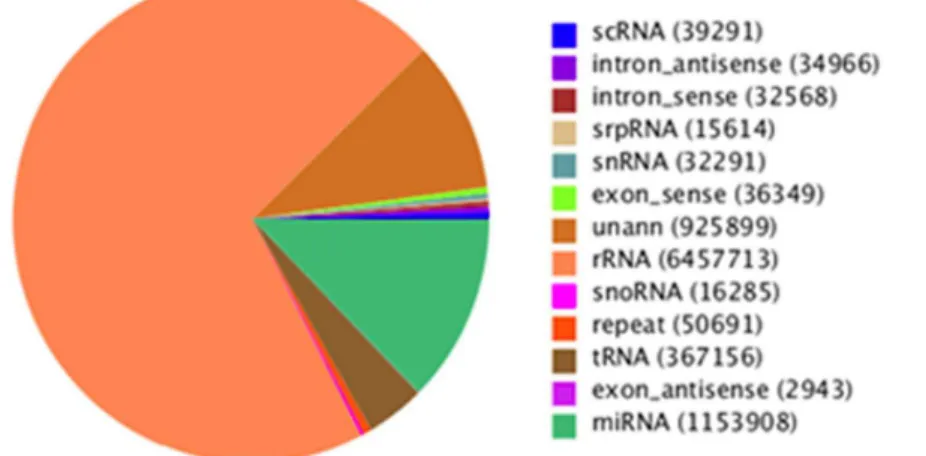
To identify miRNAs in FMDV infection in PK-15 cells, two small RNA libraries pooled from infected and uninfected groups were constructed respectively, and sequenced using an Illumina/ Solexa 1G high-throughput sequencer. As a result, a total of 18,839,052 and 18,164,583 raw reads were identified in infected and uninfected groups. After removing the low quality reads, adaptors, and insufficient tags, ultimately 9,165,674 and 9,230,378 clean reads of 18,30 nt were obtained (Table 1). The lengths of the majority of clean reads were 19,26 nt, and a large number of the distributed sequences were 22 nt, which accounted for 19.9% and 38.62% in infected and uninfected group, and are typical of the small RNA dicer-processed products and consistent with the known 18,25 nt range for miRNAs(Figure 1). After mapping to theSus crofagenome, a total of 1839602 small RNAs sequences were provided, including 438307(accounting for 2.38%) specific sequences in FMDV-infected group, 390087 ( account for 2.12%) specific sequences in FMDV-uninfected group and 17567758(95.5%) common sequences(Table 1). All identical sequence reads were grouped together to simplify the sequencing data, with a total of 770488 unique sequences identified in two groups, which contained 324849 (account for 42.16%) specific unique sequences for infected group, 339772 (account for 42.93%) specific unique sequences for uninfected group, and 114867(14.91%) for common unique sequences (Table 1). To assess the efficiency of high-throughput sequencing for small RNA detection, all sequences were annotated and classified through alignment with GenBank and Rfam databases. All of reads were annotated and classified as miRNA, rRNA, tRNA, snRNA and snoRNA. The size distribution of the miRNAs in the infected group was less than the uninfected group was found (Figure 2). The unannotated reads were further analyzed for novel miRNA candidates.
Conserved miRNAs and Differential Expression
To identify the known miRNAs in our sequenced set of small RNAs, the sequences from our libraries with the repository of mature animal miRNAs in miRBase 18.0 were compared using MIREAPv0.2 software. After BLASTN searches (number mis-Figure 1. Length distribution and abundance of sequences in peak and late lactation.Sequence length distribution of clean reads based on the abundance and distinct sequences.
doi:10.1371/journal.pone.0090865.g001
Figure 2. Distribution of small RNAs among different categories: infected and uninfected FMDV groups. The clean reads were annotated and classified as miRNA, rRNA, tRNA, snRNA and snoRNA in GenBank and Rfam databases, and partial reads were not annotated. doi:10.1371/journal.pone.0090865.g002
Table 1.Summary of Illumina Hiseq sequencing data for small RNAs in PK-15 infected FMDV and negative groups.
Categories Infection FMDV NC
Unique sRNA Total sRNAs Unique sRNAs Total sRNAs
Raw reads number 18,839,052 18,164,583
Clean reads 9,165,674 9,230,378
Perfect match to genome Percent (%)
439716 43.79% 8018737 87.49% 196095 44.0% 7815321 84.67%
Specific sequences Percent (%) 324849 42.16% 4382707 2.38% 330772 42.93% 390087 2.21%
Common Percent(%) 114867 14.91% 17567758 95.50%
Total 324849 438207 330772 390087
doi:10.1371/journal.pone.0090865.t001
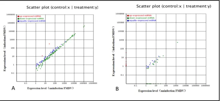
match !3) and further sequence analysis, 192556 and 196095 unique small RNAs were indicated perfect matches to known animal miRNAs deposited in miRBase 18.0, in infected and uninfected FMDV groups. According to the changes in relative miRNA abundance between the two libraries, a total of 172 differently known miRNAs were found (File S1, S2). All the different miRNAs were predominantly expressed at almost up to 1000,000 reads, suggesting abundant expression during infection. Compared with miRNA expression in uninfected group, almost all of miRNAs in infected FMDV group were significantly downreg-ulated (P,0.01), and differentially expressed miRNAs in
fold-change ranged from 0.1 to 3.0 between the two libraries (Fig. 3A, file S1). Single nucleotide polymorphism (SNP) analysis demon-strated that differences in miRNA edit positions and rates between the two libraries were identified (file S3, file S4), which may be related to FMDV infection.
Novel miRNAs Candidates and Differential Expression
In this study, 242885 and 268097 unannotated small RNAs were present in infected and uninfected groups. They were used to predict potentially novel miRNA candidates. Further, to deter-mine the relationship of these small RNAs with genuine Sus scrofa
Figure 3. Comparison of expression levels of known miRNAs (A) and novel miRNAs (B) in infected and uninfected groups.The X- and Y-axes show the expression levels of miRNAs in the two samples. The red points represent miRNAs with ratios above 2; the blue points represent miRNAs with ratios between 1/2 and 2; the green points represent miRNAs with ratios under 1/2. Ratios = miRNA expression levels in infection/miRNA expression levels in uninfected group.
doi:10.1371/journal.pone.0090865.g003
Figure 4. The endogenous novel miRNA exhibited significant base bias especially in 20, 22 and 24 nt miRNAs. doi:10.1371/journal.pone.0090865.g004
miRNA, typical secondary structures of the miRNA precursors were used to remove pseudo-miRNAs. It was predicted that 181 novel miRNAs possessed a typical stem-loop structure and free energy ranging from 25 Kcal/mol to 70 Kcal/mol (File S5, S6). According to differential analysis as discussed previously, 72 differentially expressed novel miRNA candidates were found (File S7, S8). Additionally, differential analysis showed almost all of miRNAs in the infected group were significantly downregulated (P,0.01) and differentially expressed miRNAs in fold-change ranged from 0.1 to 14.0-fold between the two libraries (Figure 3B, file S7, file S8). Additionally, novel miRNA nucleotides were detected initially at each location. Results revealed that the endogenous miRNA exhibited significant base bias, especially in 20, 23 and 24 nt miRNAs between the two groups (Fig. 4).
Validation of Differential miRNA Expression with Quantitative RT-PCR
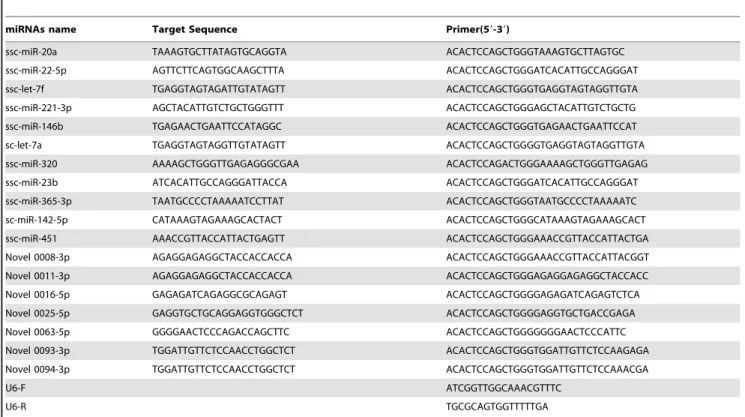
To confirm the reliability of the data, stem-loop qRT-PCR was used to compare differently expressed miRNAs and newly identified miRNAs. The three individuals in each group were subjected, to qRT-PCR. The 20 miRNAs differently expressed (11 known miRNAs and 9 novel miRNAs) were selected randomly and validated in infected and uninfected groups (file S1). The profile of 8 known miRNAs and 7 novel miRNAs was consistent with Solexa sequencing results, and 2 novel miRNAs were not detected (Figure 5), perhaps due to the deviation of qRT-PCR. Results indicated that miR-20a, miR-22-5p, let-7f, 221-3p, 146b, ssc-let-7a, 320 and ssc-miR-451 were differently expressed in two groups, and the expression levels of ssc-miR-146b, ssc-let-7a, ssc-miR-320 in uninfected group were higher than infected group.
Target Prediction, GO and KEGG Analysis
To understand the molecular function and biological processes of miRNAs during FMDV infection, target gene prediction of different miRNAs was performed. Determination of miRNA/ mRNA interactions provided molecular insight into FMDV infection mechanisms using MIREAPv0.2 software (MATERI-ALS & METHODS). About 414, 359 target genes (file S9, S10) for 244 differently expressed miRNAs (known miRNA and novel miRNA) were successfully detected. The predicted targets for these miRNAs were classified according to KEGG function annotations, and identified pathways that were actively regulated by miRNAs in FMDV infection (File S11,file S12). Results showed that about 1500 predicted genes were annotated, with the genes primarily active in endocytosis, viral myocarditis, phagosome, calcium signaling, disease and signal transduction pathways associated with cellular metabolism, innate immunity and virus infection (Figure 6). Furthermore, all targets regulated by the different miRNAs were successfully classified into three modules through GO analysis(File S13, file S14).The results showed that about 8480 genes were annotated successfully, and the majority of the genes were clustered into intracellular and membrane regions (Figure 7). Molecular functional analysis showed that the functions were Figure 5. qRT-PCR validation of the identified miRNAs using Solexa sequencing technology.Real-time RT-PCR results for 11 known miRNAs in peak and late lactation. The relative quantification of expression was calculated using the 2-DDCT method after the threshold cycle (Ct) and was normalized with the Ct of U6. The relative expression levels were presented as the 2-DDCT means 6 S+SE. * represents p,0.05, ** represents p,0.01.
doi:10.1371/journal.pone.0090865.g005
Figure 6. Pathway analysis of targets for known miRNAs according to KEGG function annotations.
doi:10.1371/journal.pone.0090865.g006
related to binding activity, with most of the genes involved in cellular or signaling processes.
Discussion
In recent years, highthroughput sequencing has become a powerful strategy for identifying novel miRNAs and studying the expression profiles of miRNA in different samples. Unlike microarray technology, Solexa highthroughput sequencing sheds light on functionally novel miRNAs [29,30]. Essentially, miRNA represents a vital component of the innate antiviral response in virus infection [31].It serves as a host gene-regulation mechanism triggered by the expression of structured miRNA molecules [11]. However, the role of host cellular miRNAs defensing against FMDV infection is still obscure. Our previous studies revealed that FMDV proliferation can reach to the maximum at the sixth hour after infection in PK-15 cells. In this study, using Solexa sequencing technology, miRNAs differentially expressed in FMDV infected and uninfected groups of PK-15 cells were identified at 6 hours point. The length distribution showed that most clean reads were primarily distributed in 19,24 nt (Fig. 1), which is consistent with the typical size of mature mammalian miRNA from dicer-processed products. The results were consis-tent with previous studies [32].
However, differences in distribution existed between the two groups (Fig. 2) showed that different levels of miRNAs perform different functions during FMDV infection. The reads were aligned against the Sus scrofagenome and annotated in GenBank and Rfam databases. It was indicated that the sequencing data in this study were highly enriched for small RNA sequences (Fig. 2). In addition to known miRNAs, functionally important novel miRNAs were initially predicted (File S5, S6), which might be undetectable using traditional methods.
The host antiviral immune response requires precise regulation [33,34], and any aberrations are associated with potentially devastating consequences. The precise activation of antiviral immunity requires exquisite regulation of host cellular miRNAs expression [35]. Changes in host cellular miRNAs expression during FMDV infection essentially disrupts the fine mechanism controlling host antiviral immunity. Differential expression anal-ysis showed 172 differential known miRNAs and 72 novel potential miRNAs. Compared with uninfected group, the down-regulated miRNAs occupied most of the differential expressed miRNAs in infected group (Fig. 3). The mechanism of down-regulation is still unclear, although researchers revealed that downregulation of cellular miRNAs during vaccine virus infection was caused by degradation of dicer enzyme [36]. Influenza virus may also downregulate cellular miRNA expression by repressing the expression of dicer [37]. Therefore it is essential to investigate the mechanism of cellular miRNAs downregulation caused by FMDV. SNP alteration analysis of edit position and rate between the two libraries (File S3, file S4) suggested that changes in SNP may be related to FMDV infection. In addition, we found that the endogenous novel miRNA exhibited significant base bias espe-cially in 20, 23, and 24 nt miRNAs (Fig. 4). It warrants additional research into miRNA editing mechanisms associated with FMDV infection, along with quantitative studies involvingt differential miRNA expression. The 20 differentially expressed miRNAs including 11 known miRNAs and 9 novel miRNAs, were selected randomly for validation with stem-loop qRT-PCR method. Results indicated that 18 miRNAs were validated and 11 miRNAs were consistent with our sequencing results, while 2 novel miRNAs were not obtained (Fig. 5), possibe reason is the decreased expression of the two miRNAs.
To further insights into the physiological function of miRNAs, target genes for differentially expressed miRNAs were predicted by aligning miRNA sequences to pig ESTs, following the rules of Figure 7. Part gene ontology classification annotated by BGI WEGO for predicted target genes.The figure shows partial GO enrichment for the predicted target genes in molecular function, cellular component and biological processes.
doi:10.1371/journal.pone.0090865.g007
target prediction published by Allen [38] and Schwab [39] using MIREAP and targetScan 6.0 software. KEGG pathway analyses and GO annotation for target genes also offer a better understanding of the target genes at molecular, cellular and biological levels. Pathway analysis suggested that these targeted genes were involved in multiple signaling pathways, such as the cytokine receptor signaling, NOD-like receptor signaling (NLRs), and Toll-like receptor (TLRs) rand pathways (Fig. 6, file S11, S12). The activation of TLRs and NLRs triggers intracellular signaling pathways that lead to effective mechanisms in innate immunity and inflammation. Researchers found the negative regulation of this signaling during posttranscriptional regulation by miRNA-155, miRNA-146, miRNA-21, miRNA-132 [40]. As the targets of Let-7f are lymphocyte-mediated, miR-142 regulates immune response via inhibition cAMP production in CD4+, CD25+ T cells and CD4+,CD25+ TREG cells by targeting AC9 mRNA [41]. In this study, ssc-miRNA-155, ssc-miRNA-146, ssc-miR-142 and miRNA-21 (file S1) were identified. It was indicated that these miRNAs primarily related to cellular immune response to FMDV infection and FMDV replication may be indirectly regulated by these miRNAs. We also found that miRNAs are associated with cell cycle, including ssc-miR-27a, and ssc-miR-27b, which are involved in apoptosis and cell death, correlating with targets of miR-29a are miR-29b related to cell division and the cell cycle [42,43]. GO analysis found that these genes are mainly involved in cell binding, transcriptional regulation, apoptosis regulation, and the response to stress (Fig. 7, file S13, S14).
Our future studies will aim to investigate the function of candidate miRNAs in PK-15 cells by altering their expression and investigating the effect on the immune response to FMDV infection by measuring levels of cytokine secretion and cell surface antigen expression.
Materials and Methods
Materials
PK-15 cells were purchased from Center for Type Culture Collection(CTCC, Wuhan, China) and cultured in modified Eagle medium (MEM, HyClone) in T-flasks (25 cm2) with 10% fetal bovine serum (FBS, HyClone) within 5% CO2incubator. FMDV
serotype Asia1 Jiangsu strain was stored at the national foot-and-mouth disease reference laboratory in China. Monolayer cells were washed three times with MEM. We added 5 mL serum-free medium containing 500mL of 104 TCID50/mL of Asia1 type
FMDV to infetecd group of cells in T-flasks. T-flasks containing the 5 mL serum-free medium were used as control. The infected cells were harvested at 6 hours post-infection (hpi) and uninfected cells were harvested at the same time. With each group in triplicates. Samples harvested from the three groups, respectively, were mixed together and used for RNA extraction.
Construction and Sequencing of small RNA libraries
Total RNA from each group of cells was extracted using the RNeasy Mini Kit (QIAGEN) according to the manufacturer’s instructions. After purification, quality control testing for RNA was conducted using an Agilent 2100 BioAnalyzer (Agilent Technol-ogies, Palo Alto, CA) and concentration analyzed using a Nanodrop ND-1000 (Nanodrop Technologies, Wilmington, DE). The small RNAs were size-fractionated from the RNA pool of two samples, purified by polyacrylamide gel electrophoresis to enrich for molecules in the range of 18–30 nt, and ligated to 59- and 39 -end RNA oligonucleotide adaptors. cDNA constructs were created by RT-PCR based on the small RNAs ligated with 39 and 59
adaptors. The PCR products (90 bp, small RNA+adaptors) were purified with 4% agarose gels and used for sequencing with Solexa sequencing technology (Illumina HiSeq 2000, San Diego, CA,
Table 2.Summary of miRNA primers used in real-time qRT-PCR.
miRNAs name Target Sequence Primer(59-39)
ssc-miR-20a TAAAGTGCTTATAGTGCAGGTA ACACTCCAGCTGGGTAAAGTGCTTAGTGC
ssc-miR-22-5p AGTTCTTCAGTGGCAAGCTTTA ACACTCCAGCTGGGATCACATTGCCAGGGAT
ssc-let-7f TGAGGTAGTAGATTGTATAGTT ACACTCCAGCTGGGTGAGGTAGTAGGTTGTA
ssc-miR-221-3p AGCTACATTGTCTGCTGGGTTT ACACTCCAGCTGGGAGCTACATTGTCTGCTG
ssc-miR-146b TGAGAACTGAATTCCATAGGC ACACTCCAGCTGGGTGAGAACTGAATTCCAT
sc-let-7a TGAGGTAGTAGGTTGTATAGTT ACACTCCAGCTGGGGTGAGGTAGTAGGTTGTA
ssc-miR-320 AAAAGCTGGGTTGAGAGGGCGAA ACACTCCAGACTGGGAAAAGCTGGGTTGAGAG
ssc-miR-23b ATCACATTGCCAGGGATTACCA ACACTCCAGCTGGGATCACATTGCCAGGGAT
ssc-miR-365-3p TAATGCCCCTAAAAATCCTTAT ACACTCCAGCTGGGTAATGCCCCTAAAAATC
sc-miR-142-5p CATAAAGTAGAAAGCACTACT ACACTCCAGCTGGGCATAAAGTAGAAAGCACT
ssc-miR-451 AAACCGTTACCATTACTGAGTT ACACTCCAGCTGGGAAACCGTTACCATTACTGA
Novel 0008-3p AGAGGAGAGGCTACCACCACCA ACACTCCAGCTGGGAAACCGTTACCATTACGGT
Novel 0011-3p AGAGGAGAGGCTACCACCACCA ACACTCCAGCTGGGAGAGGAGAGGCTACCACC
Novel 0016-5p GAGAGATCAGAGGCGCAGAGT ACACTCCAGCTGGGGAGAGATCAGAGTCTCA
Novel 0025-5p GAGGTGCTGCAGGAGGTGGGCTCT ACACTCCAGCTGGGGAGGTGCTGACCGAGA
Novel 0063-5p GGGGAACTCCCAGACCAGCTTC ACACTCCAGCTGGGGGGGAACTCCCATTC
Novel 0093-3p TGGATTGTTCTCCAACCTGGCTCT ACACTCCAGCTGGGTGGATTGTTCTCCAAGAGA
Novel 0094-3p TGGATTGTTCTCCAACCTGGCTCT ACACTCCAGCTGGGTGGATTGTTCTCCAAACGA
U6-F ATCGGTTGGCAAACGTTTC
U6-R TGCGCAGTGGTTTTTGA
doi:10.1371/journal.pone.0090865.t002
USA). All sequencing was carried out at the Beijing Genomics Institute (BGI), Shenzhen.
Analysis of Sequence Data
Low quality reads were filtered according to the base quality value. The clean reads were mapped to the sus scrofagenome to analyze their expression and distribution using SOAPv1.11 software [44]. All validated sequences were obtained for further analysis. The sequences were aligned against the known miRNAs precursors and mature miRNAs deposited in the miRBase 18.0 to identify conserved miRNAs. The unannotated sequences were used to predict potentially novel miRNA candidates. Mfold3.2 software was used to analyze the RNA secondary structures [45], and MIREAPv0.2 software (https://sourceforge.net/projects/ mireap/) [46] was used to predict the potential miRNA candidates according to established rules [47,48].
Differentially Expressed miRNAs in infected and uninfected PK-15 cells
The random variance model corrected t-test (RVM-T test) was used to calculate the significance level (P-value) of miRNA expression and the false positive rate (FDR), and miRNAs differentially expressed in infected and uninfected PK-15 cells were identified. The UCSC genome database was used to identify pri-miRNA sequences and all differentially expressed miRNAs were compared with the Sanger database to determine the specific location of pri-miRNAs in porcine genome. The presence of transcription factor binding sequences in pri-miRNA regulatory regions was used to indicate transcription factors that may regulate their transcription. This result was used to construct a regulatory network of transcription factors and miRNA molecules.
Prediction of Differential Expression of miRNA Target Genes
The target genes for differentially expressed miRNAs (known as miRNAs, novel miRNAs) were predicted by aligning the Sus scrofa EST database in NCBI, according to previously established rules [38,39].
GO Annotation and KEGG Pathway Analysis of Target Genes
According to established algorithm [48], InterProScan [49] and BGI WEGO (http://wego.genomics.org.cn/cgi-bin/wego/index. pl) [50] were used to perform GO annotation and enrichment analysis for three ontologies, molecular function, cellular compo-nent and biological process. The genes with FDR !0.5 were considered as significantly enriched in target gene candidates. Target genes involved in significant metabolic or signal transduc-tion pathways were analyzed to decipher the KEGG (Kyoto Encyclopedia of Genes and Genomes, http://www.genome.jp/ kegg/) [51].
Quantitation of Target miRNA in Infected and Uninfected PK-15 cells
Differentially expressed miRNAs were validated using relative real-time quantitative RT-PCR according to the manufacturer’s protocol. The stem-loop quantitative real-time PCR (qRT-PCR) was performed using ABI RPISM 7500 sequence detection system and SYBR Green qPCR SuperMix (Invitrogen)..RNA was treated with DNaseI (Promega) and cDNAs were synthesized using superscript III transcriptase (Invitrogen) with miRNA-specific forward primer and universal reverse primer. The miRNA-specific stem-loop RT primers were designed (Table 2) and synthesized
with the software primer 5.0. Housekeeping gene U6 was used as the internal reference. Briefly, 20 ul PCR reaction contained about 5 ng miRNA first-strand cDNA synthesis, 10 ul 26
miRcute miRNA premix, and 200 nM of each primer. The reactions were mixed gently and incubated at 94uC for 2 min, followed by 40 cycles of 95uC 15 s, 60uC 32 s and subsequent elongation at 72uC for 5 min. All samples were performed in triplicates. PCR efficiency calculations were based on the slopes of the standard curves. The absolute amount of each miRNA was calculated using the 22DDCT method [52] according to the standard curve. Each miRNA level was expressed as 22DDCT mean+SE. One-way ANOVA was used to examine the signifi-cance of differential expression level in each known/novel miRNA between infected and uninfected groups, and the difference was considered as significant when P,0.05.
Supporting Information
File S1 Differentially expressed known miRNAs. (XLSX)
File S2 Sequences of different known miRNAs. (XLSX)
File S3 Characteristics of miRNAs edit in uninfected FDMV group.
(XLSX)
File S4 Characteristics of miRNAs edit in infected FDMV group.
(XLSX)
File S5 Typical stem-loop structure and free energy range of novel miRNAs candidates in uninfected FMDV group.
(XLSX)
File S6 Typical stem-loop structure and free energy ranging of novel miRNAs candidates in infected FMDV group.
(XLSX)
File S7 Differentially expressed novel miRNA candi-dates.
(XLSX)
File S8 Sequences of different novel miRNA candidates. (XLSX)
File S9 Target genes for different known miRNAs. (XLS)
File S10 Target genes for novel miRNA candidates. (XLS)
File S11 KEGG Pathway annotations for the predicted target genes for different known miRNAs.
(XLSX)
File S12 KEGG Pathway annotations for the predicted target genes for different novel miRNA candidates. (XLSX)
File S13 GO annotations for the predicted target genes for different known miRNAs.
(XLSX)
File S14 GO annotations for the predicted target genes for different novel miRNA candidates.
(XLSX)
Acknowledgments
The authors wish to thank the journal editors and anonymous reviewers for editing and revising the manuscript.
Author Contributions
Conceived and designed the experiments: KZ YL HK XL YS. Performed the experiments: KZ YL HK. Analyzed the data: WC XL JG. Contributed reagents/materials/analysis tools: KZ HZ HT. Wrote the paper: KZ YL. Discussed the results: HZ.
References
1. Bartel DP (2004) MicroRNAs: genomics, biogenesis, mechanism, and function. Cell 116: 281–297.
2. Filipowicz W, Jaskiewicz L, Kolb FA, Pillai RS (2005) Post-transcriptional gene silencing by siRNAs and miRNAs. Current opinion in structural biology 15: 331–341.
3. Lee RC, Feinbaum RL, Ambros V (1993) The C. elegans heterochronic gene lin-4 encodes small RNAs with antisense complementarity to lin-14. Cell 75: 843–854.
4. Ghosh Z, Mallick B, Chakrabarti J (2009) Cellular versus viral microRNAs in host-virus interaction. Nucleic acids research 37: 1035–1048.
5. Zhang R, Su B (2008) MicroRNA regulation and the variability of human cortical gene expression. Nucleic acids research 36: 4621–4628.
6. Esau C, Kang X, Peralta E, Hanson E, Marcusson EG, et al. (2004) MicroRNA-143 regulates adipocyte differentiation. The Journal of biological chemistry 279: 52361–52365.
7. Inoue K (2007) [MicroRNA function in animal development]. Tanpakushitsu kakusan koso Protein, nucleic acid, enzyme 52: 197–204.
8. Ye L, Su X, Wu Z, Zheng X, Wang J, et al. (2012) Analysis of differential miRNA expression in the duodenum of Escherichia coli F18sensitive and -resistant weaned piglets. Plos One 7: e43741.
9. Scaria V, Hariharan M, Maiti S, Pillai B, Brahmachari SK (2006) Host-virus interaction: a new role for microRNAs. Retrovirology 3: 68.
10. Gottwein E, Cullen BR (2008) Viral and cellular microRNAs as determinants of viral pathogenesis and immunity. Cell Host Microbe 3: 375–387.
11. Boss IW, Plaisance KB, Renne R (2009) Role of virus-encoded microRNAs in herpesvirus biology. Trends in microbiology 17: 544–553.
12. Singh CP, Singh J, Nagaraju J (2012) A baculovirus-encoded MicroRNA (miRNA) suppresses its host miRNA biogenesis by regulating the exportin-5 cofactor Ran. Journal of virology 86: 7867–7879.
13. Ho BC, Yu SL, Chen JJ, Chang SY, Yan BS, et al. (2011) Enterovirus-induced miR-141 contributes to shutoff of host protein translation by targeting the translation initiation factor eIF4E. Cell Host Microbe 9: 58–69.
14. Jopling CL, Yi M, Lancaster AM, Lemon SM, Sarnow P (2005) Modulation of hepatitis C virus RNA abundance by a liver-specific MicroRNA. Science 309: 1577–1581.
15. Zheng Z, Ke X, Wang M, He S, Li Q, et al. (2013) Human microRNA hsa-miR-296-5p suppresses enterovirus 71 replication by targeting the viral genome. Journal of virology 87: 5645–5656.
16. Huang J, Wang F, Argyris E, Chen K, Liang Z, et al. (2007) Cellular microRNAs contribute to HIV-1 latency in resting primary CD4+ T lymphocytes. Nat Med 13: 1241–1247.
17. Sung TL, Rice AP (2009) miR-198 inhibits HIV-1 gene expression and replication in monocytes and its mechanism of action appears to involve repression of cyclin T1. Plos Pathog 5: e1000263.
18. Umbach JL, Kramer MF, Jurak I, Karnowski HW, Coen DM, et al. (2008) MicroRNAs expressed by herpes simplex virus 1 during latent infection regulate viral mRNAs. Nature 454: 780–783.
19. Wu Q, Luo Y, Lu R, Lau N, Lai EC, et al. (2010) Virus discovery by deep sequencing and assembly of virus-derived small silencing RNAs. Proc Natl Acad Sci U S A 107: 1606–1611.
20. Reese TA, Xia J, Johnson LS, Zhou X, Zhang W, et al. (2010) Identification of novel microRNA-like molecules generated from herpesvirus and host tRNA transcripts. Journal of virology 84: 10344–10353.
21. Metzker ML (2010) Sequencing technologies - the next generation. Nature reviews Genetics 11: 31–46.
22. Parameswaran P, Sklan E, Wilkins C, Burgon T, Samuel MA, et al. (2010) Six RNA viruses and forty-one hosts: viral small RNAs and modulation of small RNA repertoires in vertebrate and invertebrate systems. Plos Pathog 6: e1000764.
23. Schopman NC, Willemsen M, Liu YP, Bradley T, van Kampen A, et al. (2012) Deep sequencing of virus-infected cells reveals HIV-encoded small RNAs. Nucleic acids research 40: 414–427.
24. Wu YQ, Chen DJ, He HB, Chen DS, Chen LL, et al. (2012) Pseudorabies virus infected porcine epithelial cell line generates a diverse set of host microRNAs and a special cluster of viral microRNAs. Plos One 7: e30988.
25. Chen C, Ridzon DA, Broomer AJ, Zhou Z, Lee DH, et al. (2005) Real-time quantification of microRNAs by stem-loop RT-PCR. Nucleic acids research 33: e179.
26. Grubman MJ, Baxt B (2004) Foot-and-mouth disease. Clinical microbiology reviews 17: 465–493.
27. Sellers R, Gloster J (2008) Foot-and-mouth disease: a review of intranasal infection of cattle, sheep and pigs. Vet J 177: 159–168.
28. Davies G (2002) Foot and mouth disease. Research in veterinary science 73: 195–199.
29. Song QX, Liu YF, Hu XY, Zhang WK, Ma B, et al. (2011) Identification of miRNAs and their target genes in developing soybean seeds by deep sequencing. BMC plant biology 11: 5.
30. Li B, Qin Y, Duan H, Yin W, Xia X (2011) Genome-wide characterization of new and drought stress responsive microRNAs in Populus euphratica. Journal of experimental botany 62: 3765–3779.
31. Mahajan VS, Drake A, Chen J (2009) Virus-specific host miRNAs: antiviral defenses or promoters of persistent infection. Trends in immunology 30: 1–7. 32. Zhou Y, Tang X, Song Q, Ji Y, Wang H, et al. (2013) Identification and
characterization of pig embryo microRNAs by Solexa sequencing. Reproduction in domestic animals 48: 112–120.
33. Aldridge JR Jr, Moseley CE, Boltz DA, Negovetich NJ, Reynolds C, et al. (2009) TNF/iNOS-producing dendritic cells are the necessary evil of lethal influenza virus infection. Proc Natl Acad Sci U S A 106: 5306–5311.
34. Li Y, Chan EY, Li J, Ni C, Peng X, et al. (2010) MicroRNA expression and virulence in pandemic influenza virus-infected mice. Journal of virology 84: 3023–3032.
35. Flynt AS, Lai EC (2008) Biological principles of microRNA-mediated regulation: shared themes amid diversity. Nature reviews Genetics 9: 831–842. 36. Grinberg M, Gilad S, Meiri E, Levy A, Isakov O, et al. (2012) Vaccinia virus
infection suppresses the cell microRNA machinery. Archives of virology 157: 1719–1727.
37. Matskevich AA, Moelling K (2007) Dicer is involved in protection against influenza A virus infection. The Journal of general virology 88: 2627–2635. 38. Allen E, Xie Z, Gustafson AM, Carrington JC (2005) microRNA-directed
phasing during trans-acting siRNA biogenesis in plants. Cell 121: 207–221. 39. Schwab R, Palatnik JF, Riester M, Schommer C, Schmid M, et al. (2005)
Specific effects of microRNAs on the plant transcriptome. Developmental cell 8: 517–527.
40. Coll RC, O’Neill LA (2010) New insights into the regulation of signalling by toll-like receptors and nod-toll-like receptors. J Innate Immun 2: 406–421.
41. Huang B, Zhao J, Lei Z, Shen S, Li D, et al. (2009) miR-142-3p restricts cAMP production in CD4+CD25- T cells and CD4+CD25+TREG cells by targeting AC9 mRNA. EMBO reports 10: 180–185.
42. Tang X, Muniappan L, Tang G, Ozcan S (2009) Identification of glucose-regulated miRNAs from pancreatic {Nabetani,#896} cells reveals a role for miR-30d in insulin transcription. RNA 15: 287–293.
43. Lerner M, Lundgren J, Akhoondi S, Jahn A, Ng HF, et al. (2011) MiRNA-27a controls FBW7/hCDC4-dependent cyclin E degradation and cell cycle progression. Cell cycle 10: 2172–2183.
44. Li R, Li Y, Kristiansen K, Wang J (2008) SOAP: short oligonucleotide alignment program. Bioinformatics 24: 713–714.
45. Zuker M (2003) Mfold web server for nucleic acid folding and hybridization prediction. Nucleic acids research 31: 3406–3415.
46. Li Y, Zhang Z, Liu F, Vongsangnak W, Jing Q, et al. (2012) Performance comparison and evaluation of software tools for microRNA deep-sequencing data analysis. Nucleic acids research 40: 4298–4305.
47. Zuker M, Jacobson AB (1998) Using reliability information to annotate RNA secondary structures. RNA 4: 669–679.
48. Ji Z, Wang G, Xie Z, Wang J, Zhang C, et al. (2012) Identification of novel and differentially expressed MicroRNAs of dairy goat mammary gland tissues using solexa sequencing and bioinformatics. Plos One 7: e49463.
49. Quevillon E, Silventoinen V, Pillai S, Harte N, Mulder N, et al. (2005) InterProScan: protein domains identifier. Nucleic acids research 33: W116–120. 50. Conesa A, Gotz S, Garcia-Gomez JM, Terol J, Talon M, et al. (2005) Blast2GO: a universal tool for annotation, visualization and analysis in functional genomics research. Bioinformatics 21: 3674–3676.
51. Kanehisa M, Goto S, Sato Y, Furumichi M, Tanabe M (2012) KEGG for integration and interpretation of large-scale molecular data sets. Nucleic acids research 40: D109–114.
52. Schmittgen TD, Livak KJ (2008) Analyzing real-time PCR data by the comparative C(T) method. Nature protocols 3: 1101–1108.