Identification and Characterization of MicroRNAs in
Normal Equine Tissues by Next Generation Sequencing
Myung-Chul Kim1, Seung-Woo Lee2, Doug-Young Ryu2,3, Feng-Ji Cui1, Jong Bhak4, Yongbaek Kim1,3* 1Laboratory of Clinical Pathology, College of Veterinary Medicine, Seoul National University, Seoul, Republic of Korea,2Laboratory of Environmental Health, College of Veterinary Medicine, Seoul National University, Seoul, Republic of Korea,3Research Institute for Veterinary Science, College of Veterinary Medicine, Seoul National University, Seoul, Republic of Korea,4Theragen Bio Institute, Suwon-city, Gyeonggi-do, Republic of Korea
Abstract
The role of microRNAs (miRNAs) as a post-transcriptional gene regulator has been elucidated in a broad range of organisms including domestic animals. Characterization of miRNAs in normal tissues is an important step to investigate the functions of miRNAs in various physiological and pathological conditions. Using Illumina Next Generation Sequencing (NGS) technology, we identified a total of 292 known and 329 novel miRNAs in normal horse tissues including skeletal muscle, colon and liver. Distinct sets of miRNAs were differentially expressed in a tissue-specific manner. The miRNA genes were distributed across all the chromosomes except chromosomes 29 and 31 in the horse reference genome. In some chromosomes, multiple miRNAs were clustered and considered to be polycistronic transcript. A base composition analysis showed that equine miRNAs had a higher frequency of A+U than G+C. Furthermore, U tended to be more frequent at the 59 end of miRNA sequences. This is the first experimental study that identifies and characterizes the global miRNA expression profile in normal horse tissues. The present study enriches the horse miRNA database and provides useful information for further research dissecting biological functions of miRNAs in horse.
Citation:Kim M-C, Lee S-W, Ryu D-Y, Cui F-J, Bhak J, et al. (2014) Identification and Characterization of MicroRNAs in Normal Equine Tissues by Next Generation Sequencing. PLoS ONE 9(4): e93662. doi:10.1371/journal.pone.0093662
Editor:Ratna B. Ray, SAINT LOUIS UNIVERSITY, United States of America
ReceivedJanuary 16, 2014;AcceptedMarch 4, 2014;PublishedApril 2, 2014
Copyright:ß2014 Kim et al. This is an open-access article distributed under the terms of the Creative Commons Attribution License, which permits unrestricted use, distribution, and reproduction in any medium, provided the original author and source are credited.
Funding:This study was supported by Bio-industry Technology Development Program (Grant no. 1111594) of iPET (Korea Institute of Planning and Evaluation for Technology in Food, Agriculture, Forestry and Fisheries), Ministry of Agriculture, Food and Rural Affairs, Republic of Korea. Myung-Chul Kim and Seung-Woo Lee were additionally supported by the Brain Korea 21 Plus Program for Creative Veterinary Science Research, College of Veterinary Medicine, Seoul National University. The funders had no role in study design, data collection and analysis, decision to publish, or preparation of the manuscript.
Competing Interests:The authors have declared that no competing interests exist.
* E-mail: [email protected]
Introduction
MicroRNAs (miRNAs) play a crucial role in various physiolog-ical and pathologphysiolog-ical conditions in a broad range of organisms from viruses to animals [1–2]. The short non-coding RNAs regulate the expression of thousands of genes by partial or complementary binding to target mRNAs, resulting in transla-tional inhibition and/or degradation of the target mRNAs [3–5]. The mature miRNAs are generated from serial stepwise processing [6]. The enzyme Drosha cleaves the single stranded primary miRNA transcripts(pri-miRNAs) to produce a precursor stem-loop secondary structure(pre-miRNAs) [7]. In turn, the pre-miRNAs are cleaved by the enzyme Dicer to produce mature miRNA duplexes [8]. Only one of the two strands acts as a regulatory RNA associated with RNA-induced silencing complex (RISC) [9].
The importance of miRNAs has been recently recognized in veterinary medicine. There is increasing interest in canine miRNAs because of their clinical relevance for human breast cancers [10–11]. Down-regulation of miR-130 and miR-30 in canine heart plays an important role in the pathogenesis of chronic atrial fibrillation [12]. Distinct sets of miRNAs including miR-122, miR-193b, and miR-483 were reported to be involved in the development of type-2 diabetes mellitus in cats [13]. A large number of miRNAs have been identified in normal tissues of swine and bovine species [14–17]. However, information about equine miRNAs and their role in clinical conditions is scarce. Recently,in
silicoanalytical methodology was applied to identify and charac-terize mature 354 equine miRNAs [18]. A study revealed 82 new miRNAs in equine sperms, suggesting the role of miRNAs in the regulation of sperm function, fertility and reproduction [19]. Barrey et al. [20] described the expression profile of muscular miRNAs in healthy and myopathic horses using a direct cloning technology.
Forward genetics, bioinformatics and direct cloning have been utilized to study miRNAs [21]. Recently, next generation sequencing (NGS) technology has emerged as a major tool to scrutinize small RNAs including miRNAs [22]. An important feature of the NGS is parallel sequencing of clonally amplified or single DNA molecules that are spatially separated in a flow cell [23]. Due to its ability to generate millions of reads with determined lengths, NGS greatly improves the capacity to identify a large number of novel miRNAs on a genomic scale [24].
high-throughput NGS technology. Our study identifies a list of known miRNAs as well as candidates for novel miRNAs in horse tissues. Additionally, characteristics of horse microRNAs including expression patterns in major tissues, sequence composition and chromosomal distribution were determined. Our study provides
background data and information to facilitate research on the role of miRNAs in the pathogenesis of various conditions in horses.
Results
Library construction of small RNAs from horses
We performed high-throughput short read sequencing of small RNAs from skeletal muscle, liver, and colon tissues from eight Thoroughbred horses. A number of reads, with counts ranging from 11973300 to 21838589, were obtained from the cDNA libraries of small RNAs, of which approximately 100% were of high quality. After eliminating 39 adapter null, 59 adapter contaminants, insert null, smaller than 18 nucleotides (nts) and poly A sequences, more than 94% of clean reads in all tissue samples were subjected to further analyses using bioinformatic tools (Table S1). The resultant small RNAs ranged from 18 to 30 nts in length, with the majority having a length of 22 nt.
In general, small RNA libraries generated by NGS is complex in composition because they harbor a large number of degradation fragments derived from coding and noncoding transcripts [32]. To annotate the small RNAs generated by NGS, we performed a BLAST that search of the small RNAs against the equine genome database. Unique small RNAs from muscle, colon, and liver tissues were annotated against the NCBI Genbank and Rfam databases. The annotation results showed that non-coding RNA including exons, introns, repeats, rRNA, scRNA, snRNA, snoRNA, Figure 1. Length distribution and abundance of all miRNAs in horse muscle, colon, and liver tissues.Approximately 83% of all sequences in horse tissues are concentrated in the 20–24 nt range. The most frequent length is 23 nt in all tissues.
doi:10.1371/journal.pone.0093662.g001
Figure 2. Venn diagram representing the distribution of known and novel miRNAs in horse muscle, colon, and liver tissues. Counts in the Venn diagram are the number of miRNAs identified in each tissue. A total 292 known (A) and 329 novel miRNAs (B) are identified in horse tissues including muscle, colon, and liver.
srpRNA, and tRNA accounted for a large percentage of the unique read counts. It also revealed that miRNAs constituted a small portion of the unique read counts but a significant portion of overall read counts (Table S2). More than 70% of unique sequences in all the tissues could not be classified. The sequences of unannotated small RNAs that could be mapped to the equine genome were subject to further analyses to identify novel miRNA candidates.
Expression profile of miRNAs in major organs
To identify previously reported miRNAs in horse tissues, the small RNAs were submitted for a BLAST search against known equine miRNAs and their precursors in the latest miRBase release version 15.0 [32]. As a result, a total of 201 families including 292 known miRNAs were detected across horse samples (Table S3). The length distribution of known miRNAs was similar in all samples mainly 22–24 nts (Figure 1). The expression profile of known miRNAs in horse tissues was analyzed (Figure 2A). Subsets of tissue-specific miRNAs were identified: 36 miRNAs in muscle samples, 99 miRNAs in colon samples, and 31 miRNAs in liver samples (Table S4).
Identification of novel equine miRNAs
To identify candidates for novel equine miRNAs, a prediction software Mireap (http://sourceforge.net/projects/mireap/) was used to determine secondary structures, the Dicer cleavage sites and the minimum free energy of the unannotated small RNA tags [33]. Based on the analytical criteria, a total of 329 unannotated miRNA sequences were determined as candidates for novel miRNAs in horse (Table S5). The putative novel miRNAs exhibited a concentrated length distribution between 20 nt and
24 nt, with a peak at 21,22 nt (Figure 3). Expression profile of the novel miRNAs in horse tissues was investigated (Figure 2B). Similar to the known miRNAs, subsets of novel miRNAs were expressed in a tissue-specific manner: 31 novel miRNAs in muscle samples, 123 novel miRNAs in colon samples, 45 novel miRNAs in liver samples (Table S6). Read counts of most novel miRNAs were less than 100 (Data not shown).
Mapping to genome and miRNA transcriptional units
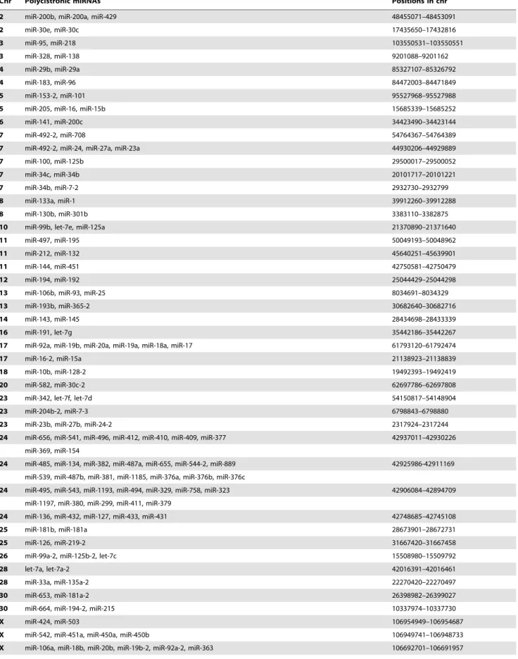
A total of 292 known miRNAs were mapped across the 32 horse chromosomes with the exception of chromosomes 29 and 31 (Figure 4). We attempted to determine whether equine miRNAs show expression patterns representing single transcriptional units. In our analysis, if two miRNAs were within 3 kb of each other, they were considered to be in the same cluster [34]. We found that up to 160 miRNAs were closely co-localized in clusters with the 3 kb threshold. The equine genome contains 51 miRNA clusters with 160 miRNAs accounting for approximately 55% of the known miRNAs (Figure 5). The clusters were variably located on individual chromosome. For example, 40 miRNA genes in 4 clusters were mapped to chromosome 24, while only 2 miRNA genes were mapped to chromosomes 6, 12, 14, 16, 18, and 20 (Table 1).
Nucleotide bias of equine miRNAs
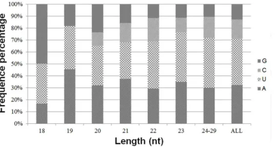
The first nucleotide at the 59end of organ-specific miRNAs of any length was predominantly U with a frequency of 40%, 35%, and 43% in muscle, colon, and liver tissues, respectively (Figure 6). Likewise, G and A were the only preferred nucleotide at the 59end of 18 and 19 nt long miRNAs in colon, although very short miRNAs that were 18 to 19 nt long were rare in horse. The base composition at each position of all mature miRNAs revealed a clear tendency for U being the most frequently observed nucleotide at specific sites (1, 9, 21, 23–26, and 28). However, C was the least used nucleotide at specific sites (3, 7, 12, 13, 15, 16, 18, 19, 21, 24, and 25) in all tissue miRNA sequences. The distribution of A+U, accounting for an average of 72%, was generally preferred to C+G in all tissue samples (Figure 7). In contrast, from third to sixth nucleotide positions, C+G was more abundant than A+U in all tissue-specific miRNAs. Furthermore, from the second to eighth nucleotide positions, which belong to the seed region of miRNAs, colon miRNAs showed prominent C+G compared to A+U (Data not shown) [35].
Discussion
Using Illumina short read sequencing technology, a total of 292 known and 329 novel miRNAs were identified in equine tissues including muscle, liver, and colon. Furthermore, it was found that subsets of miRNAs were differentially expressed in horse tissues, suggesting tissue-specific behavior of equine miRNAs. The identification and characterization of equine miRNAs was carried out using NGS technology to obtain millions of small RNA sequences and to construct small RNA differential expression profile in target organs [23]. Although NGS has the advantage of cloning small RNAs exclusively, it has several disadvantages compared to computational approaches [41]. The limitations include the difficulty of finding miRNAs that are expressed at a low level and a cloning inability due to physical properties such as sequence composition and/or post-transcriptional modifications [42]. Further studies are warranted to elucidate a comprehensive list of equine miRNAs that are biologically important.
Although horses are one of the most important domestic animals, research on equine miRNAs has been limited [36]. Figure 3. Length distribution of novel miRNAs in horse tissues.
miRNA sequences of all lengths are distributed in the 20–24 nt range. The most frequent length is 22 nt (34.65%) in horse miRNAs. doi:10.1371/journal.pone.0093662.g003
Attempts have recently been made to investigate the role of equine miRNAs in various conditions. A study using direct sequencing verified two miRNAs, miR-433 and -127, in horse, thus proving the conservation of the miRNAs throughout mammalian species [37]. Zhou et al.[18] discovered a total of 354 mature miRNAs through an integrated computational analysis of an equine genome, providing a baseline for horse miRNA research. Additionally, they presented valuable references for all major sequence characteristics of horse miRNAs, such as the contents of each nucleotide, A+U, C+G, and base composition at each position of pre-miRNA and mature miRNA sequences. Recently, expression profiles of 82 miRNAs were characterized in equine sperms, indicating their role in the regulation of sperm function, fertility, and reproduction [19]. A study revealed that 10 miRNAs were significantly expressed in equine muscles, some of which were also expressed in blood samples [20]. The present study provides the first experimental data on expression profile of global equine
miRNAs using NGS technology, some of which were previously reported in anin silicostudy [18].
In horse, the characterization of miRNAs in major organs including liver, skeletal muscle, and large intestine has significant clinical relevance to important equine diseases. Horses are more prone to develop colic, a fatal disease complex resulting from numerous factors including strangulation, obstruction and volvulus [38]. Approximately 34% of horses undergoing an exploratory laparotomy had displacement or volvulus of the large colon due to its free mobility in the abdominal cavity [38–39]. Unfortunately, despite the magnitude of the problem of equine colic, relatively little has been known about exact causes. Because of their nature and human usage, horses are affected by various types of muscle diseases including exercise-associated myopathy, post-exhaustion syndrome, and nutritional myopathy [40]. Horses frequently develop liver disease because of their grazing habits, causing significant economic losses [41]. Given that the expression profile of miRNA is specific for organs and/or tissues [3], it is clinically Figure 4. Chromosomal locations of 292 known miRNA genes in horse.Black vertical lines represent the miRNA gene, the depth of color represents the number of miRNA genes in this region. The relative locations of individual miRNAs across the 32 horse chromosomes are shown with the exception of chromosomes 29 and 31.
important to discover subsets of organ-specific miRNAs. Our study revealed that both known and novel miRNAs in colon have a larger proportion of total miRNAs than other tissues. In contrast to other mammals, the equine large intestine has relatively complicated anatomical structures and physiological functions to facilitate a steady flow of nutrient [42]. Our data may indicate that the colon-specific miRNAs have more sophisticated roles in the colon’s regulatory system [17].
Barrey et al. [20] demonstrated that muscle-specific miRNAs were also detected in blood samples of horses with heritable muscular pathology. The study suggested the potential value of the miRNAs for the development of novel and minimally invasive methodology for diagnosing muscular pathology in horses [20]. Similarly, further studies are warranted to investigate expression profile of tissue-specific miRNAs in the blood and may provide valuable biomarker to identify organ damage and various disease conditions.
Characterization of 59-end sequences of the miRNA is important because seed sequence of a miRNA is critical in miRNA-target mRNA binding [4–43]. Analysis of nucleotide sequences in eukaryotic miRNAs showed a clear bias for U or A at the 59 position [44]. Our study revealed that U was the most frequent nucleotide at the 59-end of tissue-specific miRNAs, followed by A. This finding is consistent with results from the published studies on miRNAs of other organisms [18–32–33–45]. In plant, U at the end of 59was proposed as being critical for the biogenesis of miRNAs through the recognition of targeted miRNA precursor by RISC [46]. The C+G contents in the 6thposition of the seed region were known to induce or enhance the function of a miRNA [45]. In our study, all tissue miRNAs had high percentages of C+G from the third to sixth positions in the sequence. The miRNAs in colon had more distinct sequence bias showing high percentage of C+G in all of the seed nucleotides such a bias may be responsible for distinct functions in colon compared to different organs [45]. The base bias in seed sequence may
provide a valuable reference for further study on the identification of regulatory cellular function of miRNAs.
In our study, the equine miRNAs were located on diverse chromosomes. An intriguing feature was that the distribution of miRNAs along the chromosomes was uneven; some chromosomes were relatively miRNA-rich while others were miRNA-poor. Based on chromosomal mapping, more than half of the known miRNAs that exists within 3 kb threshold of each other were considered as a polycistronic transcript. It is interesting to note that our study revealed a number of putative miRNA clusters in horse genome. Consistent with a published study on horse miRNAs [18], a miR-17-92 cluster was also detected on chromosome 17 in our study. This cluster is reportedly harboring 17, 18a, miR-19a, miR-19b, miR-20a, and miR-92a, and is a well-preserved cluster in human and all vertebrates [11–18]. In mammals, the miR-17-92 cluster is divided into two cluster paralogs: miR-106b-25 cluster and the miR-106a-363 cluster [47]. In our study, the former cluster was located on chromosome 13 and the latter cluster on chromosome X. Further studies addressing the precise role of polycistronic transcripts in horse may enhance our understanding about the mechanisms of miRNA generation.
The present study revealed that approximately 53% of the known miRNAs were observed as part of a polycistronic unit. The finding suggests that the clustering property of miRNAs is preserved in horse, although there is partial discrepancy in chromosomal location compared with human data [47]. The proportion of miRNAs in polycistronic units is similar to that of zebrafish miRNAs (50%) [48]. It is noteworthy that approximately 72% of clustered equine miRNAs in our study correspond to those detected by the computational analysis of horse [18]. However, 45 of the 160 miRNAs that are considered as a polycistron could not be identified at orthologous positions in thein silicostudy [18]. We speculated that difference in analysis methodologies is responsible for this discrepancy in miRNA clustering. In our study the miRNA clustering was made based on sequence data from NGS Figure 5. Chromosomal locations of polycistronic miRNAs in horse. Individual black horizontal lines represent polycistronic miRNA transcripts, the stars refer to multiple miRNA genes in this region. The relative locations of individual miRNAs that are considered as a polycistron are within 3 kb.
doi:10.1371/journal.pone.0093662.g005
Table 1.Polycistronic miRNAs in horse genome.
Chr Polycistronic miRNAs Positions in chr
2 miR-200b, miR-200a, miR-429 48455071–48453091
2 miR-30e, miR-30c 17435650–17432816
3 miR-95, miR-218 103550531–103550551
3 miR-328, miR-138 9201088–9201162
4 miR-29b, miR-29a 85327107–85326792
4 miR-183, miR-96 84472003–84471849
5 miR-153-2, miR-101 95527968–95527988
5 miR-205, miR-16, miR-15b 15685339–15685252
6 miR-141, miR-200c 34423490–34423144
7 miR-492-2, miR-708 54764367–54764389
7 miR-492-2, miR-24, miR-27a, miR-23a 44930206–44929889
7 miR-100, miR-125b 29500017–29500052
7 miR-34c, miR-34b 20101717–20101221
7 miR-34b, miR-7-2 2932730–2932799
8 miR-133a, miR-1 39912260–39912288
8 miR-130b, miR-301b 3383110–3382875
10 miR-99b, let-7e, miR-125a 21370890–21371640
11 miR-497, miR-195 50049193–50048962
11 miR-212, miR-132 45640251–45639901
11 miR-144, miR-451 42750581–42750479
12 miR-194, miR-192 25044429–25044298
13 miR-106b, miR-93, miR-25 8034691–8034329
13 miR-193b, miR-365-2 30682640–30682716
14 miR-143, miR-145 28434698–28433339
16 miR-191, let-7g 35442186–35442267
17 miR-92a, miR-19b, miR-20a, miR-19a, miR-18a, miR-17 61793120–61792474
17 miR-16-2, miR-15a 21138923–21138839
18 miR-10b, miR-128-2 19492393–19492419
20 miR-582, miR-30c-2 62697786–62697808
23 miR-342, let-7f, let-7d 54150817–54148904
23 miR-204b-2, miR-7-3 6798843–6798880
23 miR-23b, miR-27b, miR-24-2 2317924–2317244
24 miR-656, miR-541, miR-496, miR-412, miR-410, miR-409, miR-377 42937011–42930226
miR-369, miR-154
24 miR-485, miR-134, miR-382, miR-487a, miR-655, miR-544-2, miR-889 42925986-42911169
miR-539, miR-487b, miR-381, miR-1185, miR-376a, miR-376b, miR-376c
24 miR-495, miR-543, miR-1193, miR-494, miR-329, miR-758, miR-323 42906084–42894709
miR-1197, miR-380, miR-299, miR-411, miR-379
24 miR-136, miR-432, miR-127, miR-433, miR-431 42748685–42745108
25 miR-181b, miR-181a 28673901–28672731
25 miR-126, miR-219-2 31667420–31667458
26 miR-99a-2, miR-125b-2, let-7c 15508980–15509792
28 let-7a, let-7a-2 42016391–42016461
28 miR-33a, miR-135a-2 22270420–22270497
30 miR-653, miR-181a-2 26398982–26399027
30 miR-664, miR-194-2, miR-215 10337974–10337730
X miR-424, miR-503 106954949–106954687
X miR-542, miR-451a, miR-450a, miR-450b 106949741–106948733
technology, whereas thein silicostudy utilized a limited number of miRNAs identified by BLAST search. Zhou et al. [18] reported polycistronic miRNAs such as miR-302a, -302b, -302c, -302d, and -367 on chromosome 2 and miR-1912 and -1264 on chromosome X, these were not found in our result. In contrast, newly found polycistronic miRNAs in our study were not reported by Zhou et. al [16]. A number of questions for the clustering property for equine miRNAs remain to be clarified in further studies.
Conclusions
In the present study, we identified 292 known and 329 novel miRNAs in normal equine tissues by using NGS technology. This study significantly enriches the horse miRNA database and presents a valuable reference for equine miRNAs. In addition, the global miRNA expression profile in equine tissues revealed that distinct sets of miRNAs were expressed in a tissue-specific pattern. Clustering in chromosomes and sequence characteristics of miRNA may be useful for dissecting their biological functions in horse. Taken together, the present study suggests that equine miRNAs may play an important role in development and function of specific tissues, and therefore, they can be developed as a valuable molecular marker for various pathophysiological condi-tions.
Materials and Methods
Ethics statement
Animals were sampled with the appropriate consent from horse owners. These horses sacrificed were humanely euthanized following Institutional Animal Care Guidelines for Experimental Animal Use and approved by the Seoul National University Animal Care Committee.
Tissue collection and high-throughput sequencing of small RNAs
Before the sacrifice, the animals were determined to be healthy based on the results from physical exams, clinical observations and clinicopathologic tests. Major organs including skeletal muscle, liver, and colon were collected immediately after the sacrifice. Collected tissue samples were snap-frozen in liquid nitrogen and stored at280uC until use. Total RNAs were isolated by a phenol-chloroform method with Trizol reagent (Invitrogen, Carlsbad, CA, USA) according to the manufacturer’s recommendations. RNA quality and quantity was determined by Nanodrop (Nanodrop Technologies, Wilmington, DE, USA) and Agilent’s 2100 Bioanalyzer (Agilent Technologies, Palo Alto, CA, USA). Total RNAs with high quality were subjected to NGS analysis performed at Theragen Bio Institute (Suwon-city, Gyeonggi-do, The Republic of Korea).
Table 1.Cont.
Chr Polycistronic miRNAs Positions in chr
X miR-374a, miR-545 55500596–55500517
X miR-374b, miR-421 55435984–55435897
X miR-502, miR-660, miR-500-2, miR-501, miR-362, miR-500 40077407–40073367
X miR-188, miR-532 40068847–40068549
X miR-222, miR-221 37091426–37090756
doi:10.1371/journal.pone.0093662.t001
Figure 6. The percentage distribution of the first nucleotide at the 59end of all miRNAs of all lengths.Various nucleotides are detected at the 59end of miRNA sequences. In general, U is the predominant nucleotide at 59end of all miRNAs of all lengths except for miRNAs of lengths 19 and 21.
doi:10.1371/journal.pone.0093662.g006
The total RNAs were separated on 15% denaturation polyacrylamide gels, and the band of small RNA fragments between 18 and 32 nt in size were excised. Recovered RNAs from the gels were ligated to a 59adaptor and a 39adaptor sequentially, and reverse-transcribed to cDNA to obtain sufficient product for Illumina short read sequencing technology (Hiseq 2000). The cDNA products of the small RNA fragments were sequenced directly using Illumina HiSeq 2000 sequencer (Illumina Inc, San Diego, CA, USA) following the Illumina’s protocols.
Sequence analysis of small RNAs
The sequence tags from the NGS were subject to a data cleaning process, which removes the low quality reads, 59primer contaminants, reads without 39 primer or insert tag, reads with poly A, and reads shorter than 18 nt. Then, standard bioinfor-matics analyses were carried out to annotate the resultant clean tags into different categories. The small RNA tags were annotated using the Rfam RNA family database (http://www.sanger.ac.uk/ software/Rfam) and the NCBI GenBank noncoding RNA database (http://www.ncbi.nlm.nih.gov/). Sequences perfectly matching the equine genome along their entire length were subjected to subsequent analyses. Sequences matching known equine small RNAs such as rRNA, scRNA, snoRNA, snRNA and tRNA or degradation fragments of mRNAs were excluded in further analyses.
Identification of conserved and novel miRNAs
The small RNAs cleaned with Genbank and Rfam were aligned against the miRNA databases, miRBase15.0, to identify the conserved miRNA sequences. Whenever there was miRNA information of horse in miRBase 15.0, perfectly matched
sequences were considered as known miRNAs. The small RNAs that cannot be annotated to any category were subjected to novel miRNA prediction software, Mireap (http://sourceforge.net/ projects/mireap/), which is utilized to predict novel miRNA by exploring the secondary structure, the Dicer cleavage site and the minimum free energy of the unannotated small RNA tags that could be mapped to the horse genome. Potential novel miRNA candidates should meet the following 10 parameters according to Mireap. (1) minimal miRNA sequence length of 18 nt and maximal miRNA sequence length of 26 nt; (2) minimal miRNA reference sequence length of 20 nt and maximal miRNA reference sequence length of 24 nt; (3) minimal depth of Drosha/Dicer cutting site, 3 nt; (4) maximal copy number of miRNAs on reference, 20 nt; (5) maximal free energy allowed for a miRNA precursor,218 kcal/mol; (6) maximal space between miRNA and miRNA*, 35 nt; (7) minimal base pairs of miRNA and miRNA*, 14 nt (8) maximal bulge of miRNA and miRNA*, 4 nt; (9) maximal asymmetry of miRNA/miRNA* duplex, 5 nt; and (10) flank sequence length of miRNA precursor, 10 nt. The sequencing data of the conserved and novel miRNAs were deposited in the National Center for Biotechnology Information Sequence Read Archive (http://www.ncbi.nlm.nih.gov/Traces/sra/) under sub-mission number SRA058805.
Characterization of equine miRNAs
To analyze chromosomal distribution, the known miRNAs identified in the miRBase database were mapped to the genome by an alignment program Short Oligonucleotide Analysis Package software (SOAP, http://soap.genomics.org.cn). The miRNAs on each chromosome were grouped into clusters based on their locations in the equine genome. If two miRNAs were within 3 kb Figure 7. The percentage distribution of base composition at each position of miRNAs.MiRNAs in all combined tissues.
of each other, they were considered as being in the same cluster [34]. Using Illumina Hiseq 2000 sequencer, a more detailed analysis was also performed to further characterize the base composition of miRNA sequences. miRNA Sequences were aligned to the miRNA precursor of horse (mature miRNA if there is no precursor information in miRBase15.0) to predict the base bias in the first position of identified miRNAs of certain length and at each position of all the identified miRNAs.
Supporting Information
Table S1 Summary of small RNA sequencing data in
muscle, colon, and liver samples.
(XLSX)
Table S2 Distribution of the genome-mapped sequence
reads in small RNA libraries.
(XLSX)
Table S3 Detailed information of all 292 known miRNA
genes in horse genome.
(XLSX)
Table S4 Detailed information of 166 tissue specific
miRNA genes in horse genome.
(XLSX)
Table S5 Detailed information of all 329 novel miRNA
genes in horse genome.
(XLSX)
Table S6 Detailed information of 199 tissue specific
novel miRNA genes in horse genome.
(XLSX)
Acknowledgments
The authors are grateful to Bokyoung Yang (Theragen Bio Institute, Suwon-city, The Republic of Korea) for her excellent technical assistance.
Author Contributions
Conceived and designed the experiments: MCK YK. Performed the experiments: MCK SWL FJC. Analyzed the data: MCK YK DYR JB. Contributed reagents/materials/analysis tools: MCK YK DYR JB. Wrote the paper: MCK YK DYR.
References
1. Hwang H, Mendell J (2006) MicroRNAs in cell proliferation, cell death, and tumorigenesis. Br J Cancer 94: 776–780.
2. Carrington JC, Ambros V (2003) Role of microRNAs in plant and animal development. Sci Signal 301: 336–338.
3. Flynt AS, Lai EC (2008) Biological principles of microRNA-mediated regulation: shared themes amid diversity. Nat Rev Genet 9: 831–842.
4. Engels B, Hutvagner G (2006) Principles and effects of microRNA-mediated post-transcriptional gene regulation. Oncogene 25: 6163–6169.
5. Ambros V (2004) The functions of animal microRNAs. Nature 431: 350–355. 6. Lee Y, Jeon K, Lee J-T, Kim S, Kim VN (2002) MicroRNA maturation: stepwise processing and subcellular localization. The EMBO journal 21: 4663– 4670.
7. Bartel DP (2004) MicroRNAs: genomics, biogenesis, mechanism, and function. Cell 116: 281–297.
8. Bernstein E, Caudy AA, Hammond SM, Hannon GJ (2001) Role for a bidentate ribonuclease in the initiation step of RNA interference. Nature 409: 363–366. 9. Hammond SM, Bernstein E, Beach D, Hannon GJ (2000) An RNA-directed
nuclease mediates post-transcriptional gene silencing in Drosophila cells. Nature 404: 293–296.
10. Boggs RM, Wright ZM, Stickney MJ, Porter WW, Murphy KE (2008) MicroRNA expression in canine mammary cancer. Mamm Genome 19: 561– 569.
11. Boggs RM, Moody JA, Long CR, Tsai KL, Murphy KE (2007) Identification, amplification and characterization of miR-17-92 from canine tissue. Gene 404: 25–30.
12. Li H, Li S, Yu B, Liu S (2012) Expression of miR-133 and miR-30 in chronic atrial fibrillation in canines. Molecular medicine reports 5: 1457–1460. 13. Fleischhacker SN, Bauersachs S, Wehner A, Hartmann K, Weber K (2013)
Differential expression of circulating microRNAs in diabetic and healthy lean cats. The Veterinary Journal 197: 688–693.
14. Li G, Li Y, Li X, Ning X, Li M, et al. (2011) MicroRNA identity and abundance in developing swine adipose tissue as determined by Solexa sequencing. J Cell Biochem 112: 1318–1328.
15. Dunn W, DuRaine G, Reddi AH (2009) Profiling microRNA expression in bovine articular cartilage and implications for mechanotransduction. Arthritis Rheum 60: 2333–2339.
16. Gu Z, Eleswarapu S, Jiang H (2007) Identification and characterization of microRNAs from the bovine adipose tissue and mammary gland. FEBS Lett 581: 981–988.
17. Jin W, Grant JR, Stothard P, Moore SS, Guan LL (2009) Characterization of bovine miRNAs by sequencing and bioinformatics analysis. BMC Mol Biol 10: 90.
18. Zhou M, Wang Q, Sun J, Li X, Xu L, et al. (2009) In silico detection and characteristics of novel microRNA genes in the Equus caballus genome using an integrated ab initio and comparative genomic approach. Genomics 94: 125–131. 19. Das PJ, McCarthy F, Vishnoi M, Paria N, Gresham C, et al. (2013) Stallion Sperm Transcriptome Comprises Functionally Coherent Coding and Regula-tory RNAs as Revealed by Microarray Analysis and RNA-seq. PLoS One 8: e56535.
20. Barrey E, Bonnamy B, Barrey E, Mata X, Chaffaux S, et al. (2010) Muscular microRNA expressions in healthy and myopathic horses suffering from polysaccharide storage myopathy or recurrent exertional rhabdomyolysis. Equine Vet J 42: 303–310.
21. Wang L, Liu H, Li D, Chen H (2011) Identification and characterization of maize microRNAs involved in the very early stage of seed germination. BMC Genomics 12: 154.
22. Shendure J, Ji H (2008) Next-generation DNA sequencing. Nat Biotechnol 26: 1135–1145.
23. Voelkerding KV, Dames SA, Durtschi JD (2009) Next-generation sequencing: from basic research to diagnostics. Clin Chem 55: 641–658.
24. Kozomara A, Griffiths-Jones S (2011) miRBase: integrating microRNA annotation and deep-sequencing data. Nucleic Acids Res 39: D152–D157. 25. Vila` C, Leonard JA, Go¨therstro¨m A, Marklund S, Sandberg K, et al. (2001)
Widespread origins of domestic horse lineages. Science 291: 474–477. 26. Wade C, Giulotto E, Sigurdsson S, Zoli M, Gnerre S, et al. (2009) Genome
sequence, comparative analysis, and population genetics of the domestic horse. Science 326: 865–867.
27. Nicholas FW (2003) Online Mendelian Inheritance in Animals (OMIA): a comparative knowledgebase of genetic disorders and other familial traits in non-laboratory animals. Nucleic Acids Res 31: 275–277.
28. Chowdhary BP, Paria N, Raudsepp T (2008) Potential applications of equine genomics in dissecting diseases and fertility. Anim Reprod Sci 107: 208–218. 29. Lacourt M, Gao C, Li A, Girard C, Beauchamp G, et al. (2012) Relationship
between cartilage and subchondral bone lesions in repetitive impact trauma-induced equine osteoarthritis. Osteoarthritis Cartilage 20: 572–583. 30. Yang H, Ma Y, Li B, Dugarjaviin M (2010) Progress on horse genome project.
Yi chuan 32: 211–218.
31. McKenzie E (2011) Muscle physiology and nutrition in exercising horses. Equine Vet J 43: 637–639.
32. Wei LQ, Yan LF, Wang T (2011) Deep sequencing on genome-wide scale reveals the unique composition and expression patterns of microRNAs in developing pollen of Oryza sativa. Genome Biol 12: R53.
33. Ji Z, Wang G, Xie Z, Zhang C, Wang J (2012) Identification and characterization of microRNA in the dairy goat (Capra hircus) mammary gland by Solexa deep-sequencing technology. Mol Biol Rep 39: 9361–9371. 34. Liang Y, Ridzon D, Wong L, Chen C (2007) Characterization of microRNA
expression profiles in normal human tissues. BMC Genomics 8: 166. 35. Creighton CJ, Reid JG, Gunaratne PH (2009) Expression profiling of
microRNAs by deep sequencing. Brief Bioinform 10: 490–497.
36. Chi X, Yang Q, Chen X, Wang J, Pan L, et al. (2011) Identification and characterization of microRNAs from peanut (Arachis hypogaea L.) by high-throughput sequencing. PLoS One 6: e27530.
37. Song G, Wang L (2009) A conserved gene structure and expression regulation of miR-433 and miR-127 in mammals. PLoS One 4: e7829.
38. Mair T, Smith L (2005) Survival and complication rates in 300 horses undergoing surgical treatment of colic. Part 1: Short-term survival following a single laparotomy. Equine Vet J 37: 296–302.
39. Mair TS, Divers TJ, Ducharme NG (2002) Manual of equine gastroenterology. London: W.B. Saunders.
40. Freestone J, Carlson G (1991) Muscle disorders in the horse: a retrospective study. Equine Vet J 23: 86–90.
41. West HJ (1996) Clinical and pathological studies in horses with hepatic disease. Equine Vet J 28: 146–156.
42. Galuppo L, Snyder J, Pascoe J (1995) Laparoscopic anatomy of the equine abdomen. Am J Vet Res 56: 518–531.
43. Rajewsky N (2006) microRNA target predictions in animals. Nat Genet 38: S8– S13.
44. Frank F, Sonenberg N, Nagar B (2010) Structural basis for 5’-nucleotide base-specific recognition of guide RNA by human AGO2. Nature 465: 818–822. 45. Ai L, Xu M, Chen M, Zhang Y, Chen S, et al. (2012) Characterization of
microRNAs in Taenia saginata of zoonotic significance by Solexa deep sequencing and bioinformatics analysis. Parasitol Res 110: 2373–2378.
46. Zhang B, Pan X, Cannon CH, Cobb GP, Anderson TA (2006) Conservation and divergence of plant microRNA genes. The Plant Journal 46: 243–259. 47. Mendell JT (2008) miRiad roles for the miR-17-92 cluster in development and
disease. Cell 133: 217–222.