A case of Chagas’ disease panniculitis after kidney transplantation
Paniculite chagásica pós-transplante renal: relato de caso
Authors
Fábio Prestes de Campos 1
Henry Mor Pansard 1
Luiz Cláudio Arantes 1
Arnaldo Teixeira Rodrigues 1
Melissa Falster Daubermann 1
Marcos Felipe Azambuja 1
Laércio Cassol Argenta 1
Luiz Alberto Michet da Silva 1
1 Hospital Universitário de
Santa Maria - Universidade Federal de Santa Maria.
Submitted on: 06/15/2015. Approved on: 08/04/2015.
Correspondence to:
Luiz Alberto Michet da Silva. Hospital Universitário de Santa Maria Serviço de Nefrologia Universidade Federal de Santa Maria.
Rua Carlos Brenner, nº 100, Bairro Nossa Senhora de Lourdes, Santa Maria, RS, Brazil.
CEP: 97050-100.
E-mail: [email protected]
DOI: 10.5935/0101-2800.20160018
Chagas’ disease carries high morbidity and mortality due to acute parasitemia or cardiac, digestive, cutaneous or neurolo-gic chronic lesions. Latin American coun-tries have the majority of infected or at risk people. Transplanted patients using immunosuppressive agents may develop severe and even fatal forms of the disease. The available treatment causes frequent severe side-effects. A 59 years-old woman with end stage renal disease and positive serology for Chagas` disease, but without any clinical manifestation of this patho-logy, underwent kidney transplantation from a cadaveric donor and displayed three months later a thigh panniculitis from which a biopsy unveiled amastigo-te forms of Trypanosoma cruzi. The skin lesions disappeared following treatment with benzonidazole, but the drug was discontinued due to severe pancytopenia. Along with this, infection with E. faeca-lis and cytomegalovirus were treated with vancomicin and ganciclovir. The patient kept very well afterwards, with no new skin lesions and with good graft func-tion. One year and three months after the transplant, she had an emergency surgery for an aortic dissecting aneurysm. Irre-versible shock and death occurred in the immediate post-surgical period. It was not possible to establish or to rule out a rela-tionship between the trypanosomiasis and the aortic lesions. Chagas` disease must be remembered in differential diagnosis of several clinical situations in transplant patients, mainly in endemic areas. The treatment can yeld good clinical respon-se, but serious side-effects from the drugs may ensue. More effective and better tole-rated options are in need for treatment or prophylaxis.
A
BSTRACTKeywords: Chagas disease; immunosup-pressive agents; kidney transplantation.
A doença de Chagas acarreta grande mor-bimortalidade, por parasitemia aguda ou por lesões cardíacas, digestivas, cutâneas ou neurológicas crônicas. Os países latino-americanos apresentam a maioria das pes-soas infectadas ou em risco. Pacientes trans-plantados em uso de imunossupressores po-dem desenvolver formas graves da doença, muitas vezes fatais. As drogas disponíveis para o tratamento causam frequentemente efeitos colaterais graves. Uma paciente de 59 anos, com insuficiência renal crônica avançada e sorologia positiva para doença de Chagas, mas sem qualquer manifestação clínica dessa patologia, recebeu transplante renal de doador cadáver e apresentou três meses depois paniculite na coxa, tendo a biópsia das lesões mostrado formas amas-tigotas de Trypanosoma cruzi. Foi tratada com benzonidazol, observando-se o desa-parecimento das lesões, mas a droga teve que ser suspensa por pancitopenia grave. Simultaneamente, apresentou infecção por E. faecalis e por citomegalovírus, tratadas com vancomicina e ganciclovir. Manteve-se depois muito bem clinicamente, sem novas lesões cutâneas e com boa função do enx-erto. Um ano e três meses após o trans-plante, foi submetida à cirurgia de urgência por aneurisma dissecante da aorta. Evoluiu com choque irreversível e óbito no pós-oper-atório imediato. Não foi possível estabelecer ou afastar alguma relação entre as lesões aórticas e a tripanossomíase. A doença de Chagas deve ser lembrada no diagnóstico diferencial de várias situações clínicas em pacientes transplantados, principalmente em zonas endêmicas. Pode haver resposta clínica à medicação, mas são possíveis para-efeitos graves com as drogas utilizadas. O tratamento ou a profilaxia ainda aguardam por opções mais efetivas e melhor toleradas.
R
ESUMOI
NTRODUCTIONChagas disease or American trypanosomiasis is caused in humans by the flagellate protozoan Trypanosoma cruzi. In the acute phase, the parasite predominates circulating in the bloodstream, bringing manifestations such as fever, headache, lymphadenopathy, pallor, shortness of breath, abdominal or chest pain. Severe acute forms are potentially the cause of death. Signs and symptoms may disappear spontaneously in other cases, evolving into the chronic phase, with rare parasites found in the bloodstream. This phase is initially asymptomatic, but it can progress to cardiac, digestive and neurologic involvement.1,2
Worldwide, it is estimated that there are 10 million people infected by Trypanosoma cruzi, and more than 25 million individuals at risk of acquiring the disease, with most living in Latin America. Among those chronically ill, 30% have cardiac disorders and up to 10% have digestive disorders, neurological disorders or combined diseases.1
The modes of transmission to humans are the vector, the T. infestans (“kissing bugs”), transfusion and transplacental infection; more recently an oral transmission has been reported, by eating food contaminated with T. cruzi. Less common mechanisms involve laboratory accidents, handling of infected animals, solid organ transplants and maternal milk.1,2
It should be remembered as one of the infectious diseases that can affect immunocompromised patients.3,4
Chagas panniculitis is a skin lesion caused by reactivation of chronic Chagas disease. It occurs predominantly in immunocompromised patients, such as those with human immunodeficiency syndrome and transplanted under regular use of immunosuppressants, while it may appear isolated or associated with changes in other organs or systems.5,6
C
ASE REPORTFemale patient, 59, brown skin, with chronic renal failure due to advanced polycystic kidney disease, on a regular program of hemodialysis and investigation for kidney transplantation. She had no cardiac or digestive abnormalities, but showed positive serology for Chagas disease - positive IgG and negative IgM against the 23% panel (Class I) and 8% (Class II). After a year on hemodialysis, she was transferred to continuous ambulatory peritoneal dialysis (CAPD)
due to vascular access difficulties. Two weeks later she received a cadaveric renal transplantation, having two mismatches in locus A, two mismatches in locus B and no mismatch in locus DR. She received induction with antithymocyte globulin and maintenance with tacrolimus, mycophenolate mofetil, and prednisone. She continued oligoanuric after transplantation, and remained on CAPD. She required reintervention for ureteral necrosis, being submitted to graft pelvis anastomosis with the native ureter and she received a Double J catheter. As of the 14th postoperative
day, she was started on allopurinol at a dose of 100 mg per day. A graft biopsy on the 17th day showed
findings consistent with acute tubular necrosis and no elements indicative of cellular or antibody-mediated rejection; C4d was negative. From the 25th day, she
developed a progressive increase in diuresis and reduction in plasma creatinine, having her peritoneal dialysis suspended, but keeping the peritoneal catheter. Her plasma creatinine was 3.6 mg/dl in the 30th postoperative day.
Two months after the transplant, she presented clinical symptoms of graft pyelonephritis; urine culture identified E. faecalis. The Double-J was removed and she received vancomycin treatment, with excellent clinical response.
Her serum creatinine remained between 2.5 and 3.0 mg/dl in that period. We also decided to maintain her peritoneal catheter, since she did not have any evidence of abdominal infection or catheter-related infection, and had no hemodialysis vascular access.
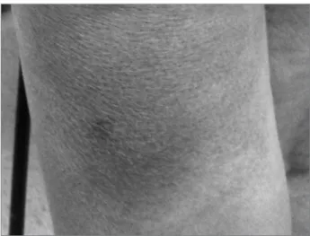
At three months’ post-transplant, she reported dark spots on the left thigh, without fever or other symptoms. She had erythematous-violet plaques of infiltrative aspect, slightly painful, of 3 to 4 cm in diameter, on the described location (Figure 1). Biopsy of the lesions showed dermis and hypodermis with multiple histiocytes of large volume cytoplasm, phagocytosing microorganisms morphologically consistent with T. cruzi amastigotes (Figures 2, 3 and 4).
She was started on benznidazole (7 mg/kg/ day, divided into two daily doses), with treatment planned for 60 days. Her skin lesions improved progressively.
Figure 1. erythematous-violet iniltrative plaque on her left leg.
Figure 2. intracellular amastigote forms (hematoxylin & eosin).
Figure 3. Details of intracellular amastigotes (hematoxylin & eosin).
vancomycin used for treatment. We removed the peritoneal dialysis catheter. There was complete resolution of the peritonitis. Simultaneously, she had pancytopenia (= 2.9 million red blood cells/ mm3, WBC = 2150/mm3, platelets = 8,000/mm3)
Figure 4. Details of intracellular amastigotes (hematoxylin & eosin).
and positive antigenemia for cytomegalovirus (7/50,000 cells). Benznidazole and mycophenolate mofetil were suspended, and she was started on ganciclovir. There was clinical improvement and gradual recovery of erythrocytes, leukocytes and platelets. Mycophenolate mofetil was progressively reintroduced. She no longer received benznidazole.
Tacrolimus serum levels were between 3.4 and 7.5 ng/dl, and 4.4 ng/dl at the time of the skin lesions.
She remained very well clinically, without any other skin lesion. She had progressive improvement in graft function with plasma creatinine of 1.3 mg/ dl at six months post-transplant. In her last visit at 14 months’ post-transplantation, she had no complaints and no significant changes on physical examination (BP = 128/80; normal heart and lung auscultation; a mild lower-limbs edema). She was on tacrolimus, mycophenolate mofetil, prednisone, furosemide, clonidine, amlodipine and enalapril. On that date, she had hemoglobin = 12 g/dL, hematocrit = 38%, WBC = 9100/mm3; platelets
= 217,000/mm3, serum creatinine = 1.6 mg/dl,
normal qualitative urinalysis and blood tacrolimus = 6.1 ng/dl.
D
ISCUSSIONAsymptomatic reactivation of Chagas disease may occur in patients using immunosuppressants. Most cases are related to kidney or heart transplant, especially in the first year post-transplant.3-5 There may
be fever, malaise, myocarditis, meningoencephalitis and skin lesions, and it can also mimic allograft rejection. Hardened erythematous plaques, areas of necrosis, nodules, panniculitis and ulcers are among the possible skin lesions.3-7 The patient described here
had panniculitis in her left leg.
Serological tests have low sensitivity in immunocompromised patients. Parasitemia confirmation can be obtained by direct methods such as Strout, or indirect as xenodiagnosis or blood cultures. However, the sensitivity of these methods has also been lower than desired.8 Polymerase chain
reaction techniques have shown greater sensitivity than traditional methods, even in immunocompromised patients.1,9 With signs and symptoms of reactivation,
the finding of trypomastigotes parasites in the peripheral blood or cerebrospinal fluid confirms the diagnosis,3 just like amastigotes in compromised
tissue biopsies.1,5,6
Treatment can be done with nifurtimox (8 mg/kg/ day) or benznidazole (5-7 mg/kg/day) for 60 days. For its lower toxicity, benznidazole is suggested as first choice treatment. Treatment should be discontinued when there is leucopenia (WBC < 2500/mm³) and neutropenia (<500/mm³). Transaminase increase (AST and ALT > 3 times the upper limit of normal) and severe leukopenia (leukocytes < 1000/mm³) led to intensive monitoring.7
Although there are immunosuppressed patients with clinical improvement with treatment, it is known that Chagas disease brings high morbidity and mortality in such conditions.4
Some authors suggest that similarly to diseases such as leprosy, tuberculosis and AIDS, whose only control was obtained with the combination of drugs, it would be interesting to study the effectiveness of two or more concurrent medications for the treatment of tripanossomyasis.10,11
The antiTrypanosoma effect of allopurinol alone or in combination with other medications has been suggested.6,10 However, there is no conclusive evidence
or definitive recommendation in regards of its use as treatment or prophylaxis. An attractive point is their much lower toxicity profile when compared to
benznidazole and nifurtimox. The described patient received allopurinol intended for prophylaxis at a dose of 100 mg per day, for she was still dependent on dialysis and then in the early stages of recovery from graft function with plasma creatinine around 3 mg/dl.
Panniculitis occurred despite the use of allopurinol in the aforementioned dose. There was complete resolution of skin lesions with benznidazole, but pancytopenia, with combined viral and bacterial infections, caused the suspension of the drug after 30 days. Still, she remained without clinical signs of Chagas disease reactivation during the subsequent 11 months.
Her death resulted from a dissecting aortic aneurysm. Experimental studies show that Trypanosoma cruzi infection can cause vasculitis with aorta involvement.12,13 However, human studies
have found no alterations in aortic distensibility14 and
endothelial function15 in patients with Chagas disease.
In our case, autopsy was not performed, thus being impossible to know whether there was a relationship between the aortic changes and the Trypanosoma cruzi.
Chagas disease must be considered in various clinical situations in immunosuppressed patients, such as renal transplant, especially in endemic areas.
The drugs used can lead to severe-effects, which is complicated by the need for simultaneous use of other drugs, such as immunosuppressants.
In transplant patients, disease treatment or its prophylaxis in cases with positive serology only, still await more effective and better tolerated options.
R
EFERENCES1. Rassi A Jr, Rassi A, Marin-Neto JA. Chagas disease. Lancet 2010;375:1388-402. PMID: 20399979 DOI:http://dx.doi. org/10.1016/S0140-6736(10)60061-X
2. Brasil. Ministério da Saúde. Doenças infecciosas e parasitárias: guia de bolso. Ministério da Saúde, Secretaria de Vigilância em Saúde, Departamento de Vigilância Epidemiológica. Brasília; 2010.
3. Bern C. Chagas disease in the immunosuppressed host. Curr Opin Infect Dis 2012;25:450-7. DOI:http://dx.doi.org/10.1097/ QCO.0b013e328354f179
4. Silva AE, Silva AC, Faleiros AC, Guimarães CS, Corrêa RR, Oli-veira FA, et al. Acute Chagas' disease in postrenal transplant and treatment with benzonidazole. Ann Diagn Pathol 2010;14:199-203. DOI:http://dx.doi.org/10.1016/j.anndiagpath.2009.06.008 5. Gallerano V, Consigli J, Pereyra S, Gómez Zanni S, Danielo C,
Gal-lerano RH, et al. Chagas' disease reactivation with skin symptoms in a patient with kidney transplant. Int J Dermatol 2007;46:607-10. DOI: http://dx.doi.org/2007;46:607-10.1111/j.1365-4632.2007.03127.x 6. Kocher C, Segerer S, Schleich A, Caduff R, Wyler LG, Müller
7. Bacal F, Silva CP, Pires PV, Mangini S, Fiorelli AI, Stolf NG, et al. Transplantation for Chagas' disease: an overview of immu-nosuppression and reactivation in the last two decades. Clin Transplant 2010;24:E29-34. DOI:http://dx.doi.org/10.1111/ j.1399-0012.2009.01202.x
8. Riarte A, Luna C, Sabatiello R, Sinagra A, Schiavelli R, De Rissio A, et al. Chagas' disease in patients with kidney transplants: 7 years of experience 1989-1996. Clin Infect Dis 1999;29:561-7. PMID: 10530448 DOI:http://dx.doi. org/10.1086/598634
9. Diez M, Favaloro L, Bertolotti A, Burgos JM, Vigliano C, Las-tra MP, et al. Usefulness of PCR sLas-trategies for early diagnosis of Chagas' disease reactivation and treatment follow-up in heart transplantation. Am J Transplant 2007;7:1633-40. DOI: http:// dx.doi.org/10.1111/j.1600-6143.2007.01820.x
10. Perez-Mazliah DE, Alvarez MG, Cooley G, Lococo BE, Ber-tocchi G, Petti M, et al. Sequential combined treatment with allopurinol and benznidazole in the chronic phase of Trypa-nosoma cruzi infection: a pilot study. J Antimicrob Chemo-ther 2013;68:424-37. DOI: http://dx.doi.org/10.1093/jac/ dks390
11. Coura JR. Present situation and new strategies for Chagas disease chemotherapy: a proposal. Mem Inst Oswaldo Cruz 2009;104:549-54. PMID: 19722074 DOI: http://dx.doi. org/10.1590/S0074-02762009000400002
12. Rossi MA. Aortic endothelial cell changes in the acute septi-cemic phase of experimental Trypanosoma cruzi infection in rats: scanning and transmission electron microscopic study. Am J Trop Med Hyg 1997;57:321-7. PMID:9311644
13. Sunnemark D, Frostegård J, Orn A, Harris RA. Cellular and cytokine characterization of vascular inflammation in CBA/J mice chronically infected with Trypanosoma cruzi. Scand J Im-munol 1998;48:480-4. PMID: 9822255
14. Villacorta H, Bortolotto LA, Arteaga E, Mady C. Aortic distensi-bility measured by pulse-wave velocity is not modified in patients with Chagas' disease. J Negat Results Biomed 2006;5:9. PMID: 16768804 DOI:http://dx.doi.org/10.1186/1477-5751-5-9
15. Consolim-Colombo FM, Lopes HF, Rosetto EA, Rubira