330
BILATERAL TESTICULAR NEOPLASIA Case Report
International Braz J Urol
Official Journal of the Brazilian Society of Urology
Vol. 29 (4): 330-331, July - August, 2003
BILATERAL AND SYNCHRONIC SEMINOMATOUS TESTICULAR
NEOPLASIA
ADEMAR SCHMITZ
Maicé Hospital, Caçador, Santa Catarina, Brazil
ABSTRACT
Testicular neoplasia is rare, especially when it is bilateral, and even more when it is synchronic, with its incidence being only 0.17% of germinative tumors of testicles.
We present here the case of a male, 32-year old patient, without children. Patient underwent a bilateral radical orchiectomy, following previous sperm harvest, in a sperm bank. Surgery was per-formed in 2 stages, with a 12-day interval, with implantation of a silicone testicular prosthesis. The result of anatomicopathological examination revealed bilateral classical seminoma, pT2 on the right side andT1 on the left. He was submitted to bilateral complementary radiotherapy, with 2,500 cGy on each side.
Patient had a good outcome from a medical and oncologic perspective, but a follow-up with psychotherapy was needed.
Key words: testis; testicular neoplasms; seminoma; synchronous neoplasms Int Braz J Urol. 2003; 29: 330-1
INTRODUCTION
The incidence of germinative testicular tu-mors in the general population is 0.005%, and ac-counts for 1% of all tumors that affect men. The risk of a patient having a successive (metachronic) bilat-eral tumor ranges between 1 and 5% (1). As for synchronic tumors, they are even more rare. Recently a report was described on 2,431 germinative testicu-lar tumors diagnosed and treated from 1978 to 1999, and only 24 of these cases were bilateral, and among them 20 were metachronic, that is, 1% of all tumors. Synchronic tumors were described in only 4 cases (0.17%) (2). The histological type is usually the same in both testicles, however in metachronic ones, his-tological types can be different.
Synchronous testicular tumors require a bilat-eral surgical therapy, leading to the patient’s sterility.
CASE REPORT
Male, 32-year old patient, married with no children. He had 5-month history of bilateral,
hard-ened, slightly painful, testicular mass, involving prac-tically the entire glands. Ultra-sonography revealed a right testicle measuring 7.8 x 8.4 x 4.2 cm and left testicle measuring 5.4 x 4.0 x 2.7 cm. The main hy-pothesis was bilateral neoplasia, without excluding a granulomatous process. Tumoral markers were nor-mal. Thorax radiography and abdominal computer-ized tomography did not reveal metastases. The mag-netic resonance imaging of scrotum suggested bilat-eral testicular neoplasia.
331
BILATERAL TESTICULAR NEOPLASIA
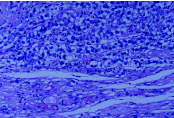
cord (Figure-2). The anatomicopathological diagnosis was seminoma as well (I-A stage or pT1N0M0), with focal invasion of the tunica albuginea, without trans-posing the capsule. The lymph node was negative for neoplasia.
As a complementary treatment, the patient was submitted to a conformational radiotherapy, with 2.500 cGy at each side (bilateral iliac and para-aortic chains), 60 days after surgery. He presented a good outcome, and is receiving hormone replacement.
DISCUSSION
Bilateral germinative testicular tumors are
rare and, when diagnosed synchronically, they are
even rarer. Seminoma is the most common
his-tological type. Current indication for treating such
tumors begins with the clinical staging, with
or
chiectomy being the first step (3). It is hard to evalu-ate whether a conservative surgery, in bilevalu-ateral cases,followed by chemotherapy and/or radiotherapy, could provide good results without bilateral testicular abla-tion, due mainly to the reduced number of cases of studied synchronic tumors. In this particular case, such procedure could not have been possible, since both testicles were involved by the neoplasia, in more than 90% of the normal tissue, with only a small band of tubules with normal aspect (Figure-2). In this case there was a great resistance from patient and his wife, in accepting the bilateral surgical treatment, with a freeze biopsy only, choosing thus to perform a sur-gery in 2 times, even though we had the clinical diag-nosis of neoplasia pre-operatively. Complementary treatment was radiotherapy, due to staging and bilat-erality.
Figure-1 was supplied by Drs. Maria CN Zerbini and Claudia RGCM Oliveira, from Fleury Laboatory, SP
REFERENCES
1. Yokomizo S, Tsujimura A, Nonomura N, Kirime S, Takahara S, Okuyama A: Metachronous bilateral tes-ticular tumors in a child. J Urol. 2001; 166: 2341. 2. Che M, Tamboli P, Ro JY, Park DS, Ro JS, Amato RJ,
et al.: Bilateral testicular germ cell tumors: twenty-year experience at M.D. Anderson Cancer Center. Cancer 2002; 95: 1288-33.
3. Richie JP: Neoplasms of the Testis. In: Walsh PC, Retik AB, Vaughan ED Jr, Wein AJ (eds.), Campbell’s Urol-ogy, 7th ed. New York, WB Saunders. l998; pp. 2411-52.
Received: February 28, 2003 Accepted after revision: May 9, 2003
Correspondence address: Dr. Ademar Schmitz
Rua Herculano C. de Souza, 727 / 202 Caçador, SC, 89500-000, Brazil Fax: + 55 49 563-0678
E-mail: [email protected] Figure 2 – Surgical specimen of left side. Note the minimal
thick-ness of normal tissue in the testicle periphery.