Complications Related to Carotid Sinus Massage in 502
Ambulatory Patients
Gustavo de Castro Lacerda
1,2, Roberto Coury Pedrosa
2, Renato Côrtes de Lacerda
1, Marcela Cedenilla dos
Santos
1, Alfredo Teixeira Brasil
1, Aristarco Gonçalves de Siqueira-Filho
2Hospital Geral de Bonsucesso - Ministério da Saúde1, Universidade Federal do Rio de Janeiro2, Rio de Janeiro, RJ - Brazil
Summary
Background: The carotid sinus massage (CSM) is a simple and low-cost technique with many indications.
Objective: To determine the safety of CSM in outpatients with high prevalence of atherosclerotic disease and cardiopathy.
Methods: A transversal study. Inclusion criteria: Outpatients aged ≥50 years, referred for ECG. Exclusion criteria: Individuals that refused to participate in a study on the prevalence of the cardioinhibitory response to CSM, patients with dementia, patients with pacemakers, individuals with carotid murmur or history of stroke or AMI in the last three months. The CSM was carried out in the supine position during 10 seconds. The occurrence of sustained arrhythmias or the occurence of neurological deficit during the CSM or in the first 24 hours after its end were considered complications of the CSM.
Results: 562 patients were randomly selected from a total of 1,686 individuals that met the inclusion criteria. Sixty individuals met the exclusion criteria. The remaining 502 patients (52% males, 69% with cardiopathies and 50% with atherosclerotic disease) were submitted to 1,053 CSM. Two patients presented complications (0.4%; 95%CI:0%-0.9%). A 71-yr-old man developed left arm monoparesis with complete regression within 30 minutes. Another 56-yr-old man presented left homonymous hemianopsia, with regression after 7 days.
Conclusion: The incidence of CSM-related complications was small, particularly when considering that the population submitted to the maneuver was elderly, with high incidence of structural heart disease and atherosclerotic disease. (Arq Bras Cardiol 2009;92(2):78-83)
Key words: Carotid sinus; syncope; arrhythmias, cardiac/complications.
Mailing address: Gustavo de Castro Lacerda •
R. Hadock Lobo, 369/Sl. 308, Estácio, 20260-131, Rio de Janeiro, RJ - Brazil E-mail: [email protected]
Manuscript received March 08, 2008; revised manuscript received April 22, 2008; accepted April 28, 2008.
years, the CSM has been scarcely employed to revert PSVT and in the diagnosis of CSH4. There seems to be an irrational fear regarding its risks, which decreases the acquaintance with its use and contributes to its relinquishment.
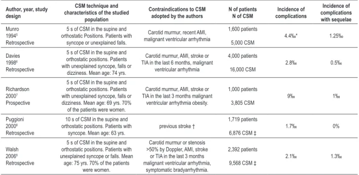
Case reports seems to be responsible for a large part of the excessive and unjustified fear that involves the practice of CSM. There is no scientific basis for the allegation that CSM should not be used due to the risks it involves. In five large case series (Table 1), no cardiologic complication related to the procedure were observed5-9. In these series, the incidence of cerebrovascular complications of the CSM was also small, varying from 1.7‰ to 9‰5-9. The present study is the first one carried out of the European continent with the objective of evaluating the incidence of CSM-related complications in
outpatients aged ≥50 years and with a high prevalence of
structural cardiopathy and atherosclerotic disease.
Methods
Design
Transversal study carried out at the Outpatient Clinic of
Hospital Geral de Bonsucesso (HGB), a tertiary public hospital. The studied population was recruited during a study that
Introduction
aimed at evaluating the prevalence and the predictors of the cardioinhibitory response to CSM10. This study was approved by the Ethics Committee in Research of the institution. The study sample consisted of 562 randomly selected outpatients
aged ≥50 years, referred for electrocardiogram (ECG)
recording before a routine medical visit and patients that had been referred for ECG at the request of physicians from other specialties, either clinical or surgical.
The patients excluded from the study were those that did not sign the free and informed consent form, obese individuals, those with tracheotomies or neck wounds, patients with dementia, individuals with symptomatic bradyarrhythmia, those with pacemakers and patients with a history of complex ventricular arrhythmia. Patients with carotid murmur or carotid
stenosis ≥50%, documented by previous carotid Doppler,
patients with a history of acute myocardial infarction (AMI) in the previous 3 months and patients with a history of cerebrovascular accident (CVA) or transient ischemic attack (TIA) in the previous three months were also excluded.
The CSM was carried out in an environment with cardiac defibrillator, transcutaneous pacemaker and all the material necessary to perform a cardiopulmonary resuscitation. The first author of the present study performed all CSM in all cases.
The CSM was initially performed only in the supine position, firstly on the right side. Pressure was exercised with longitudinal movements at the point where the maximum impulse of the carotid pulse is palpated, immediately above the thyroid cartilage and below the angle of the mandible, for a period of 10 seconds. The procedure was repeated after one minute on the left side, in the cases with no immediate complications.
After the CSM, all individuals were re-evaluated and questioned regarding the presence of symptoms. Before they
were released, the patients were instructed to seek the first author of the study at the Heart Service of HGB either in person or by telephone, in case of doubts or complications related to the study or the CSM. The individuals with CSM-related complications were identified and treated at the Cardiology Service of HGB.
The CSM complications were defined before the start of the study as the occurrence of sustained ventricular or supraventricular arrhythmias (those lasting more than 30 seconds or the ones that needed medical intervention for control), the occurrence of prolonged asystole that required resuscitation maneuvers and the onset of neurological deficit (stroke or TIA) during the CSM or within the first 24 hours after its end.
Results
Description of the population
During the period of recruitment, 1,686 outpatients aged
≥50 years recorded an ECG at the HGB, of which 562 (33.3%)
patients were randomly selected. Sixty patients were excluded from the study (Figure 1).
The characteristics of the 502 patients submitted to the CSM are summarized in Table 2. The cardioinhibitory response
to the CSM was present in 52 patients (prevalence: 10.4%; 95%CI: 7.7%-13%). The characteristics of these 52 patients
and the predictors of the cardioinhibitory response can be found in an article published in this same journal10.
The mortality and the CSM-related cardiologic complications
were 0%. Two patients presented neurological deficit (0.4%; 95%CI: 0%-0.9%).
Table 1 - Studies performed with the objective of evaluating the safety of CSM
Author, year, study design
CSM technique and characteristics of the studied
population
Contraindications to CSM adopted by the authors
N of patients N of CSM
Incidence of complications Incidence of complications with sequelae Munro 19945 Retrospective
5 s of CSM in the supine and orthostatic Positions. Patients with
syncope or unexplained falls.
Carotid murmur, recent AMI, malignant ventricular arrhythmia
1,600 patients 5,000 CSM 4.4‰* 1.25‰ Davies 19986 Retrospective
5 s of CSM in the supine and orthostatic positions. Patients with unexplained syncope, falls or
dizziness. Mean age: 74 yrs.
Carotid murmur, AMI, stroke or TIA in the last 6 months, malignant
ventricular arrhythmia 4,000 patients 16,000 CSM 2.8‰ 0.5‰ Richardson 20007 Prospective
5 s of CSM in the supine and orthostatic positions. Patients with unexplained syncope, falls or dizziness. Mean age: 69 yrs. 70%
of the patients were women.
Carotid murmur, AMI, stroke or TIA in the last 3 months malignant
ventricular arrhythmia obesity.
1,000 patients 3,805 CSM 9‰ 1‰ Puggioni 20008 Retrospective
10 s of CSM in the supine and orthostatic positions. Patients with
syncope. Mean age: 63 yrs.
previous stroke †
1,719 patients
6,876 CSM ‡
1.7‰ 0%
Walsh 20069
Retrospective
5 s of CSM in the supine and orthostatic positions. Patients with unexplained syncope or falls. Mean age: 75 yrs. 70% of the patients
were women.
Carotid murmur or stenosis >50% by Doppler, AMI, stroke
or TIA in the last 3 months malignant ventricular arrhythmia,
symptomatic bradyarrhythmia.
2,392 patients
9,568 CSM ‡
2.1‰ 1.3‰
A 71-year-old hypertensive and dyslipidemic man, with a past history of surgical myocardial revascularization in December 2005 and a carotid/vertebral duplex scan performed at that time with no significant lesions, presented asystole of 4,380 ms during a right CSM. The maneuver was interrupted 9 seconds after its start, when the patient referred “sluggishness and numbness” all over the body. Less than one minute after the end of the maneuver, the presence of dysarthria with slight decrease in the strength of the left Table 2 - Characteristics of the 502 patients submitted to CSM
Mean age ± SD / range 65 ± 9.6 / 50 to 93 years
Age ≥ 65 yrs 254 (50.6%)
Male sex 259 (51.6%)
Healthy patients 35 (6.9%)
Arterial Hypertension 380 (75.7%)
Diabetes 117 (23.3%)
Dyslipidemia 270 (53.8%)
Smoking 58 (11.6%)
Known coronary disease 238 (47.4%)
History of myocardium infarction 166 (33.1%)
Previous myocardial revascularization 113 (22.5%)
Atrial ibrillation 24 (4.8%)
Structural heart disease 347 (69.1%)
Coronary, cerebrovascular or vascular
peripheral atherosclerosis 253 (50.4%)
Known cerebrovascular disease 13 (2.5%)
Normal ECG 133 (26.5%)
First degree heart block or bundle branch block 66 (13.1%)
ECG with ibrosis or with ischemia 110 (21.9%)
Use of cardiovascular medication 428 (85.3%)
Use of negative chronotropic medication 303 (60.4%)
History of syncope in the previous year 41 (8.2%)
History of syncope or fall in the previous year 71 (14.1%)
Cardioinhibitory response to CSM 52 (10.4%)
upper limb was observed. The patient was medicated with acetylsalicylic acid (ASA), the speech disorder reverted in less than 5 minutes and the monoparesis disappeared in 30 minutes. A new carotid duplex scan showed the presence of diffuse parietal irregularities. Thirty days after the CSM the patients remained asymptomatic, with no neurological sequelae (Figure 2).
Another male patient, a 56-year-old hypertensive, dyslipidemic individual, with a past history of AMI and a Doppler echocardiogram showing severe ventricular dysfunction, complained of a feeling of drunkenness that started 30 minutes after the end of the CSM. The neurological examination revealed the presence of left homonimous hemianopsia, with no motor, sensitivity or balance alteration. He was medicated with ASA and admitted at the Infirmary of Cardiology of HGB.
The computed tomography (CT) of the head confirmed the presence of an ischemic cerebrovascular accident (small infarction in the right occipital region). Seven days after the ictus, the patient did not present any visual field deficit. The duplex scan of the carotids showed the presence of mixed
plaque with 50% obstruction of the right internal carotid
and the Doppler echocardiogram confirmed the presence of severe left ventricular dysfunction. A coronary angiography was ordered. The procedure was carried out 14 days after the CSM, disclosing a moderate stenotic lesion in the circumflex artery. There was no atherosclerotic disease in the anterior descending artery or in the right coronary artery. Immediately after the catheterism, the presence of monoplegia was observed in the left upper limb. Thirty days after the coronary angiography, the patient recovered normal strength in the proximal region of the arm, but remained with left hand monoparesis (Figure 3).
Discussion
The complications of the CSM are well known1-3. It is the responsibility of the physician performing the maneuver to inform its indications and risks. Severe and life-threatening arrhythmic complications are considered extremely rare1-3. They are mostly secondary to the effects of the CSM on the sinus and atrioventricular nodes. The maneuver can cause prolonged periods of asystole, interrupted by escape beats, Figure 1 -Selection of the 502 patients submitted to the CSM.
Figure 2 -ECG of a 71-year-old man undergoing treatment with statins, ASA, diuretics and betablockers, showing normal sinus rhythm, HR of 75 bpm, irst-degree atrioventricular block (PR 220ms), anteroseptal ibrosis with inverted T waves from V2 to V6. The RCSM (lead V1) provoked asystole of 4,380 ms. The maneuver was
interrupted 9 seconds after its start, when the patients reported “sluggishness and numbness” in the entire body.
,
Figure 3 -ECG of a 56-year-old man undergoing treatment with captopril, diuretics, nitrates, ASA, statins and betablockers, showing sinus rhythm, HR of 51 bpm and left bundle branch block. The RCSM (lead V1) provoked sinus interruption, 2:1 atrioventricular The RCSM (lead V1) provoked sinus interruption, 2:1 atrioventricular block with 1 ventricular escape beat and asystole of 3,420 ms associated with pre-syncope. Thirty minutes after the end of the CSM, the presence of left homonimous
hemianopsia was detected.
,
which rarely degenerate into more severe arrhythmias. Many case series have been published on the safety of the CSM5-9 and more than 10,000 patients were submitted to the CSM in these studies. None of these series reported episodes of malignant ventricular arrhythmia or prolonged asystole that needed medical intervention5-9.However, isolated cases of patients that developed ventricular fibrillation have been described11. These cases led to the recommendations of having a cardiac defibrillator available in the places where the procedure is performed2,3.
The cerebrovascular complications of the CSM are much feared. The first case was reported by Weiss and Baker in the 30s12. In the 60s, Lown and Levine1 presented their experience with the technique and reported that its complications were extremely rare1. These authors reported having performed the maneuver in thousands of patients and described only one complication: 1 episode of facial paralysis with no sequelae1.
Other isolated cases of patients with neurological deficit caused by the CSM have been described13. Many publications do not have any record of the technique employed to stimulate the carotid sinus. It is possible that, in certain occasions, the duration of the massage was overly long, and that, in some situations, the procedure was performed in patients that, nowadays, would have been considered as presenting contraindications to the maneuver.
In the present study, the mortality and the cardiologic
complications related to the maneuver were 0%. Two patients
presented neurological deficit. This incidence of complications (4‰) did not significantly differ from the one described in 5 large case series5,9 (Table 1). In these studies, 10,711 patients were submitted to approximately 42,000 CSM and only 35 presented complications (3.2 neurological complications per 1,000 patients)5-9. The incidence of complications with sequelae was even lower (0.74‰)5-9.
Richardson et al7 believe that the incidence of complications described in some of these studies might have been underestimated7. The retrospective design of 4 of them raises doubts about that5,6,8,9.This difference in study design, the distinct characteristics of the assessed populations and the differences between the techniques employed to perform the CSM might have contributed to the difference in the incidence of complications, which varied from 1.7‰ to 9‰.
When the CSM is performed in elderly, or dyslipidemic individuals or those with atherosclerotic disease, the risk of neurological complications is higher. The possibility of embolic infarction is higher in this population. Theoretically, the risk of complications should also be higher in patients with intracranial atherosclerotic disease and in those with previous lacunar infarctions. In these individuals, an episode of prolonged arterial hypotension could cause brain anoxia and neurological deficit9. It is possible that this mechanism had an effect on the genesis of the complication presented by one of our patients from HGB, in whom the duplex-scan of the carotids showed only diffuse parietal irregularities.
In the other patient, with a mixed plaque of 50% in the
right internal carotid, the cerebrovascular complication had a clear embolic origin. In this individual, the left
homonymous hemianopsia appeared 30 minutes after the end of the CSM.
The real incidence of the CSM related complications can be underestimated by the presence of neurological deficits of late onset. In the study by Davies and Kenny, 10 of 11 individuals with complications were identified immediately after the end of the CSM. However, in one case, the neurological deficit appeared only two hours later6. After the CSM, the patients from the HGB were instructed to contact the author of the present study in person or by phone in case of new symptoms. It is unlikely that patients with a late neurological deficit sought assistance at another service, as the population treated at the HGB has little access to other healthcare services.
Patients with carotid murmur or previous duplex-scan of
carotids documenting carotid stenosis ≥50% were excluded
from the present study. It is noteworthy that most of the individuals had not undergone a previous carotid duplex-scan. This was the case of the patient that developed left homonymous hemianopsia. This individual did not have carotid murmur and had never been submitted to an ultrasonographic assessment of the carotids. The routine performance of the duplex-scan of the carotids, preceding the CSM, and the exclusion of the patients with stenosis
> 50% of the lumen of the artery would have reduced in 50% the incidence of complications observed in the present
study. Indeed, most researchers don’t recommend the routine performance of the duplex scan of carotids before the CSM2,3,5-9.
Richardson et al14 demonstrated that the CSM can be considered safe even in patients with carotid lesions documented by the duplex-scan14. These authors identified 167 patients that presented recurrent or unexplained falls and carotid murmur. All of them were submitted to the ultrasonographic assessment of the carotids. Forty-six patients
(28%) were excluded from the study (34 with carotid stenosis ≥70%, 4 with a history of CVA in the previous three months
and 8 for other reasons). The remaining 121 patients were submitted to the CSM. In the 18 individuals with stenosis
≥50% and <70%, the CSM was performed exclusively in the supine position. In the patients with stenosis <50%, the
maneuver was performed in the usual manner, i.e., in the supine and orthostatic positions during 5 seconds. None of the 121 patients submitted to the CSM developed persistent neurological deficit14.
Conclusion
In the present study, the performance of the duplex-scan of carotids prior to the CSM and the exclusion of patients
with stenoses > 50% would have decreased the incidence
References
1. Lown B, Levine SA. The carotid sinus: clinical value of its stimulation. Circulation. 1961; 23: 766-89.
2. Guidelines on management (diagnosis and treatment of syncope – update 2004 ) The Task force on Syncope, European Society of Cardiology. Europace. 2004; 6 (6): 467-537.
3. Sociedade Brasileira de Cardiologia. Diretrizes para avaliação e tratamento de pacientes com arritmias cardíacas. Arq Bras Cardiol. 2002; 79 (supl 5): 1-50.
4. Disertori M, Brignole M, Menozzi C, Raviele A, Rizzon P, Santini M, et al. Management of patients with syncope referred urgently to general hospitals. Europace. 2003; 5 (3): 283-91.
5. Munro N, McIntosh S, Lawson J, Morley C, Sutton R, Kenny R. The incidence of complications after carotid sinus massage in older patients with syncope. J Am Geriatr Soc. 1994; 42: 1248-51.
6. Davies G, Kenny R. Frequency of neurological complications following carotid sinus massage. Am J Cardiol. 1998; 81: 1256-7.
7. Richardson D, Bexton R, Shaw F, Steen N, Bond J, Kenny R. Complications of carotid sinus massage – a prospective series of older patients. Age Ageing. 2000; 29: 413-7.
8. Puggioni E, Guiducci V, Brignole M, Menozzi C, Oddone D, Donateo P, et al. Results and complications of the carotid sinus massage performed according to the “method of symptoms”. Am J Cardiol. 2002; 89 (5): 599-601.
9. Walsh T, Clinch D, Costelloe A, Moore A, Sheehy T, Watts M, et al. Carotid sinus massage – how safe is it? Age Ageing. 2006; 35 (5): 518-20.
10. Lacerda G, Pedrosa R, Lacerda R, Santos M, Perez M, Teixeira A, et al. Prevalência e preditores da resposta cardioinibitória à massagem do seio carotídeo em 502 pacientes ambulatoriais. Arq Bras Cardiol. 2008; 90 (3); 163-71.
11. Deepak M, Jenkins N, Davidson N, Bennett D. Ventricular fibrillation induced by carotid sinus massage without preceding bradycardia. Europace. 2005; 7 (6): 638-40.
12. Weiss S, Baker J. The carotid sinus reflex in health and disease: its role in the causation of faitining and convulsions. Medicine. 1933; 12: 297.
13. Beal F, Park S, Fisher M. Cerebral atheromatous embolism following carotid sinus pressure. Arch Neuro. 1981; 38: 310-2.
14. Richardson D, Shaw F, Bexton R, Steen N, Kenny R. Presence of a carotid bruit in adults with unexplained or recurrent falls: implications for carotid sinus massage. Age Ageing. 2002; 31: 379-84.
Acknowledgements
We thank our colleagues from Serviço do Coração
of Hospital Geral de Bonsucesso for their incentive and collaboration during the study.
Potential Conflict of Interest
No potential conflict of interest relevant to this article was reported.
Sources of Funding
There were no external funding sources for this study.
Study Association